Full text PDF (3.9MB) - Jurnalul de Chirurgie
Full text PDF (3.9MB) - Jurnalul de Chirurgie
Full text PDF (3.9MB) - Jurnalul de Chirurgie

Create successful ePaper yourself
Turn your PDF publications into a flip-book with our unique Google optimized e-Paper software.
Anatomie şi tehnici chirurgicale <strong>Jurnalul</strong> <strong>de</strong> <strong>Chirurgie</strong>, Iaşi, 2008, Vol. 4, Nr. 2 [ISSN 1584 – 9341]<br />
ANATOMIA LAPAROSCOPICĂ A JONCŢIUNII ESO-GASTRICE<br />
E. Târcoveanu, C. Bra<strong>de</strong>a, R. Moldovanu, A. Vasilescu<br />
Clinica I <strong>Chirurgie</strong> „I. Tănăsescu-Vl. Buţureanu” Iaşi<br />
Universitatea <strong>de</strong> Medicină şi Farmacie „Gr.T. Popa” Iaşi<br />
LAPAROSCOPIC ANATOMY OF ESO-GASTRIC REGION (Abstract): Successful surgery starts in<br />
the anatomy laboratory. Unfortunately for surgeons, anatomy is not constant and operations are <strong>de</strong>signed<br />
for the most common anatomical consi<strong>de</strong>ration. Even more, in some regions the laparoscopic anatomy<br />
seems to be „different” as in open approach. During the laparoscopic surgery the anatomical landmarks<br />
are more important than in open surgery (due to the lack of palpation). We <strong>de</strong>scribe the anatomical<br />
landmarks of the diaphragm, liver and eso-cardial junction important for different operations such Nissen,<br />
Toupet, dor or Heller surgical procedures. Some data from literature are also reviewed.<br />
Corespon<strong>de</strong>nţă: Prof. Dr. E. Târcoveanu, Clinica I <strong>Chirurgie</strong>, Spitalul „Sf. Spiridon” Iaşi, Bd.<br />
In<strong>de</strong>pen<strong>de</strong>nţei Nr. 1, 700111; e-mail: etarco@iasi.mednet.ro *<br />
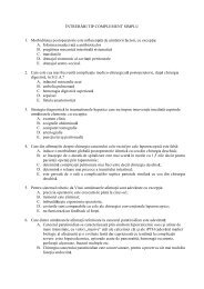
EXPLORAREA LAPAROSCOPICĂ INIŢIALĂ ŞI EXPUNEREA REGIUNII<br />
Explorarea laparoscopică iniţială a cadranului abdominal superior stâng<br />
urmăreşte spaţiul subfrenic stâng, faţa inferioară a diafragmului, faţa diafragmatică a<br />
lobului stâng hepatic, stomacul, marele epiploon şi colonul. Splina este vizibilă în<br />
recesusul lateral stâng (Fig. 1). Ligamentul rotund al ficatului este i<strong>de</strong>ntificat cu uşurinţă<br />
urmărindu-se şi ligamentul falciform care separă spaţiul subfrenic drept <strong>de</strong> cel<br />
stâng. (Fig. 2).<br />
Fig. 1 Imagine laparoscopică <strong>de</strong><br />
ansamblu a spaţiului subfrenic<br />
stâng, în care se observă faţa<br />
superioară a lobului stâng hepatic,<br />
polul superior gastric şi splina,<br />
acoperită parţial <strong>de</strong> epiplon.<br />
Pentru intervenţiile pe esofag, stomac şi regiunea hiatală, pacientul este aşezat în<br />
anti-Tren<strong>de</strong>lenburg, cu membrele inferioare în<strong>de</strong>părtate, între care se va aşeza chirurgul.<br />
Un laparoscop <strong>de</strong> 30º sau 45º oferă o lumină mai bună pe hiatus, situat înalt şi posterior,<br />
în special la obezi [1]. Vizibilitatea este mai bună în câmpul operator atunci când<br />
laparoscopul este introdus în hipocondrul stâng <strong>de</strong>cât atunci când este trecut<br />
supraombilical. Nu întot<strong>de</strong>auna este necesară secţiunea ligamentului triunghiular.<br />
* received date: 21.02.2008<br />
accepted date: 25.03.2008<br />
114