

Turkish Journal of Hematology Volume: 31 - Issue: 2

Create successful ePaper yourself
Turn your PDF publications into a flip-book with our unique Google optimized e-Paper software.
Letter to the Editor<br />
DOI: 10.4274/tjh.2013.0280<br />
An Unusual Cause <strong>of</strong> Thigh Swelling: Extramedullary<br />
Myeloid Tumor<br />
Uylukta Şişliğin Nadir Rastlanan Bir Sebebi: Ekstramedullar<br />
Myeloid Tümör<br />
Memiş Hilmi Atay1, Engin Kelkitli2, Piltan Büyükkaya3, Kubilay Ekiz4, Levent Yıldız5, Mehmet Turgut3<br />
1Van Training and Research Hospital, Department <strong>of</strong> Internal Medicine, Division <strong>of</strong> <strong>Hematology</strong>, Van, Turkey<br />
2Erzurum Training and Research Hospital, Department <strong>of</strong> Internal Medicine, Division <strong>of</strong> <strong>Hematology</strong>, Erzurum, Turkey<br />
3Ondokuz Mayıs University Faculty <strong>of</strong> Medicine, Department <strong>of</strong> Internal Medicine, Division <strong>of</strong> <strong>Hematology</strong>, Samsun, Turkey<br />
4Ondokuz Mayıs University Faculty <strong>of</strong> Medicine, Department <strong>of</strong> Internal Medicine, Samsun, Turkey<br />
5Ondokuz Mayıs University Faculty <strong>of</strong> Medicine, Department <strong>of</strong> Pathology, Samsun, Turkey<br />
To the Editor,<br />
Extramedullary myeloid tumor (EMMT) is a rare<br />
neoplasm <strong>of</strong> immature myeloid cells that arises at an<br />
extramedullary site [1]. The most common sites <strong>of</strong> EMMT<br />
are the bone, lymph nodes, skin, and s<strong>of</strong>t tissue [2]. EMMT<br />
rarely infiltrates the lower extremities.<br />
A 47-year-old male patient was admitted to the orthopedics<br />
clinic because <strong>of</strong> swelling and pain in the right thigh for 1<br />
month. His past medical history was unremarkable. Physical<br />
examination indicated a 10 cm-long solid s<strong>of</strong>t tissue lesion<br />
in the anterior and lateral parts <strong>of</strong> the right thigh. Complete<br />
blood count results were as follows: white blood cell count<br />
<strong>of</strong> 15.1x10 9 /L, hemoglobin level <strong>of</strong> 11.9 g/dL, and platelet<br />
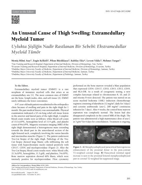
count <strong>of</strong> 94x10 9 /L. Magnetic resonance imaging (MRI) <strong>of</strong> the<br />
right thigh demonstrated a heterogeneous mass extending<br />
towards the distal part in the anterolateral section <strong>of</strong> the<br />
right femoral neck, completely involving the vastus lateralis<br />
and intermedius muscles (Figure 1). The patient underwent<br />
Tru-Cut biopsy <strong>of</strong> the right thigh. Pathology <strong>of</strong> the Tru-<br />
Cut biopsy showed large blastic cells infiltrating the s<strong>of</strong>t<br />
tissue with hyperchromatic nuclei stained positively with<br />
CD117, CD34, and myeloperoxidase (Figure 2). After the<br />
Tru-Cut biopsy, blood count results were: white blood cells,<br />
21.5x10 9 /L, hemoglobin, 11.5 g/dL, and platelets, 74x10 9 /L.<br />
Bone marrow aspiration showed 60% blasts, which were<br />
intensely positive for myeloperoxidase. Flow cytometry<br />
performed on the bone marrow revealed a blast population<br />
that expressed CD34, CD117, CD33, CD15, CD13, CD19,<br />
and HLA-DR. As a result <strong>of</strong> cytogenetic testing, a new<br />
complex karyotype related to chromosomes 8, 10, and 21<br />
and trisomy 8 were detected. The patient was started on an<br />
acute myeloid leukemia (AML) induction chemotherapy<br />
regimen consisting <strong>of</strong> idarubicin (12 mg/m2, daily for 3 days)<br />
and cytosine arabinoside (ara-C; 200 mg/m2 continuous<br />
infusion for 7 days). After 4 weeks, the control bone marrow<br />
aspiration was completely normal. The lesion had also<br />
disappeared completely in the control MRI <strong>of</strong> the thigh. The<br />
patient was administered a high-maintenance dose <strong>of</strong> ara-C<br />
at 3 g/m2 for 6 days for consolidation. Treatment is ongoing.<br />
A<br />
B<br />
Figure 1. A) Metaphysodiaphyseal paracortical heterogeneous<br />
enhancement <strong>of</strong> the proximal femur in the post-contrast<br />
T1-weighted coronal image. B) Paracortical minimal signal<br />
increase is seen in the proximal femoral metaphysodiaphyseal<br />
part in coronal T2-weighted fat-suppressed image.<br />
Address for Correspondence: Memiş Hilmi Atay, M.D.,<br />
Van Training and Research Hospital, Department <strong>of</strong> Internal Medicine, Division <strong>of</strong> <strong>Hematology</strong>, Van, Turkey<br />
Phone: +90 432 215 76 00 E-mail: dr.atay@mynet.com<br />
Received/Geliş tarihi : August 19, 2013<br />
Accepted/Kabul tarihi : November 16, 2013<br />
201


