

Turkish Journal of Hematology Volume: 31 - Issue: 2

Create successful ePaper yourself
Turn your PDF publications into a flip-book with our unique Google optimized e-Paper software.
Sepehrizadeh Z, et al: Cytokine Gene Pr<strong>of</strong>ile in AML Patients<br />
Turk J Hematol 2014;<strong>31</strong>:149-154<br />
Table 4. Patients’ chemotherapy outcome.<br />
Frequency Percentage Valid<br />
percentage<br />
Cumulative<br />
percentage<br />
Unknown 6 13 13 13<br />
Complete remission 24 52.2 52.2 65.2<br />
Expired 10 21.7 21.7 87<br />
No remission 5 10.9 10.9 97.8<br />
No treatment 1 2.2 2.2 100<br />
Total 46 100 100<br />
Clinical criteria for complete remission: bone marrow blasts <strong>of</strong> 10 g/dL, platelets <strong>of</strong><br />
>100,000, absolute neutrophil count <strong>of</strong> >1500.<br />
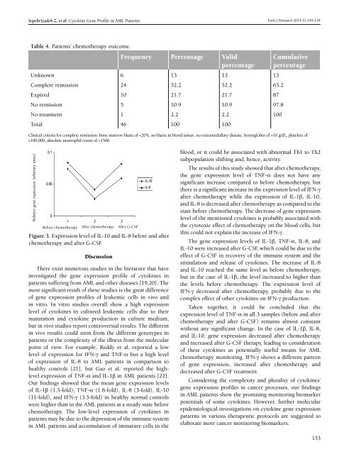
Relative gene expression (arbitrary units)<br />
Before chemotherapy After chemotherapy After G-CSF<br />
Figure 3. Expression level <strong>of</strong> IL-10 and IL-8 before and after<br />
chemotherapy and after G-CSF.<br />
Discussion<br />
There exist numerous studies in the literature that have<br />
investigated the gene expression pr<strong>of</strong>ile <strong>of</strong> cytokines in<br />
patients suffering from AML and other diseases [19,20]. The<br />
most significant result <strong>of</strong> these studies is the great difference<br />
<strong>of</strong> gene expression pr<strong>of</strong>iles <strong>of</strong> leukemic cells in vivo and<br />
in vitro. In vitro studies overall show a high expression<br />
level <strong>of</strong> cytokines in cultured leukemic cells due to their<br />
maturation and cytokine production in culture medium,<br />
but in vivo studies report controversial results. The different<br />
in vivo results could stem from the different genotypes in<br />
patients or the complexity <strong>of</strong> the illness from the molecular<br />
point <strong>of</strong> view. For example, Reddy et al. reported a low<br />
level <strong>of</strong> expression for IFN-γ and TNF-α but a high level<br />
<strong>of</strong> expression <strong>of</strong> IL-8 in AML patients in comparison to<br />
healthy controls [21], but Gao et al. reported the highlevel<br />
expression <strong>of</strong> TNF-α and IL-1β in AML patients [22].<br />
Our findings showed that the mean gene expression levels<br />
<strong>of</strong> IL-1β (1.5-fold), TNF-α (1.8-fold), IL-8 (3-fold), IL-10<br />
(11-fold), and IFN-γ (3.5-fold) in healthy normal controls<br />
were higher than in the AML patients at a steady state before<br />
chemotherapy. The low-level expression <strong>of</strong> cytokines in<br />
patients may be due to the depression <strong>of</strong> the immune system<br />
in AML patients and accumulation <strong>of</strong> immature cells in the<br />
blood, or it could be associated with abnormal Th1 to Th2<br />
subpopulation shifting and, hence, activity.<br />
The results <strong>of</strong> this study showed that after chemotherapy,<br />
the gene expression level <strong>of</strong> TNF-α does not have any<br />
significant increase compared to before chemotherapy, but<br />
there is a significant increase in the expression level <strong>of</strong> IFN-γ<br />
after chemotherapy while the expression <strong>of</strong> IL-1β, IL-10,<br />
and IL-8 is decreased after chemotherapy as compared to the<br />
state before chemotherapy. The decrease <strong>of</strong> gene expression<br />
level <strong>of</strong> the mentioned cytokines is probably associated with<br />
the cytotoxic effect <strong>of</strong> chemotherapy on the blood cells, but<br />
this could not explain the increase <strong>of</strong> IFN-γ.<br />
The gene expression levels <strong>of</strong> IL-1β, TNF-α, IL-8, and<br />
IL-10 were increased after G-CSF, which could be due to the<br />
effect <strong>of</strong> G-CSF in recovery <strong>of</strong> the immune system and the<br />
stimulation and release <strong>of</strong> cytokines. The increase <strong>of</strong> IL-8<br />
and IL-10 reached the same level as before chemotherapy,<br />
but in the case <strong>of</strong> IL-1β, the level increased to higher than<br />
the levels before chemotherapy. The expression level <strong>of</strong><br />
IFN-γ decreased after chemotherapy, probably due to the<br />
complex effect <strong>of</strong> other cytokines on IFN-γ production.<br />
Taken together, it could be concluded that the<br />
expression level <strong>of</strong> TNF-α in all 3 samples (before and after<br />
chemotherapy and after G-CSF) remains almost constant<br />
without any significant change. In the case <strong>of</strong> IL-1β, IL-8,<br />
and IL-10, gene expression decreased after chemotherapy<br />
and increased after G-CSF therapy, leading to consideration<br />
<strong>of</strong> these cytokines as potentially useful means for AML<br />
chemotherapy monitoring. IFN-γ shows a different pattern<br />
<strong>of</strong> gene expression, increased after chemotherapy and<br />
decreased after G-CSF treatment.<br />
Considering the complexity and plurality <strong>of</strong> cytokines’<br />
gene expression pr<strong>of</strong>iles in cancer processes, our findings<br />
in AML patients show the promising monitoring biomarker<br />
potentials <strong>of</strong> some cytokines. However, further molecular<br />
epidemiological investigations on cytokine gene expression<br />
patterns in various therapeutic protocols are suggested to<br />
elaborate more cancer monitoring biomarkers.<br />
153


