

Turkish Journal of Hematology Volume: 31 - Issue: 2

You also want an ePaper? Increase the reach of your titles
YUMPU automatically turns print PDFs into web optimized ePapers that Google loves.
Turk J Hematol 2014;<strong>31</strong>:128-135<br />
El-Menshawy N, et al: Clinical Significance <strong>of</strong> LYL1 Gene Expression<br />
Chambers et al. found that LYL1 is not essential for<br />
embryonic development; however, deletion <strong>of</strong> LYL1 together<br />
with its paralog, the stem-cell leukemia (SCL) gene, causes<br />
rapid apoptosis <strong>of</strong> hematopoietic progenitors in adult<br />
mice [11]. The upregulation <strong>of</strong> LYL1 has been linked to<br />
a subtype <strong>of</strong> T-ALL defined by a stem-like phenotype and<br />
an unfavorable prognosis [12]. Excess LYL1 blocked the<br />
dimerizations <strong>of</strong> E2A and thus inhibited the regulatory<br />
activity <strong>of</strong> E2A on the CD4 promoter, leading to increased<br />
proliferation and suppressed apoptosis <strong>of</strong> the progenitor<br />
cells [13]. Furthermore, overexpression <strong>of</strong> LYL1 in mouse<br />
bone marrow causes expansion <strong>of</strong> the hematopoietic<br />
progenitors and the mature T-cells. These effects were most<br />
likely due to the anti-apoptotic and proliferative roles <strong>of</strong> the<br />
LYL1 overexpression in the hematopoietic system [14].<br />
Ferrando et al. found that expression <strong>of</strong> LYL1 was increased<br />
in immature T cell precursor cells and was associated with<br />
unfavorable prognosis in T-ALL cases [15]. Zhong et al.<br />
studied the effect <strong>of</strong> LYL1 in transgenic mice and observed<br />
that a significant proportion <strong>of</strong> the mice developed B and T<br />
cell lymphoma after an average latent period <strong>of</strong> 1 year [16].<br />
However, few studies have looked for the LYL1 expression<br />
<strong>of</strong> leukemic cell lines and clinical cases <strong>of</strong> myeloid leukemia.<br />
The aim <strong>of</strong> the present study was to investigate the expression<br />
rate <strong>of</strong> the oncogene LYL1 in primary and high-risk myeloid<br />
leukemia and to assess its impact on prognosis.<br />
Materials and Methods<br />
Patients<br />
Thirty-nine patients with primary or secondary acute<br />
myeloid leukemia (AML) being followed at the medical<br />
oncology center <strong>of</strong> Mansoura University and 10 healthy<br />
individuals were included in this case-control study. Twentytwo<br />
patients were diagnosed with de novo AML (Group I), 8<br />
patients were in the accelerated and myeloid blast phases <strong>of</strong><br />
chronic myeloid leukemia (CML) (Group II), and 9 patients<br />
had AML transformed from myelodysplastic syndrome<br />
(MDS) (Group III). The control group consisted <strong>of</strong> 10<br />
healthy individuals with normal peripheral blood counts<br />
and morphology. Peripheral blood and bone marrow (BM)<br />
samples were obtained from healthy controls and patients<br />
in accordance with the protocols <strong>of</strong> the local institutional<br />
ethics committee.<br />
All patients were subjected to complete clinical<br />
examination. Diagnosis was based on complete blood count,<br />
BM examination, and immunohistochemical, morphological,<br />
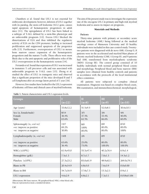
Table 1. Patient characteristics and LYL1 expression levels.<br />
Groups<br />
I<br />
(n=22)<br />
II<br />
(n=8)<br />
III<br />
(n=9)<br />
IV<br />
(n=10)<br />
Age (years) 33.8±12.2 51.5±9.5 52.0±8.1 30.6±10.1<br />
Sex (n, female/male)<br />
Female<br />
Male<br />
8/14<br />
36.4%<br />
63.6%<br />
3/5<br />
37.5%<br />
62.5%<br />
3/6<br />
33.4%<br />
66.6%<br />
4/6<br />
40.0%<br />
60.0%<br />
Splenomegaly (n, +ve/-ve)<br />
+ve: improve as positive<br />
-ve: improve as negative<br />
15/7<br />
69.2%<br />
30.8%<br />
8/0<br />
100%<br />
00.0%<br />
0/0<br />
00.0%<br />
00.0%<br />
0/10<br />
00.0%<br />
100%<br />
Lymphadenopathy (n, +ve/-ve)<br />
14/8<br />
2/6<br />
0/9<br />
0/10<br />
+ve: improve as positive<br />
-ve: improve as negative<br />
63.6%<br />
36.4%<br />
25.0%<br />
75.0%<br />
00.0%<br />
100.0%<br />
00.0%<br />
100.0%<br />
WBCs (x109/L) 61.9±45.0 55.5±47.4 40.2±76.4 6.9±1.4<br />
Hemoglobin (g/dL) 7.5±1.3 8.7±1.7 7.0±1.3 14.5±1.2<br />
Platelets (x10 9 /L) 27.5±23.2 83.0±61.9 48.9±62.1 269.0±79.1<br />
Blasts in PB 20.4±14.9 6.8±5.0 7.6±3.9 0.0±0.00<br />
Blasts in BM 54.7±16.9 17.8±7.3 15.3±2.1 0.9±1.1<br />
LYL1 expression 4.6±2.9 6.8±2.1 7.3±3.1 0.018±0.106<br />
Abbreviations; BM: bone marrow, PB: peripheral blood, WBCs: white blood cells.<br />
Data are represented as mean ± standard deviation.<br />
130


