

Turkish Journal of Hematology Volume: 31 - Issue: 2

You also want an ePaper? Increase the reach of your titles
YUMPU automatically turns print PDFs into web optimized ePapers that Google loves.
Letter to the Editor<br />
DOI: 10.4274/tjh.2013.0270<br />
Isolated Breast Relapse Mimicking Breast Cancer in<br />
Elderly Patient with Acute Lymphoblastic Leukemia<br />
Akut Lenfoblastik Lösemi Tanısı Olan İleri Yaş Hastada Meme<br />
Kanserini Taklit Eden İzole Meme Nüksü<br />
Ajay Gogia1, Prashant Mehta1, Raja Pramanik1, Rajive Kumar2<br />
1Dr. B.R.A. Institute Rotary Cancer Hospital All India Institute <strong>of</strong> Medical Sciences, Department <strong>of</strong> Medical Oncology, New Delhi, India<br />
2Dr. B.R.A. Institute Rotary Cancer Hospital All India Institute <strong>of</strong> Medical Sciences, Lab <strong>of</strong> Oncology, New Delhi, India<br />
To the Editor,<br />
Acute lymphoblastic leukemia (ALL) in adults is associated<br />
with high relapse rates. Isolated extramedullary relapse other<br />
than in the central nervous system is rare in adult females<br />
with ALL. We present a case <strong>of</strong> isolated breast relapse in a<br />
65-year-old female with ALL, mimicking breast cancer.<br />
A 65-year old female presented with a 2-month<br />
history <strong>of</strong> fever and generalized lymphadenopathy in<br />
December 2006. Physical examination revealed generalized<br />
lymphadenopathy and hepatosplenomegaly. Her hemoglobin<br />
was 11.9 g/dL, total leukocyte count 9.2x10 9 /L, and platelet<br />
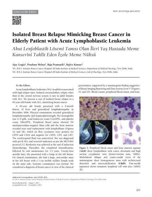
count 106x10 9 /L. Peripheral blood smear showed 5%<br />
myeloperoxidase-negative blast cells and the bone marrow<br />
revealed near total replacement with lymphoblasts (Figures<br />
1A and 1B), which on flow cytometry were positive for<br />
CD19 and CD22 and negative for CD10, CD5, and CD7.<br />
The cerebrospinal fluid was uninvolved. She was diagnosed<br />
with pre-B ALL and received induction as per the MCP-841<br />
protocol [1]. Remission was achieved at the end <strong>of</strong> induction<br />
chemotherapy. Thereafter, she completed intensification,<br />
followed by oral maintenance for 1.5 years. Twenty-four<br />
months later, she presented with a lump in the left breast.<br />
On clinical examination, she had a large, non-tender mass<br />
in her left breast with a 1-cm mobile axillary lymph node<br />
on the same side. Systemic examination was normal. We<br />
considered a diagnosis <strong>of</strong> breast cancer in view <strong>of</strong> the classical<br />
presentation, supported by a mammogram finding suggestive<br />
<strong>of</strong> Breast Imaging Reporting and Data System Score V (Figures<br />
1C and 1D). Blood counts, peripheral blood smear, and bone<br />
Figure 1. Peripheral blood smear and bone marrow aspirate<br />
(A&B) show lymphoblasts with coarse chromatin and high<br />
nuclear cytoplasmic ratio (Jenner-Giemsa stain, 1000x).<br />
Mediolateral oblique and cranio-caudal views <strong>of</strong> the<br />
mammogram show homogeneous mass with architectural<br />
distortion and microcalcification (C&D). Fine-needle<br />
aspiration shows immature lymphoid cell with duct (400X)<br />
(E).<br />
Address for Correspondence: Ajay Gogİa, M.D.,<br />
Dr. B.R.A. Institute Rotary Cancer Hospital All India Institute <strong>of</strong> Medical Sciences, Department <strong>of</strong> Medical Oncology, New Delhi, India<br />
Received/Geliş tarihi : August 7, 2013<br />
Accepted/Kabul tarihi : November 5, 2013<br />
203


