Leben nach Stammzelltransplantation
Leben nach Stammzelltransplantation
Leben nach Stammzelltransplantation

Sie wollen auch ein ePaper? Erhöhen Sie die Reichweite Ihrer Titel.
YUMPU macht aus Druck-PDFs automatisch weboptimierte ePaper, die Google liebt.
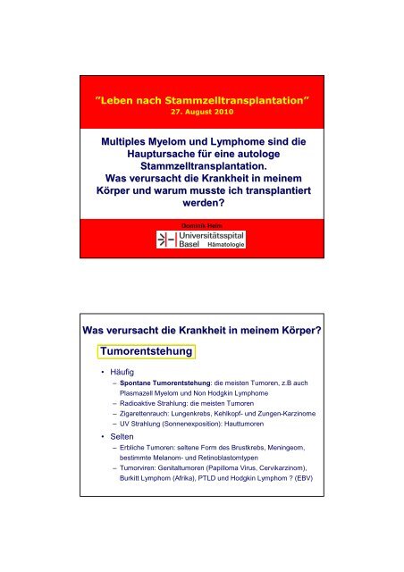
”<strong>Leben</strong> <strong>nach</strong> <strong>Stammzelltransplantation</strong>”<br />
27. August 2010<br />
Multiples Myelom und Lymphome sind die<br />
Hauptursache für f r eine autologe<br />
<strong>Stammzelltransplantation</strong>.<br />
Was verursacht die Krankheit in meinem<br />
Körper und warum musste ich transplantiert<br />
werden?<br />
Dominik Heim<br />
Hämatologie<br />
Was verursacht die Krankheit in meinem Körper? K<br />
Tumorentstehung<br />
• Häufig<br />
– Spontane Tumorentstehung: die meisten Tumoren, z.B auch<br />
Plasmazell Myelom und Non Hodgkin Lymphome<br />
– Radioaktive Strahlung: die meisten Tumoren<br />
– Zigarettenrauch: Lungenkrebs, Kehlkopf- und Zungen-Karzinome<br />
– UV Strahlung (Sonnenexposition): Hauttumoren<br />
• Selten<br />
– Erbliche Tumoren: seltene Form des Brustkrebs, Meningeom,<br />
bestimmte Melanom- und Retinoblastomtypen<br />
– Tumorviren: Genitaltumoren (Papilloma Virus, Cervikarzinom),<br />
Burkitt Lymphom (Afrika), PTLD und Hodgkin Lymphom ? (EBV)
Tumorentstehung<br />
Onkogene und Tumor Suppressor Gene<br />
Verlust der Funktion eines Tumor Suppressor Gens<br />
oder verstärkte Aktivität eines Onkogens<br />
führt zum Verlust des kontorllierten Zellwachstums<br />
Bsp. Tumor Suppressor Gen p53<br />
Auswirkungen der Aktivierung des Bcr-Abl Onkoproteins<br />
Anti-apoptosis
Was verursacht die Krankheit in meinem Körper? K<br />
Tumorentstehung<br />
Erworbene genetische Veränderungen in den Tumorzellen<br />
• t(4;14) – typischerweise Multiples Myelom<br />
• t(14;16) – typisch Multiples Myelom<br />
• t(8;14) – typischerweise bei Burkitt-Lymphom, diffus-großzelligem B-NHL,<br />
selten Multiples Myelom<br />
• t(11;14) – typisch für das Mantelzelllymphom, gelegentlich Multiples<br />
Myelom oder chronische lymphatische Leukämie<br />
• t(14;18) – typisch für das follikuläre Lymphom<br />
• Del 17p – bei CLL, Multiplem Myelom<br />
• Del 11q – bei CLL<br />
DIFFERENZIERUNG UND PROLIFERATION (EXPANSION)<br />
ERYTHROZYTEN<br />
THROMBOZYTEN<br />
SELBST-<br />
ERNEUERUNG<br />
GRANULOZYTEN<br />
MONOZYTEN<br />
STAMMZELLE<br />
T-LYMPHOZYTEN<br />
B-LYMPHOZYTEN
DIFFERENZIERUNG DER B-ZELLEN<br />
CLL<br />
D-J<br />
VDJ<br />
IgM<br />
IgM<br />
IgD<br />
IgM<br />
IgG<br />
IgG/A<br />
IgG/A<br />
pro-B pre-B “unreife<br />
”<br />
B-Zelle<br />
“reife”<br />
B-Zelle<br />
Memory<br />
B-Zelle<br />
Plasmazelle<br />
Plasmablast<br />
Lymphoblast<br />
Stammzelle<br />
Ig Gen-Rearrangement<br />
Ig class switch<br />
terminale<br />
Differenzierung<br />
Antigen unabhängig<br />
Antigen abhängig<br />
MITOSE<br />
Zweitkontakt<br />
Autologe <strong>Stammzelltransplantation</strong>
Lymphome<br />
• Mature B cell neoplasms<br />
– Chronic lymphocytic leukemia<br />
– B-cell prolymphocytic leukemia<br />
– Lymphoplasmacytic lymphoma (such as Waldenström macroglobulinemia)<br />
– Splenic marginal zone lymphoma<br />
– Plasma cell neoplasms:<br />
• Plasma cell myeloma<br />
• Plasmacytoma<br />
• Monoclonal immunoglobulin deposition diseases<br />
• Heavy chain diseases<br />
– Extranodal marginal zone B cell lymphoma, also called MALT lymphoma<br />
– Nodal marginal zone B cell lymphoma (NMZL)<br />
– Follicular lymphoma<br />
– Mantle cell lymphoma<br />
– Diffuse large B cell lymphoma<br />
– Mediastinal (thymic) large B cell lymphoma<br />
– Intravascular large B cell lymphoma<br />
– Primary effusion lymphoma<br />
– Burkitt lymphoma/leukemia<br />
• Mature T cell and natural killer (NK) cell neoplasms<br />
– T cell prolymphocytic leukemia<br />
– T cell large granular lymphocytic leukemia<br />
– Aggressive NK cell leukemia<br />
– Adult T cell leukemia/lymphoma<br />
– Extranodal NK/T cell lymphoma, nasal type<br />
– Enteropathy-type T cell lymphoma<br />
– Hepatosplenic T cell lymphoma<br />
– Blastic NK cell lymphoma<br />
– Mycosis fungoides / Sezary syndrome<br />
– Primary cutaneous CD30-positive T cell lymphoproliferative disorders<br />
• Primary cutaneous anaplastic large cell lymphoma<br />
• Lymphomatoid papulosis<br />
– Angioimmunoblastic T cell lymphoma<br />
– Peripheral T cell lymphoma, unspecified<br />
– Anaplastic large cell lymphoma<br />
WHO-Klassifikation<br />
Hodgkin lymphoma<br />
•Classical Hodgkin lymphomas:<br />
Nodular sclerosis<br />
Mixed cellularity<br />
Lymphocyte-rich<br />
Lymphocyte depleted or not depleted<br />
•Nodular lymphocyte-predominant Hodgkin lymphoma<br />
Therapie des Plasmazell Myeloms<br />
Historical perspective: 1962–2004<br />
1962 Melphalan<br />
1964 Cyclophosphamide<br />
1967 Corticosteroids<br />
1969 MP<br />
1975 Durie-Salmon staging system<br />
1982 Twin transplants for MM<br />
1984 First allogeneic transplants; VAD<br />
1985 IFN-α2<br />
1996 Bisphosphonates<br />
2000 Bortezomib; lenalidomide; other<br />
IMiDs ® ; Adriamycin ® analogues;<br />
betathine; anti-angiogenics; arsenic<br />
1976 First trials of complex<br />
chemotherapy combinations<br />
1983 High-dose melphalan; serum<br />
β 2<br />
-microglobulin for prognosis<br />
1986 High-dose Dex; HDT with ACST<br />
1996 first randomized study shows<br />
benefit of HDT-ASCT vs standard chemo<br />
1999 Thalidomide; mini-allogeneic<br />
transplants<br />
2001 New classification system<br />
2004 International Staging System
HDT with ASCT vs<br />
conventional chemotherapy<br />
Four published trials compared conventional chemotherapy (CC)<br />
with HDT in newly diagnosed Durie-Salmon stage II or III MM<br />
Study<br />
Age,<br />
years<br />
Tx<br />
n<br />
CR, %<br />
Median EFS,<br />
months<br />
Median OS,<br />
months<br />
Attal et al. 1<br />
(IFM90)<br />
< 65<br />
CC<br />
HDT<br />
100<br />
100<br />
5*<br />
22*<br />
18*<br />
28*<br />
44*<br />
57*<br />
Fermand et al. 2<br />
(MAG91)<br />
55–65<br />
CC<br />
HDT<br />
96<br />
94<br />
–<br />
–<br />
19<br />
25<br />
47.6<br />
47.8<br />
Blade et al. 3<br />
(PETHEMA)<br />
Median<br />
56<br />
CC<br />
HDT<br />
83<br />
81<br />
11*<br />
30*<br />
33<br />
42<br />
66<br />
61<br />
Child et al. 4<br />
(MRC7)<br />
< 65<br />
CC<br />
HDT<br />
200<br />
201<br />
8*<br />
44*<br />
20*<br />
32*<br />
42*<br />
54*<br />
* Significant p value.<br />
1. Attal M, et al. N Engl J Med. 1996;335:91-97. 2. Fermand J, et al. J Clin Oncol. 2005;23:9227-33.<br />
3. Blade J, et al. Blood. 2005;106:3755-59. 4. Child JA, et al. N Engl J Med. 2003;348:1875-83.<br />
Complete response rates with<br />
high-dose treatment<br />
How do the different stages of therapy contribute to remission?<br />
100<br />
Induction<br />
High-dose therapy<br />
CR rate (%)<br />
90<br />
80<br />
70<br />
60<br />
50<br />
40<br />
30<br />
ASCT<br />
Improved response rates<br />
Low treatment related mortality (1-2%)<br />
Prolonged EFS compared to standard therapy<br />
Prolonged OS (IFM and MRC trials)<br />
20<br />
10<br />
0<br />
Model of predicted response.
Novel agents as induction therapy for<br />
patients eligible for a transplant<br />
Conventional<br />
Thalidomide Bortezomib Lenalidomide<br />
VAD<br />
Dex<br />
Thal + Dex<br />
TAD<br />
CTD<br />
VTD<br />
Vel + Dex<br />
PAD<br />
VCD<br />
RVD<br />
RD<br />
Rd<br />
RAD<br />
Stem cell harvest<br />
High-dose melphalan<br />
Stem cell infusion<br />
C = cyclophosphamide; Rd = lenalidomide + low-dose dexamethasone;<br />
RD = lenalidomide + standard-dose dexamethasone.<br />
Complete response rates with<br />
high-dose treatment<br />
How do the different stages of therapy contribute to remission?<br />
CR rate (%)<br />
100<br />
90<br />
80<br />
70<br />
60<br />
50<br />
40<br />
30<br />
20<br />
10<br />
0<br />
Induction High-dose therapy Maintenance<br />
Novel<br />
agents in<br />
induction<br />
Model of predicted response.
Plasmazell Myelom<br />
Klinik - Symptome<br />
Symptome bedingt durch<br />
Paraprotein<br />
Gewebeinfiltration<br />
Knochenkrankheit<br />
Symptomatisches Plasmazell-<br />
Myelom definiert durch<br />
<br />
<br />
<br />
Paraprotein<br />
Klonale Plasmazellen<br />
CRAB<br />
C - hypercalcemia<br />
R - renal insufficiency<br />
A - anemia<br />
B - bone lesions<br />
+<br />
•hyperviscosity<br />
•amyloidosis<br />
•recurrent infections
Hyperviskosität<br />
Sehstörungen<br />
Neurologische Sympt.<br />
Blutungen<br />
Amyloidose<br />
Niereninsuffizienz<br />
Herzinsuffizienz<br />
andere Organe<br />
1. Paraprotein<br />
Niereninsuffizienz<br />
Neuropathie<br />
Gerinnungsstörung<br />
Cast Nephropathy<br />
CRAB<br />
Polyneuropathie<br />
Schmerzen<br />
Blutungen<br />
Thrombosen<br />
Solitäres<br />
Plasmozytom<br />
Kompression<br />
Schmerzen<br />
Anämie<br />
CRAB<br />
Verdrängung<br />
der Hämopoiese<br />
im Knochenmark<br />
Thrombopenie<br />
Neutropenie<br />
2. Gewebe-Infiltration<br />
Hypogammaglobulinämie
Knochenkrankheit<br />
<br />
<br />
Das MM ist der Tumor der am häufigsten das<br />
Skelett involviert<br />
– 90% der MM Patienten entwickeln<br />
Knochenläsionen<br />
– 60% der MM Patienten präsentieren sich mit<br />
Knochenschmerzen<br />
– 60% der MM Patienten entwickeln<br />
pathologische Frakturen<br />
MM Patienten verlieren rasch an<br />
Knochendichte<br />
– Patienten welche eine Steroid basierte Therapie<br />
erhalten verlieren in 1 Jahr bis zu 10% der<br />
Knochenmineralisation<br />
Osteolysen<br />
CRAB<br />
Schmerzen<br />
Pathologische<br />
Frakturen<br />
3. Knochenkrankheit<br />
Querschnitt<br />
Symptomatik<br />
CRAB<br />
Hyperkalzämie<br />
Polydipsie<br />
Polyurie<br />
Erbrechen<br />
Neurologische<br />
Symptome<br />
Koma<br />
Niereninsuffizienz