Institut für Diagnostische und Interventionelle Radiologie, JW ...
Institut für Diagnostische und Interventionelle Radiologie, JW ...
Institut für Diagnostische und Interventionelle Radiologie, JW ...

Erfolgreiche ePaper selbst erstellen
Machen Sie aus Ihren PDF Publikationen ein blätterbares Flipbook mit unserer einzigartigen Google optimierten e-Paper Software.
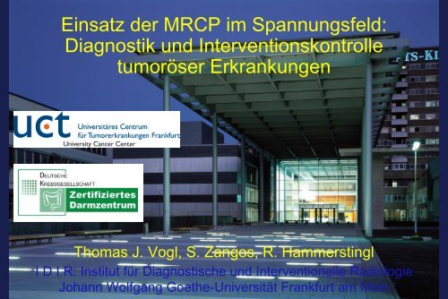
Einsatz der MRCP im Spannungsfeld:<br />
Diagnostik <strong>und</strong> Interventionskontrolle<br />
tumoröser Erkrankungen<br />
Thomas J. Vogl, S. Zangos, R. Hammerstingl<br />
I D I R: <strong>Institut</strong> für <strong>Diagnostische</strong> <strong>und</strong> <strong>Interventionelle</strong> <strong>Radiologie</strong><br />
Johann Wolfgang Goethe-Universität Frankfurt am Main
Gliederung<br />
• Klinische Fragestellung & Definition<br />
• Bildgebende Modalitäten: US<br />
CT<br />
MRT/MRCP<br />
Angiographie<br />
• <strong>Diagnostische</strong> & differentialdiagnostische Ergebnisse<br />
• <strong>Interventionelle</strong> Verfahren<br />
• Ausblick & Schlussfolgerungen<br />
<strong>Institut</strong> für <strong>Diagnostische</strong> <strong>und</strong> <strong>Interventionelle</strong> <strong>Radiologie</strong>, J.W. Goethe-Universität, Frankfurt
Ultra-Hochfeld-MRT: Leber<br />
Ultra-Hochfeld-MRT: Leber<br />
Guter Weichteilkontrast: Darstellung von kleinen Läsionen<br />
3 Tesla<br />
<strong>Institut</strong> für <strong>Diagnostische</strong> <strong>und</strong> <strong>Interventionelle</strong> <strong>Radiologie</strong>, J.W. Goethe-Universität, Frankfurt
Klassifikation maligner Tumoren der Leber<br />
Klassifikation maligner Tumoren der Leber<br />
• Primäre Malignome:<br />
Hepatozelluläres Karzinom<br />
Cholangiozelluläres Karzinom<br />
- solides Karzinom<br />
- hiläres Gallengangskarzinom<br />
der Hepatikusgabel<br />
• Sek<strong>und</strong>äre Malignome mit Kompression<br />
oder Infiltration der Gallenwege:<br />
Metastasen: CRC, Mammakarzinom<br />
<strong>Institut</strong> für <strong>Diagnostische</strong> <strong>und</strong> <strong>Interventionelle</strong> <strong>Radiologie</strong>, J.W. Goethe-Universität, Frankfurt
Cholangiokarzinome: Klassifikation<br />
Cholangiokarzinome: Klassifikation<br />
Intrahepatische Cholangiokarzinome:<br />
10%<br />
Hiläre Cholangiokarzinome: 25%<br />
Extrahepatische Cholangiokarzinome: 65%<br />
<strong>Institut</strong> für <strong>Diagnostische</strong> <strong>und</strong> <strong>Interventionelle</strong> <strong>Radiologie</strong>, J.W. Goethe-Universität, Frankfurt
Cholangiokarzinome: Klassifikation<br />
Cholangiokarzinome: Klassifikation<br />
Klassifikation anhand von<br />
Wachstumsmuster & Schittbildmorphologie<br />
Intrahepatische Cholangiokarzinome<br />
Extrahepatische Cholangiokarzinome<br />
→ Raumfordernd solide Tumore<br />
→ Periduktal wachsende Tumore<br />
→ Intraduktal wachsende Tumore<br />
Lim JH, AJR 2003; 181:819-827<br />
<strong>Institut</strong> für <strong>Diagnostische</strong> <strong>und</strong> <strong>Interventionelle</strong> <strong>Radiologie</strong>, J.W. Goethe-Universität, Frankfurt
Hiläre Gallengangstumore<br />
Hiläre Gallengangstumore<br />
• Schwierige bildgebende Diagnostik<br />
• Klatskin-Tumore:<br />
Gallengangskarzinome im Bereich<br />
der Hepatikusgabel<br />
• Auftreten als Spätkomplikation bei:<br />
Primär sklerosierender Cholangitis<br />
Caroli-Syndrom<br />
• Einteilung nach der Bismuth-Klassifikation<br />
<strong>Institut</strong> für <strong>Diagnostische</strong> <strong>und</strong> <strong>Interventionelle</strong> <strong>Radiologie</strong>, J.W. Goethe-Universität, Frankfurt
Einteilung der Klatskin-Tumoren nach Bismuth <strong>und</strong><br />
chirurgische Therapie nach den Leitlinien der<br />
Deutschen Gesellschaft für Chirurgie<br />
Tumortyp<br />
n. Bismuth<br />
I<br />
II<br />
III<br />
IV<br />
Definition<br />
Tumor betrifft den D. hepaticus<br />
communis (nicht die<br />
Hepatikusgabel)<br />
Tumor betrifft zusätzlich Hepatikusgabel,<br />
nicht die sek<strong>und</strong>äre<br />
Aufzweigung<br />
Tumor reicht einseitig bis an die<br />
Segmentabgänge; IIIa – Infiltration<br />
des rechten Hepatikusastes, IIIb –<br />
Infiltration d. linken Hepatikusastes<br />
Tumorbefall der sek<strong>und</strong>ären rechts<strong>und</strong><br />
linkshepatischen Gallenwege<br />
Therapie<br />
Resektion des extrahepatischen<br />
Gallengangs<br />
+ reg. Lymphadenektomie<br />
Resektion wie Typ I<br />
Resektion wie Typ I + Hemihepatektomie<br />
unter Mitnahme<br />
des Lobus caudatus a<br />
+ erw. Lymphadenektomie<br />
Keine kurative Resektion<br />
möglich<br />
a<br />
Ggf. zusätzlich Gefäßresektion zur Erzielung einer R0 Resektion<br />
<strong>Institut</strong> für <strong>Diagnostische</strong> <strong>und</strong> <strong>Interventionelle</strong> <strong>Radiologie</strong>, J.W. Goethe-Universität, Frankfurt
Bismuth-Klassifikation<br />
<strong>Institut</strong> für <strong>Diagnostische</strong> <strong>und</strong> <strong>Interventionelle</strong> <strong>Radiologie</strong>, J.W. Goethe-Universität, Frankfurt
Mehrschicht-CT<br />
<strong>Institut</strong> für <strong>Diagnostische</strong> <strong>und</strong> <strong>Interventionelle</strong> <strong>Radiologie</strong>, J.W. Goethe-Universität, Frankfurt
Computertomographie / Volumetrie<br />
MeVis Bremen HepaVision 2 / InterventionPlanner<br />
Leber mit Tumor V. portae V. hepatica A. hepatica<br />
3D-Segmentation 2D-Segmentation 3D-Hemihepatektomie 2D-Hemihepatektomie<br />
<strong>Institut</strong> für <strong>Diagnostische</strong> <strong>und</strong> <strong>Interventionelle</strong> <strong>Radiologie</strong>, J.W. Goethe-Universität, Frankfurt
MRCP: Technik<br />
MRCP: Technik<br />
• Turbo-Spinechoverfahren<br />
• Stehende Flüssigkeit<br />
• Hintergr<strong>und</strong>unterdrückung (orales KM)<br />
• Atemanhaltetechnik<br />
• Nicht invasiv – ambulant<br />
<strong>Institut</strong> für <strong>Diagnostische</strong> <strong>und</strong> <strong>Interventionelle</strong> <strong>Radiologie</strong>, J.W. Goethe-Universität, Frankfurt
MRCP: Technik<br />
MRCP: Technik<br />
Nativtechnik:<br />
Sequenzen<br />
Mehrschichtverfahren<br />
(3D-FSE)<br />
Projektionsverfahren<br />
(RARE-Sequenz)<br />
Mehrschichtverfahren<br />
(HASTE-Sequenz)<br />
axial <strong>und</strong> parakoronar<br />
Repetitionszeit (TR) 2000 ms 4500 ms 1040 ms<br />
Echozeit (TE) 889 ms 635 ms 114 ms<br />
Sichtfeld (FOV) 400 mm 320 mm 380 mm<br />
Schichtdicke (TH) 1,5 mm 50 mm 3 mm<br />
Schichten: Anzahl 40 1 36<br />
Pulswinkel 1,0x1,0x1,5 1,1x0,9x50 1,4x1,4x3,0<br />
Auflösung 180 180 180<br />
Matrix 384 384 320<br />
Akquisitionszeit 2,21 min 0,5 s 2-3x18-15 s<br />
Atmungsmodus Atemtriggerung: freie<br />
Atmung + Navigator<br />
Atemanhaltetechnik<br />
Atemtriggerung: Atemanhaltetechnik<br />
+ Navigator<br />
KM-verstärkte Technik: - anatomische/funktionelle Information<br />
- Mangafodipir Trisodium-enhanced MRCP<br />
<strong>Institut</strong> für <strong>Diagnostische</strong> <strong>und</strong> <strong>Interventionelle</strong> <strong>Radiologie</strong>, J.W. Goethe-Universität, Frankfurt
Funktionelle hepatobiliäre MRT<br />
Funktionelle hepatobiliäre MRT<br />
• Selektive Aufnahme von Kontrastmitteln durch die<br />
Zellsysteme selbst:<br />
Hepatozyten<br />
→ hepatobiliäre Kontrastmittel<br />
Positives Enhancement:<br />
• T1-gewichtete Kontrastmittel<br />
• Lebergewebe: hyperintens<br />
• FNH: KM-Aufnahme<br />
• Metastasen: keine KM-Aufnahme<br />
<strong>Institut</strong> für <strong>Diagnostische</strong> <strong>und</strong> <strong>Interventionelle</strong> <strong>Radiologie</strong>, J.W. Goethe-Universität, Frankfurt
Klatskin-Tumore: Kontrastverhalten - MRT<br />
Klatskin-Tumore: Kontrastverhalten - MRT<br />
• Kontrastverstärkte MRT:<br />
Abgrenzung der tumorösen Infiltration: 94%<br />
• T2-gewichtete Sequenz:<br />
Hyperintenses Signalverhalten<br />
• T1-gewichtete Sequenz nativ:<br />
Hypointenses Signalverhalten<br />
• T1-gewichtete Sequenz Gd-verstärkt:<br />
Signifikanter Anstieg der SI in der frühen Phase<br />
in infiltrierten Gallenwegsstrukturen<br />
<strong>Institut</strong> für <strong>Diagnostische</strong> <strong>und</strong> <strong>Interventionelle</strong> <strong>Radiologie</strong>, J.W. Goethe-Universität, Frankfurt
Cholangiokarzinom<br />
• Wanddicke: > 3 mm<br />
• T2-w: Hyperintens<br />
• T1-w: Hypointens<br />
• Mäßiges KM-Enhancement<br />
• Wachstum entlang der<br />
Gallenwege<br />
<strong>Institut</strong> für <strong>Diagnostische</strong> <strong>und</strong> <strong>Interventionelle</strong> <strong>Radiologie</strong>, J.W. Goethe-Universität, Frankfurt
Cholangiokarzinom<br />
<strong>Institut</strong> für <strong>Diagnostische</strong> <strong>und</strong> <strong>Interventionelle</strong> <strong>Radiologie</strong>, J.W. Goethe-Universität, Frankfurt
Polypoides CCC –<br />
distaler Ductus choledochus<br />
<strong>Institut</strong> für <strong>Diagnostische</strong> <strong>und</strong> <strong>Interventionelle</strong> <strong>Radiologie</strong>, J.W. Goethe-Universität, Frankfurt
Ampulläre Stenose<br />
Ampulläre Stenose<br />
SSFSE – coronar:<br />
• Gallengangserweiterung > 7 mm<br />
• Kinematische MRCP:<br />
- 20 single images<br />
- FOV 20x20 cm<br />
- Slice thickness 30 mm<br />
- Matrix 256x256<br />
- TE 800-1200 ms<br />
• ce MRT:<br />
- Kein Tm<br />
<strong>Institut</strong> für <strong>Diagnostische</strong> <strong>und</strong> <strong>Interventionelle</strong> <strong>Radiologie</strong>, J.W. Goethe-Universität, Frankfurt
Hiläre Gallengangskarzinome:<br />
MRC vs ERCP<br />
• Vergleichende prospektive Studie bei Patienten mit<br />
dem klinischen Verdacht auf einen Klatskin-Tumor<br />
• Durchführung der MRC <strong>und</strong> ERCP/PTC (4 W-Zeitraum)<br />
• Getrennte Auswertung der Gangsysteme durch<br />
Radiologen <strong>und</strong> Endoskopiker<br />
• Klinisches Follow-up bzw. Histopathologie zur<br />
Diagnosesicherung<br />
• Vorhandensein von tumorösen Läsionen, Stenosen, Dilatation<br />
<strong>Institut</strong> für <strong>Diagnostische</strong> <strong>und</strong> <strong>Interventionelle</strong> <strong>Radiologie</strong>, J.W. Goethe-Universität, Frankfurt
Klatskin Tumors: MRI & MRC vs ERCP<br />
Klatskin Tumors: MRI & MRC vs ERCP<br />
• Prospective Study<br />
• Patients:<br />
n = 41<br />
Type I n = 7<br />
Type II n = 4<br />
Type III n = 12<br />
Type IV n = 10<br />
• Materials & methods:<br />
MRI & MRC: n = 41<br />
ERC: n = 37<br />
PTC: n = 4<br />
Vogl T et al, European Radiology 2006; 16(10):2317-25<br />
<strong>Institut</strong> für <strong>Diagnostische</strong> <strong>und</strong> <strong>Interventionelle</strong> <strong>Radiologie</strong>, J.W. Goethe-Universität, Frankfurt
Klatskin Tumors: MRI & MRC vs ERCP<br />
Klatskin Tumors: MRI & MRC vs ERCP<br />
• Results:<br />
malignant lesions → characteristic signal intensity<br />
T1-w → hypointense<br />
T2-w → slightly hyperintense<br />
T1-ce → substantial enhancement<br />
MRC<br />
ERC/PTC<br />
sensitivity specificity diagn. accuracy<br />
94%<br />
84%<br />
100%<br />
97%<br />
95%<br />
• Conclusion:<br />
MRC combined with MRI → reliable noninvasive<br />
diagnostic method for the pretherapeutic staging of Klatskin tumors;<br />
MRC superior for completely obstructed peripheral systems<br />
Vogl T et al, European Radiology 2006; 16(10):2317-25<br />
<strong>Institut</strong> für <strong>Diagnostische</strong> <strong>und</strong> <strong>Interventionelle</strong> <strong>Radiologie</strong>, J.W. Goethe-Universität, Frankfurt
Hiläre Gallengangskarzinome:<br />
Bismuth Grad I<br />
MRC (3D-MIP coronar post)<br />
ERC<br />
<strong>Institut</strong> für <strong>Diagnostische</strong> <strong>und</strong> <strong>Interventionelle</strong> <strong>Radiologie</strong>, J.W. Goethe-Universität, Frankfurt
Cholangiokarzinom<br />
Vogl et al, Eur Rad 2006; 2317-245<br />
<strong>Institut</strong> für <strong>Diagnostische</strong> <strong>und</strong> <strong>Interventionelle</strong> <strong>Radiologie</strong>, J.W. Goethe-Universität, Frankfurt
Hiläre Gallengangskarzinome:<br />
Bismuth Grad II<br />
MRC (3D-MIP coronar post)<br />
ERC<br />
<strong>Institut</strong> für <strong>Diagnostische</strong> <strong>und</strong> <strong>Interventionelle</strong> <strong>Radiologie</strong>, J.W. Goethe-Universität, Frankfurt
Hiläre Gallengangskarzinome:<br />
Bismuth Grad III a<br />
MRC (3D-MIP coronar post)<br />
ERC<br />
<strong>Institut</strong> für <strong>Diagnostische</strong> <strong>und</strong> <strong>Interventionelle</strong> <strong>Radiologie</strong>, J.W. Goethe-Universität, Frankfurt
Hiläres Gallengangskarzinom:<br />
Bismuth Grad III<br />
<strong>Institut</strong> für <strong>Diagnostische</strong> <strong>und</strong> <strong>Interventionelle</strong> <strong>Radiologie</strong>, J.W. Goethe-Universität, Frankfurt
Cholangiokarzinom<br />
Vitellas al, Radiographics 2000; 939-957<br />
<strong>Institut</strong> für <strong>Diagnostische</strong> <strong>und</strong> <strong>Interventionelle</strong> <strong>Radiologie</strong>, J.W. Goethe-Universität, Frankfurt
Hiläre Gallengangskarzinome:<br />
Bismuth Grad IV<br />
MRC (3D-MIP coronar post)<br />
T1-GE Gd-DTPA<br />
<strong>Institut</strong> für <strong>Diagnostische</strong> <strong>und</strong> <strong>Interventionelle</strong> <strong>Radiologie</strong>, J.W. Goethe-Universität, Frankfurt
MRCP versus ERCP: Biliary Pathologies<br />
- Review of Current Literature -<br />
Group<br />
Positive<br />
predictive<br />
value [%]<br />
Negative<br />
predictive<br />
value [%]<br />
Sensitivity<br />
[%]<br />
Specificity<br />
[%]<br />
Total<br />
accuracy<br />
Stone<br />
disease<br />
(group II)<br />
100<br />
99,2<br />
88,9<br />
100<br />
99,2<br />
Tumor<br />
(group III)<br />
100<br />
100<br />
100<br />
100<br />
100<br />
Others<br />
100 100 100 100<br />
Hekimoglu K et al, J Dig Dis 2008; 9(3):162-9<br />
100<br />
<strong>Institut</strong> für <strong>Diagnostische</strong> <strong>und</strong> <strong>Interventionelle</strong> <strong>Radiologie</strong>, J.W. Goethe-Universität, Frankfurt
Pitfalls: Pneumobilie<br />
Pitfalls: Pneumobilie<br />
Irie al, Radiographics 2001; 21-37<br />
<strong>Institut</strong> für <strong>Diagnostische</strong> <strong>und</strong> <strong>Interventionelle</strong> <strong>Radiologie</strong>, J.W. Goethe-Universität, Frankfurt
Aerobilie nach ERCP<br />
Aerobilie nach ERCP<br />
<strong>Institut</strong> für <strong>Diagnostische</strong> <strong>und</strong> <strong>Interventionelle</strong> <strong>Radiologie</strong>, J.W. Goethe-Universität, Frankfurt
Pitfalls: Op-Clips<br />
Pitfalls: Op-Clips<br />
<strong>Institut</strong> für <strong>Diagnostische</strong> <strong>und</strong> <strong>Interventionelle</strong> <strong>Radiologie</strong>, J.W. Goethe-Universität, Frankfurt
Klatskin-Tumore: Differentialdiagnose<br />
Klatskin-Tumore: Differentialdiagnose<br />
• Gallenblasenkarzinom mit Befall der Hepatikusgabel<br />
• Primäre <strong>und</strong> sek<strong>und</strong>äre maligne Lebertumoren:<br />
→ Infiltration der Hepatikusgabel<br />
• Lokalisierte sklerosierende Cholangitis<br />
• Caroli-Syndrom<br />
Verbeek et al, 1992:<br />
Bei 13,4% der Patienten, die aufgr<strong>und</strong> eines V.a.<br />
Klatskin-Tumor operiert wurden, wurde eine<br />
benigne PSC entdeckt.<br />
<strong>Institut</strong> für <strong>Diagnostische</strong> <strong>und</strong> <strong>Interventionelle</strong> <strong>Radiologie</strong>, J.W. Goethe-Universität, Frankfurt
Primär sklerosierende Cholangitis (PSC)<br />
Primär sklerosierende Cholangitis (PSC)<br />
• Bei 70% der Patienten im Rahmen<br />
entzündlicher Darmerkrankungen<br />
• Früh: MRCP Sensitivität 85%<br />
• Spät: MRCP Staging: MRT >CT<br />
• Perlschnurartige Dilatation der intra-<br />
& extrahepatischen Gallenwege<br />
• Wandverdickung mit<br />
KM-Enhancement<br />
• Häufig portale Lymphadenopathie<br />
Vitellas et al, Radiographics 2000; 939-957<br />
<strong>Institut</strong> für <strong>Diagnostische</strong> <strong>und</strong> <strong>Interventionelle</strong> <strong>Radiologie</strong>, J.W. Goethe-Universität, Frankfurt
PSC: Irreguläre Gallenwege<br />
PSC: Irreguläre Gallenwege<br />
Oral-kontrastierte MRC<br />
Native MRC<br />
<strong>Institut</strong> für <strong>Diagnostische</strong> <strong>und</strong> <strong>Interventionelle</strong> <strong>Radiologie</strong>, J.W. Goethe-Universität, Frankfurt
Primär sklerosierende Cholangitis<br />
Primär sklerosierende Cholangitis<br />
<strong>Institut</strong> für <strong>Diagnostische</strong> <strong>und</strong> <strong>Interventionelle</strong> <strong>Radiologie</strong>, J.W. Goethe-Universität, Frankfurt
Caroli-Syndrom<br />
• Autosomal rezessiv vererbt<br />
• Sacculäre Dilatationen der<br />
Gallenwege<br />
• Perlschnurartig oder<br />
polyzystisch erweiterte IHGW<br />
• Nachweis von Sludge oder<br />
Konkrementen<br />
<strong>Institut</strong> für <strong>Diagnostische</strong> <strong>und</strong> <strong>Interventionelle</strong> <strong>Radiologie</strong>, J.W. Goethe-Universität, Frankfurt
Caroli-Syndrom: Zystisch irreguläre<br />
Gallenwege mit Konkrementen<br />
<strong>Institut</strong> für <strong>Diagnostische</strong> <strong>und</strong> <strong>Interventionelle</strong> <strong>Radiologie</strong>, J.W. Goethe-Universität, Frankfurt
Caroli-Syndrom<br />
Post Primovist<br />
<strong>Institut</strong> für <strong>Diagnostische</strong> <strong>und</strong> <strong>Interventionelle</strong> <strong>Radiologie</strong>, J.W. Goethe-Universität, Frankfurt
Hepatolithiasis bei Caroli-Syndrom<br />
Hepatolithiasis bei Caroli-Syndrom<br />
<strong>Institut</strong> für <strong>Diagnostische</strong> <strong>und</strong> <strong>Interventionelle</strong> <strong>Radiologie</strong>, J.W. Goethe-Universität, Frankfurt
CRC Lebermetastasen mit<br />
Gallenwegsinfiltration<br />
<strong>Institut</strong> für <strong>Diagnostische</strong> <strong>und</strong> <strong>Interventionelle</strong> <strong>Radiologie</strong>, J.W. Goethe-Universität, Frankfurt
Diagnose?<br />
KM-verstärkte CT ven.<br />
KM-verstärkte MRT ven.<br />
T2-w MRT<br />
MRCP<br />
<strong>Institut</strong> für <strong>Diagnostische</strong> <strong>und</strong> <strong>Interventionelle</strong> <strong>Radiologie</strong>, J.W. Goethe-Universität, Frankfurt
Biliäre Hamartome<br />
Biliäre Hamartome<br />
→ von-Meyenburg-Komplex<br />
→ Mikrohamartome: Darstellung als 2-15 mm<br />
große, grauweiße Herde proliferierter<br />
Gallengänge – ausgekleidet mit Zylinderepithel<br />
innerhalb eines kollagenen Bindegewebes im<br />
Rahmen einer Cholangiodysplasie<br />
→ CT: hypo- oder isointens zum umgebenden<br />
Lebergewebe<br />
→ MRT: T1-w → hypointens, T2-w → hypo-, isooder<br />
hyperintens – je nach Binnenstruktur<br />
→ KM-Enhancement: bei soliden Binnenanteilen<br />
→ DD: Leberzysten, Lebermetastasierung,<br />
kongenitale polyzystische Lebererkrankung,<br />
Caroli-Syndrom<br />
<strong>Institut</strong> für <strong>Diagnostische</strong> <strong>und</strong> <strong>Interventionelle</strong> <strong>Radiologie</strong>, J.W. Goethe-Universität, Frankfurt
Klinischer Fall<br />
Klinischer Fall<br />
Anamnese<br />
• 31-jährige adipöse Patientin<br />
• Oberbauchbeschwerden<br />
• Erhöhung der Leberwerte, AFP im Normbereich,<br />
Bilirubin im oberen Normbereich<br />
• Sono / MRT auswärts: V.a. RF im Segment 1<br />
• Weitere Abklärung <strong>und</strong> Therapieplanung<br />
<strong>Institut</strong> für <strong>Diagnostische</strong> <strong>und</strong> <strong>Interventionelle</strong> <strong>Radiologie</strong>, J.W. Goethe-Universität, Frankfurt
Zentraler Leberherd – unklare Morphologie:<br />
Biopsie ? Operation primär ?<br />
<strong>Institut</strong> für <strong>Diagnostische</strong> <strong>und</strong> <strong>Interventionelle</strong> <strong>Radiologie</strong>, J.W. Goethe-Universität, Frankfurt
Initially Unresectable Hilar Cholangiocarcinoma:<br />
Hepatic Regeneration after<br />
Transarterial Embolization<br />
• Purpose: CT assessment of hepatic regeneration after<br />
transarterial embolization of Klatskin tumors<br />
• Patients:<br />
n = 13<br />
• Materials & methods:<br />
- preoperative embolization of the right hepatic lobe<br />
- CT volumetry (entire liver, left lobe, spleen)<br />
• Results: right lobe – volume decrease: 2-33% (10% mean)<br />
left lobe – volume increase: 11-68% (37% mean)<br />
• Conclusion: right lobar embolization results in enlargement<br />
of the left hepatic lobe, thus allowing right hemihepatectomy<br />
Vogl T et al, Radiology 1998; 208(1):217-22<br />
<strong>Institut</strong> für <strong>Diagnostische</strong> <strong>und</strong> <strong>Interventionelle</strong> <strong>Radiologie</strong>, J.W. Goethe-Universität, Frankfurt
Chemoembolisation (TACE):<br />
CCC mit hilärer Infiltration<br />
<strong>Institut</strong> für <strong>Diagnostische</strong> <strong>und</strong> <strong>Interventionelle</strong> <strong>Radiologie</strong>, J.W. Goethe-Universität, Frankfurt
Hiläres Gallengangskarzinom pT2N1:<br />
Verlauf nach Chemoembolisation mit Gemzar<br />
03/06/05<br />
17/03/06<br />
<strong>Institut</strong> für <strong>Diagnostische</strong> <strong>und</strong> <strong>Interventionelle</strong> <strong>Radiologie</strong>, J.W. Goethe-Universität, Frankfurt
Pankreaskopf-Karzinom<br />
Fulcher et al, Radiographics 1999; 5-24<br />
<strong>Institut</strong> für <strong>Diagnostische</strong> <strong>und</strong> <strong>Interventionelle</strong> <strong>Radiologie</strong>, J.W. Goethe-Universität, Frankfurt
MRCP vs ERCP: Problemfälle<br />
MRCP vs ERCP: Problemfälle<br />
• Patient nicht nüchtern, Artefakte<br />
• Überschätzung von Stenosen<br />
• Erkennen von funktionellen Stenosierungen<br />
• Fehlen von endoskopisch-dynamischer Zusatzinformation<br />
• Stenosierungen ohne Dilatation<br />
• Darstellung zarter Gangsysteme<br />
<strong>Institut</strong> für <strong>Diagnostische</strong> <strong>und</strong> <strong>Interventionelle</strong> <strong>Radiologie</strong>, J.W. Goethe-Universität, Frankfurt
MRCP vs ERCP: Vorteile der MRCP<br />
MRCP vs ERCP: Vorteile der MRCP<br />
Nicht-invasive untersucherunabhängige Methode<br />
Darstellung von retrograd-endoskopisch nicht<br />
konstrastierbaren Gangabschnitten<br />
Einsatz im Follow-up<br />
Verwendung als Zusatzsequenz in der bildgebenden<br />
Diagnostik des hepatobiliären Systems mittels MRT<br />
<strong>Institut</strong> für <strong>Diagnostische</strong> <strong>und</strong> <strong>Interventionelle</strong> <strong>Radiologie</strong>, J.W. Goethe-Universität, Frankfurt
Interdisziplinäres Arbeiten<br />
Interdisziplinäres Arbeiten<br />
Chirurgie<br />
<strong>Radiologie</strong><br />
Onkologie<br />
Interne Medizin<br />
<strong>Institut</strong> für <strong>Diagnostische</strong> <strong>und</strong> <strong>Interventionelle</strong> <strong>Radiologie</strong>, J.W. Goethe-Universität, Frankfurt