Atemwegsultraschall - eine praktische Übersicht
Hier können Sie sich über das Thema umfassend informieren. Es sind zudem Filme innerhalb des Dokuments verlinkt. Es ist auch auf der Webseite des Publishers frei online verfügbar "Editor´s pick". https://authors.elsevier.com/a/1b66X7si0yR9sx
Hier können Sie sich über das Thema umfassend informieren. Es sind zudem Filme innerhalb des Dokuments verlinkt. Es ist auch auf der Webseite des Publishers frei online verfügbar "Editor´s pick".
https://authors.elsevier.com/a/1b66X7si0yR9sx

Erfolgreiche ePaper selbst erstellen
Machen Sie aus Ihren PDF Publikationen ein blätterbares Flipbook mit unserer einzigartigen Google optimierten e-Paper Software.
22<br />
R. Breitkreutz et al. / Trends in Anaesthesia and Critical Care 32 (2020) 13e32<br />
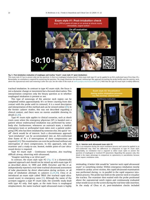
Fig. 5. Post-intubation evaluation of esophagus and trachea “tracts”, exam style #1 (post-intubation).<br />
This exam style #1 has to answer only one key question: Is there an esophageal misplacement? Only exam style type #1 can be applied in an ALS-conformed way of less than 10 s.<br />
Remarkably, no ventilation is required for answering the question. The image illustrates the dynamic stethoscope-like approach by putting the probe briefly onto the anterior neck,<br />
above the suprasternal notch and removing it after the scan. See the video of the exam at www.yumpu.com/en/SonoABCD [41]. Note that the type #2 exam style is widely different.<br />
tracheal intubation. In contrast to type #2 exam style, the focus is<br />
not a dynamic change or movement but a focused observation. This<br />
interpretation comprises only the binary question as to whether<br />
esophageal intubation is present or not.<br />
This type of sonoscopy of the anterior neck region can be<br />
completed within approximately 10 s or fewer (starting from skin<br />
contact with the probe until its removal). It is a novel description<br />
and interpretation of this method and can be viewed online [40]. In<br />
the former cadaver studies, this was not described regarding a<br />
clinical context, and there were no movies available showing its<br />
details [2,30].<br />
Type #1 exam style applies to clinical scenarios, such as shock<br />
room cases when the emergency physician (EP) is handed over a<br />
patient whose endotracheal intubation was performed by somebody<br />
else. Furthermore, whenever an outreach team, a medical<br />
emergency team or prehospital team takes over a patient undergoing<br />
CPR, who has been intubated by someone else, this quick “onoff”<br />
check would be of interest. Such a discontinuous approach<br />
“post-intubation” can be accommodated into an ALS-conformed<br />
time frame of 10 s if interruptions of chest compressions are<br />
required [40]. This examination can even be performed without any<br />
interruption of chest compressions. In this approach, only one<br />
examiner and a ready-to-use, booted, mobile point-of-care ultrasound<br />
device is required.<br />
Type #2 exam style - Continuous evaluation, also teaching<br />
difficult airway, observing with ultrasound.<br />
“Imagine watching a car drive past”.<br />
In contrast, the exam style type #2 (Fig. 6) is a diametrically<br />
opposed approach and should not be mixed up with exam type #1<br />
as described above. In 1999 and 2000, Drescher and Ma et al.<br />
independently reported a real-time tracheal ultrasound assessment<br />
of tube placements by direct observation during sections, or all<br />
steps of intubation attempts in cadavers [2,29,30]. Chou et al.<br />
introduced an exam style called TRUE (the tracheal rapid ultrasound<br />
exam) in emergency cases [3]. Although the name of the<br />
acronym suggests a type #1 exam style, the scientific study dealt<br />
with type #2 only. And again, as the main focus is esophageal<br />
misplacement, the name tracheal rapid ultrasound exam is highly<br />
Fig. 6. Anterior neck ultrasound exam style #2.<br />
This is an evaluation during the entire intubation process and cannot be applied in an<br />
ALS-conformed way. Note the differences in comparison to type #1 exam style.<br />
Remarkably, no ventilation trial is required in either exam styles to answer the<br />
question of esophageal or tracheal ETT placement. Regarding this purpose only, this<br />
detail is the major advantage in comparison to capnometry or auscultation, because<br />
these require ventilation trials.<br />
misleading. A better title would be “anterior neck rapid ultrasound<br />
exam” or something similar. Within emergency intubation mostly<br />
related to cardiac arrest victims, this rapid ultrasound examination<br />
was performed during, i.e. in parallel to the rapid sequence intubation<br />
process. The probe was held on the anterior neck to visualize<br />
the trachea. The examiner then waited with the probe positioned<br />
above the suprasternal notch region and continuously evaluated<br />
what would occur on the screen during the real time tube insertion.<br />
In the study of Chou et al., post-intubation checks included