Atemwegsultraschall - eine praktische Übersicht
Hier können Sie sich über das Thema umfassend informieren. Es sind zudem Filme innerhalb des Dokuments verlinkt. Es ist auch auf der Webseite des Publishers frei online verfügbar "Editor´s pick". https://authors.elsevier.com/a/1b66X7si0yR9sx
Hier können Sie sich über das Thema umfassend informieren. Es sind zudem Filme innerhalb des Dokuments verlinkt. Es ist auch auf der Webseite des Publishers frei online verfügbar "Editor´s pick".
https://authors.elsevier.com/a/1b66X7si0yR9sx

Sie wollen auch ein ePaper? Erhöhen Sie die Reichweite Ihrer Titel.
YUMPU macht aus Druck-PDFs automatisch weboptimierte ePaper, die Google liebt.
R. Breitkreutz et al. / Trends in Anaesthesia and Critical Care 32 (2020) 13e32 17<br />
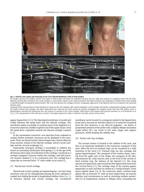
Fig. 2. Anterior neck region and sonoscopy of the Crico-Thyroid Membrane (CTM) of both genders.<br />
The images represent the (middle) anterior neck region of both genders and depict the landmark (left grey oval area, right pale remnant of a palpation action with the index<br />
fingertip), directly above and below the cricoid cartilage (c). Linear probe, median cranio-caudal orientation. The haptic sensation when palpating is a flexible-elastic touch leading<br />
to superficial depth surrounded by harder borders. This is the area between the cartilages, however, sonographic appearance of the ligament does not correspond to the measured<br />
introitus space.<br />
Sonogram shows sonoanatomy of a male individual in a long axis slice, left cephalad, right caudad. Cephalad is cricoid cartilage, then further caudad thyroid cartilage and thereafter<br />
the smaller tracheal ring cartilages. The white, hyperechoic line represents the inner mucosa-air interface throughout the complete airway. Note that CTM (yellow arrow) is<br />
hypoechogenic and above the hyperechoic line, also anterior of a portion of the cricoid cartilage. Yellow letters TACA indicate both puncture positions (A, A) of Kristensen et al.<br />
[26,27]. (For interpretation of the references to colour in this figure legend, the reader is referred to the Web version of this article.)<br />
appear hypoechoic [9,10]. The thyrohyoid membrane is located and<br />
visible between the hyoid bone and the thyroid cartilage. This<br />
membrane makes it possible to examine parts of the epiglottis in a<br />
transverse plane by a midline sagittal scan of the upper larynx, from<br />
the hyoid bone (cephalad) towards the thyroid cartilage (caudad)<br />
[2,4].<br />
In the paramedian transverse scan direction from cephalad to<br />
caudad, further anatomic structures can be displayed in the sonogram.<br />
Those are faucial tonsils, lateral tongue base, lateral vallecula,<br />
strap muscles, lamina of the thyroid cartilage, lateral cricoid cartilage<br />
and the cervical esophagus [4].<br />
Importantly, laryngeal cartilage is uncalcified in children but<br />
shows an increasing calcification with ageing [11]. At the age of 60,<br />
all humans show signs of ossification of the cartilage, visualized as a<br />
stronger echogenic structure within the cartilage, and with posterior<br />
acoustic shadow [11]. In a transverse slice, the cartilage has a<br />
shape like an inverted letter “V” with visible vocal cords [4].<br />
2.5. Thyroid and cricoid cartilage<br />
Thyroid and cricoid cartilage are hypoechogenic, can have bony<br />
structures and can be distinguished passing by from cephalad to<br />
caudad, best sliding the probe in longitudinal midline slices (Fig. 2).<br />
In between thyroid and cricoid cartilage, the cricothyroid<br />
membrane can be located in a sonogram related to the hyperechoic<br />
tissue and a mucosal-air interface deep to it. It cannot be visualized<br />
from the skin, however, it can often be palpated - see below. A<br />
paramedian position of the probe with a slightly oblique insonation<br />
(angle below 20 ) can result in the same image and support<br />
punctures, whilst keeping the midline free.<br />
2.6. Trachea and ring cartilages<br />
The normal trachea is located in the midline of the neck, and<br />
thus is an important landmark in the transverse sonogram of the<br />
neck. Often the first six tracheal rings can be visualized in a mild<br />
extension of the neck [12]. Tracheal rings are also cartilage and<br />
appear hypoechogenic. The sonogram of the trachea shows, starting<br />
from the upper part of the image, the sonogram of the skin,<br />
subcutaneous fat, strap muscles and, at the level of the second or<br />
third tracheal ring, the isthmus of the thyroid [12]. The strap<br />
muscles are hypoechoic and encased by thin hyperechoic lines from<br />
the cervical fascia [12]. The cartilage rings of the trachea appear<br />
hypoechoic as well, and they are similar to a “string of beads” in the<br />
(para)-sagittal plane [4]. In the transverse plane, tracheal-rings<br />
appear like an inverted “U” with an inner hyperechoic air-mucosa<br />
interface and reverberation as well as mirroring artifacts posteriorly<br />
[4]. In longitudinal, median or oblique slices, insonating the