Atemwegsultraschall - eine praktische Übersicht
Hier können Sie sich über das Thema umfassend informieren. Es sind zudem Filme innerhalb des Dokuments verlinkt. Es ist auch auf der Webseite des Publishers frei online verfügbar "Editor´s pick". https://authors.elsevier.com/a/1b66X7si0yR9sx
Hier können Sie sich über das Thema umfassend informieren. Es sind zudem Filme innerhalb des Dokuments verlinkt. Es ist auch auf der Webseite des Publishers frei online verfügbar "Editor´s pick".
https://authors.elsevier.com/a/1b66X7si0yR9sx

Sie wollen auch ein ePaper? Erhöhen Sie die Reichweite Ihrer Titel.
YUMPU macht aus Druck-PDFs automatisch weboptimierte ePaper, die Google liebt.
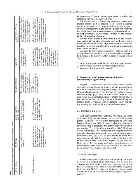
R. Breitkreutz et al. / Trends in Anaesthesia and Critical Care 32 (2020) 13e32 15<br />
Table 1<br />
Advantages and disadvantages of the different methods to evaluate airway.<br />
Fiberoptic intubation Capnometry Anterior neck and Airway<br />
Ultrasound<br />
Method Grading Stethoscope Laryngoscopy Video-<br />
Laryngoscopy<br />
no ventilation required to check if<br />
ETT is in the esophagus, provides a<br />
direct result, quick evaluation<br />
within 10 s possible, can identify<br />
main stem intubation (20 s)<br />
easy to apply, continuous<br />
measurements available/<br />
well established<br />
direct view, no ventilation<br />
required to check if ETT is in<br />
place, provides a direct result,<br />
can identify main stem<br />
intubation, established with<br />
specialists<br />
direct view on upper airway<br />
vocal cords, well established<br />
Advantage easy to handle, low cost, well<br />
established<br />
only provides a result, costs of an<br />
ultrasound machine $$$, need for<br />
another method to correct in case of<br />
esophageal misplacement, an<br />
esophageal misplacement can not<br />
be safely ruled out in a posterior<br />
position of the esophagus<br />
ventilation required to<br />
check if ETT is in place in<br />
unfasted individuals or CPR,<br />
CO2 does not provide an<br />
“on-off” result (and is not<br />
immediately ¼ zero in<br />
esophageal misplacements<br />
or cardiac standstill, can not<br />
safely identify main stem<br />
intubation, dependent on<br />
cardiac motion/bloodalveolar<br />
capnography<br />
transportation, costs $<br />
vulnerable endoscope<br />
extensive training required,<br />
time to result, costs $$<br />
if >/ ¼ C/L 3 states, not sufficient<br />
for a safe procedure, in<br />
unsecure attempts, it does not<br />
provide a direct result, only,<br />
costs $<br />
Limitation/Disadvantage tracheal auscultation cannot<br />
identify tube, can only be used<br />
after intubation for gastric or<br />
pulmonal sounds, unsafe<br />
method for mainstem<br />
intubation in many clinical<br />
scenarios<br />
interpretation of distinct sonographic patterns, within the<br />
respective clinical context, in real-time.<br />
The stethoscope is a historically established assessment<br />
method mostly used in addition to the above-mentioned<br />
methods. However, this is the only device that, in the context<br />
of airway management, might well be replaced completely by<br />
the anterior neck and airway ultrasound combined with parts<br />
of lung ultrasound, in the future - except for the acoustic<br />
finding of wheezing, of course.<br />
The aim of this narrative review is to explain and review a<br />
systematic understanding of anterior neck and airway ultrasound<br />
and, where no further high-level evidence is available,<br />
provides theoretical considerations and training suggestions<br />
from the author group.<br />
We describe three major categories of anterior neck and<br />
airway ultrasound exam methods mainly to serve the purpose<br />
to distinguish its usability within a defined clinical context<br />
(Table 2):<br />
1) to study sonoanatomy of anterior neck and upper airway<br />
2) in the context of airway management procedures<br />
3) as part of “Resuscitation Ultrasound”<br />
2. Anterior neck and airway ultrasound to study<br />
sonoanatomy of upper airway<br />
In general, anterior neck and airway ultrasound contains<br />
systematic examinations, as in conventional sonography, or<br />
focused assessments, following the concept of point-of-care<br />
ultrasound and sonoscopy. This first category is closer to conventional<br />
sonography. The exam style is used to obtain standard<br />
views, measurements and review images. This all takes at<br />
least several minutes, making it rather unfeasible if any immediate<br />
action is required. Thus, the practice context could be<br />
any learning and teaching for any planned assessment.<br />
2.1. Transducers and modes<br />
With conventional ultrasonography (US) main anatomical<br />
structures of the human airway can be visualized in sonograms.<br />
A curved low-frequency transducer is advocated<br />
because of its wider field of view [4] also to assess lateral<br />
structures such as esophagus and central neck vessels within<br />
one window (Figs. 1e4). A linear transducer (20-5 MHz) may<br />
also be sufficient [5,6] for sonoanatomy as it supports near field<br />
resolution, however it can require more movements to assess<br />
lateral structures (Figs. 1 and 2). Furthermore, in adults more<br />
penetration depth will decrease the width of the image obtained.<br />
In the inferior front of neck region, this can limit visibility<br />
e.g. of the esophagus, and make more transverse to<br />
lateral probe movements necessary. B-Mode scans and additional<br />
Colour-Mode overlays can be applied.<br />
2.2. Mouth and tongue<br />
For the mouth and tongue ultrasound exam the transducer<br />
is placed in a coronal plane posterior to the mentum [7].<br />
Subsequently the transducer is moved posteriorly and caudad<br />
towards the hyoid bone, so the dorsal surface of the tongue can<br />
be distinguished [8]. On either side the image is limited by the<br />
acoustic shadow of the mandible. Tonsils appear hypoechogenic<br />
[4].