

complete issue - IMA Fungus
complete issue - IMA Fungus
complete issue - IMA Fungus

You also want an ePaper? Increase the reach of your titles
YUMPU automatically turns print PDFs into web optimized ePapers that Google loves.
REPORTs<br />
A B<br />
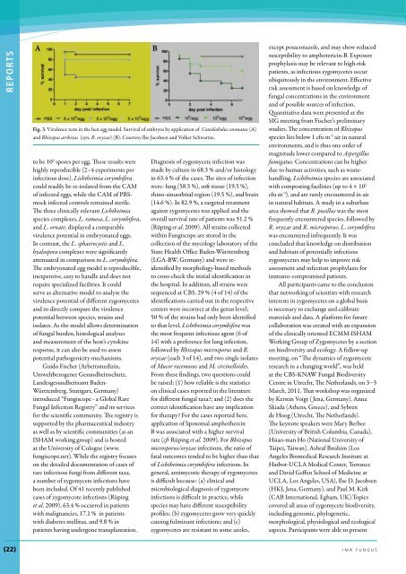
Fig. 3. Virulence tests in the hen egg model. Survival of embryos by application of Conidiobolus coronatus (A)<br />
and Rhizopus arrhizus (syn. R. oryzae) (B). Courtesy Ilse Jacobsen and Volker Schwartze.<br />
to be 10 2 spores per egg. These results were<br />
highly reproducible (2–4 experiments per<br />
infectious dose). Lichtheimia corymbifera<br />
could readily be re-isolated from the CAM<br />
of infected eggs, while the CAM of PBSmock<br />
infected controls remained sterile.<br />
The three clinically relevant Lichtheimia<br />
species complexes, L. ramosa, L. corymbifera,<br />
and L. ornate, displayed a comparable<br />
virulence potential in embryonated eggs.<br />
In contrast, the L. sphaerocystis and L.<br />
hyalospora complexes were significantly<br />
attenuated in comparison to L. corymbifera.<br />
The embryonated egg model is reproducible,<br />
inexpensive, easy to handle and does not<br />
require specialized facilities. It could<br />
serve as alternative model to analyse the<br />
virulence potential of different zygomycetes<br />
and to directly compare the virulence<br />
potential between species, strains and<br />
isolates. As the model allows determination<br />
of fungal burden, histological analyses<br />
and measurement of the host’s cytokine<br />
response, it can also be used to assess<br />
potential pathogenicity mechanisms.<br />
Guido Fischer (Arbeitsmedizin,<br />
Umweltbezogener Gesundheitsschutz,<br />
Landesgesundheitsamt Baden-<br />
Württemberg, Stuttgart, Germany)<br />
introduced “Fungiscope - a Global Rare<br />
Fungal Infection Registry” and its services<br />
for the scientific community. The registry is<br />
supported by the pharmaceutical industry<br />
as well as by scientific communities (as an<br />
ISHAM working group) and is hosted<br />
at the University of Cologne (www.<br />
fungiscope.net). While the registry focuses<br />
on the detailed documentation of cases of<br />
rare infectious fungi from different taxa,<br />
a number of zygomycete infections have<br />
been included. Of 41 recently published<br />
cases of zygomycote infections (Rüping<br />
et al. 2009), 63.4 % occurred in patients<br />
with malignancies, 17.1 % in patients<br />
with diabetes mellitus, and 9.8 % in<br />
patients having undergone transplantation.<br />
Diagnosis of zygomycete infection was<br />
made by culture in 68.3 % and/or histology<br />
in 63.4 % of the cases. The sites of infection<br />
were: lung (58.5 %), soft t<strong>issue</strong> (19.5 %),<br />
rhino-sinuorbital region (19.5 %), and brain<br />
(14.6 %). In 82.9 %, a targeted treatment<br />
against zygomycetes was applied and the<br />
overall survival rate of patients was 51.2 %<br />
(Rüping et al. 2009). All strains collected<br />
within Fungiscope are stored in the<br />
collection of the mycology laboratory of the<br />
State Health Office Baden-Württemberg<br />
(LGA-BW, Germany) and were reidentified<br />
by morphology-based methods<br />
to cross-check the initial identification in<br />
the hospital. In addition, all strains were<br />
sequenced at CBS. 29 % (4 of 14) of the<br />
identifications carried out in the respective<br />
centers were incorrect at the genus level;<br />
50 % of the strains had only been identified<br />
to that level. Lichtheimia corymbifera was<br />
the most frequent infectious agent (6 of<br />
14) with a preference for lung infection,<br />
followed by Rhizopus microsporus and R.<br />
oryzae (each 3 of 14), and two single isolates<br />
of Mucor racemosus and M. circinelloides.<br />
From these findings, two questions could<br />
be raised: (1) how reliable is the statistics<br />
on clinical cases reported in the literature<br />
for different fungal taxa?; and (2) does the<br />
correct identification have any implication<br />
for therapy? For the cases reported here,<br />
application of liposomal amphothercin<br />
B was associated with a higher survival<br />
rate (cfr Rüping et al. 2009). For Rhizopus<br />
microsporus/oryzae infections, the ratio of<br />
fatal outcomes tended to be higher than that<br />
of Lichtheimia corymbifera infections. In<br />
general, antimycotic therapy of zygomycetes<br />
is difficult because: (a) clinical and<br />
microbiological diagnosis of zygomycete<br />
infections is difficult in practice, while<br />
species may have different susceptibility<br />
profiles; (b) zygomycetes grow very quickly<br />
causing fulminant infections; and (c)<br />
zygomycetes are resistant to some azoles,<br />
except posaconazole, and may show reduced<br />
susceptibility to amphotericin-B. Exposure<br />
prophylaxis may be relevant to high-risk<br />
patients, as infectious zygomycetes occur<br />
ubiquitously in the environment. Effective<br />
risk assessment is based on knowledge of<br />
fungal concentrations in the environment<br />
and of possible sources of infection.<br />
Quantitative data were presented at the<br />
SIG meeting from Fischer’s preliminary<br />
studies. The concentration of Rhizopus<br />
species lies below 1 cfu m -3 air in natural<br />
environments, and is thus one order of<br />
magnitude lower compared to Aspergillus<br />
fumigatus. Concentrations can be higher<br />
due to human activities, such as wastehandling.<br />
Lichtheimia species are associated<br />
with composting facilities (up to 4 × 10 2<br />
cfu m -3 ), and are rarely encountered in air<br />
in natural habitats. A study in a suburban<br />
area showed that R. pusillus was the most<br />
frequently encountered species, followed by<br />
R. oryzae and R. microsporus; L. corymbifera<br />
was encountered infrequently. It was<br />
concluded that knowledge on distribution<br />
and habitats of potentially infectious<br />
zygomycetes may help to improve risk<br />
assessment and infection prophylaxis for<br />
immuno-compromised patients.<br />
All participants came to the conclusion<br />
that networking of scientists with research<br />
interests in zygomycetes on a global basis<br />
is necessary to exchange and calibrate<br />
materials and data. A platform for future<br />
collaboration was created with an expansion<br />
of the clinically oriented ECMM-ISHAM<br />
Working Group of Zygomycetes by a section<br />
on biodiversity and ecology. A follow-up<br />
meeting, on “The dynamics of zygomycete<br />
research in a changing world”, was held<br />
at the CBS-KNAW Fungal Biodiversity<br />
Centre in Utrecht, The Netherlands, on 3−5<br />
March, 2011. That workshop was organized<br />
by Kerstin Voigt ( Jena, Germany), Anna<br />
Skiada (Athens, Greece), and Sybren<br />
de Hoog (Utrecht, The Netherlands).<br />
The keynote speakers were Mary Berbee<br />
(University of British Columbia, Canada),<br />
Hsiao-man Ho (National University of<br />
Taipei, Taiwan), Ashraf Ibrahim (Los<br />
Angeles Biomedical Research Institute at<br />
Harbor-UCLA Medical Center, Torrance<br />
and David Geffen School of Medicine at<br />
UCLA, Los Angeles, USA), Ilse D. Jacobsen<br />
(HKI, Jena, Germany), and Paul M. Kirk<br />
(CAB International, Egham, UK).Topics<br />
covered all areas of zygomycete biodiversity,<br />
including genomic, phylogenetic,<br />
morphological, physiological and ecological<br />
aspects. Participants were able to present<br />
(22) ima funGuS


