Supplementum 163 - Swiss Medical Weekly
Supplementum 163 - Swiss Medical Weekly
Supplementum 163 - Swiss Medical Weekly

You also want an ePaper? Increase the reach of your titles
YUMPU automatically turns print PDFs into web optimized ePapers that Google loves.
17 S SWISS MED WKLY 2008;138(Suppl <strong>163</strong>) · www.smw.ch<br />
Free Communications 6 – SSAI / SGAI<br />
84<br />
Heterozygous hypomorphic STAT3 mutations in 4 <strong>Swiss</strong> patients<br />
of 3 unrelated families with classic autosomal dominant hyper<br />
IgE syndrome<br />
B. Drexel 1 , E.D. Renner 2 , T.R. Torgerson 2 , R.A. Seger 1 , H.D. Ochs 2 ,<br />
J. Reichenbach 1 . 1 University Children’s Hospital Zurich (Zurich, CH);<br />
2 University of Washington School of Medicine and Children’s<br />
Hospital (Seattle, USA)<br />
Background: Hyper-immunoglobulin E syndrome (HIES) is an<br />
autosomal dominant disorder characterized by a highly elevated<br />
serum IgE, eczema, recurrent staphylococcal skin abscesses and<br />
cyst-forming pneumonia, with disproportionately milder inflammatory<br />
responses, referred to as cold abscesses, and skeletal abnormalities.<br />
Therapy involves lifelong antibiotic and antimycotic prophylaxis, and<br />
occasionally surgical abscess drainage. The molecular defect,<br />
heterozygous mutations of STAT3 has recently been identified<br />
Methods: We enrolled 4 patients from 3 unrelated families with the<br />
classic symptoms of HIES and looked for mutations in the gene<br />
encoding human signal transducer and activator of transcription 3<br />
(STAT3) by sequence analysis.<br />
Findings: We found hypomorphic heterozygous STAT3 mutations in<br />
all four patients with HIES suggesting a dominant negative effect. We<br />
identified a novel R335W mutation within the DNA-binding domain in<br />
two related patients from Sri Lanka, and a V637M mutation in the<br />
other two non-related <strong>Swiss</strong> patients. The V637M mutation is one of<br />
the four known STAT3 hotspot mutations and is located within the<br />
SH2 domain. There was no difference in the clinical phenotype<br />
between patients with mutations in the DNA binding or SH2 domain.<br />
Conclusion: For four decades the molecular basis of HIES has<br />
remained elusive. Here we show that dominant-negative mutations in<br />
the STAT3 gene result in classic multisystem HIES in four patients<br />
followed in Switzerland. The identification of STAT3 as the major<br />
causative gene of HIES will facilitate early and definitive diagnosis as<br />
well as treatment, hopefully leading to the prevention of serious<br />
infectious complications and sequelae.<br />
85<br />
Tumour antigen specific CD8+ T-cells from melanoma patients<br />
exert strong ex-vivo cytolytic activity<br />
Y. Mahnke1 , E. Devêvre2 , P. Baumgaertner2 , M. Matter3 , N. Rufer4 ,<br />
P. Romero2 , D. Speiser2 . 1 NIH (Bethesda, USA); 2 Ludwig Institute<br />
for Cancer Research (Lausanne, CH); 3 CHUV (Lausanne, CH);<br />
(4)Multidisciplinary Oncology Center (Lausanne, CH)<br />
Direct analysis of ex vivo cytotoxicity by tumor Ag-specific T-cells has<br />
long been precluded, because standard laboratory methods are not<br />
suited to analyze small numbers of T-cells available from PBMC and<br />
tumor infiltrated lymph nodes (TILN) from cancer patients. Here we<br />
applied the recently described LiveCount Assay which allows to<br />
determine the cytotoxic function of small numbers of isolated T-cells.<br />
We analyzed Melan-A/HLA-A2 tetramer+ CD8+ T-cells from eight<br />
melanoma patients. Surprisingly, strong cytotoxic activity was exerted<br />
by T-cells derived from TILN or PBMC isolated after peptide<br />
vaccination. Contrary to previous reports showing that TILN produce<br />
low to no tumor-specific responses in vitro, our results illustrate that<br />
they can be quite effective if isolated from the tumor<br />
microenvironment, and thus separated from suppressor and/or<br />
regulatory cells that might be contained therein. T-cells isolated from<br />
TILN showed a trend to display lower killing activity than those from<br />
PBMC, though the difference was not significant. As expected,<br />
concomitant measurement of specific lysis and degranulation<br />
revealed that only a subset of the cells were responsible for the<br />
observed strong cytotoxicity. A direct correlation was observed<br />
between the proportion of CD107a+ T-cells that were potentially<br />
involved in the killing activity and the frequency of Granzyme B+ Tcells<br />
detected ex vivo, although not all Granzyme B+ cells<br />
participated in the lytic activity. We conclude that tumor antigen<br />
specific T-cells are highly functional outside of the tumor<br />
microenvironment. Studies are underway to elucidate the factors that<br />
repress the effectiveness of tumor-specific T-cells locally, and how to<br />
promote activation and avoid inhibition of protective immune<br />
mechanisms.<br />
86<br />
Depletion of B-cells by rituximab therapy improves atopic<br />
eczema<br />
D. Simon1 , S. Hösli2 , G. Kostylina2 , N. Yawalkar1 , H-U. Simon2 .<br />
1 Inselspital (Berne, CH); 2 University of Berne (Berne, CH)<br />
Background: Atopic eczema (AE) is a chronic inflammatory skin<br />
disorder characterized by eczematous skin lesions, pruritus and<br />
typical histopathological features. Although T cells play a key role,<br />
B cells are also found in the dermal infiltrate. In 80% of AE patients,<br />
elevated total and specific IgE levels can be detected. This study<br />
aimed to investigate the effect of a monoclonal anti-CD20 antibody<br />
therapy (rituximab) in AE.<br />
Methods: Six patients (2 males; mean age 39 ± 7 years) with severe<br />
AE received two applications of rituximab, each 1000 mg IV two<br />
weeks apart. To evaluate the efficacy of rituximab, we monitored<br />
clinical parameters (EASI, pruritus), total and specific IgE levels, and<br />
skin histology. Inflammatory cells and cytokine expression in skin<br />
lesions were assessed by immunofluorescence analysis before and<br />
after therapy.<br />
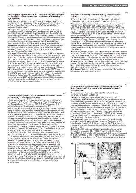
Results: All patients showed an improvement of their skin symptoms<br />
within 4 to 8 weeks. The EASI significantly decreased (before therapy:<br />
29.4 ± 4.3; week 8: 8.4 ± 3.6; p