Dye Separation - Leica Microsystems
Dye Separation - Leica Microsystems
Dye Separation - Leica Microsystems

You also want an ePaper? Increase the reach of your titles
YUMPU automatically turns print PDFs into web optimized ePapers that Google loves.
10 Confocal Application Letter<br />
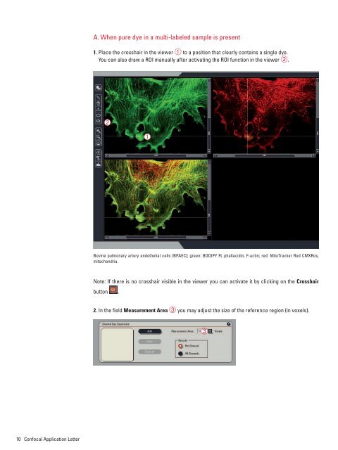
A. When pure dye in a multi-labeled sample is present<br />
1. Place the crosshair in the viewer ➀ to a position that clearly contains a single dye.<br />
You can also draw a ROI manually after activating the ROI function in the viewer ➁.<br />
➁<br />
➀<br />
Bovine pulmonary artery endothelial cells (BPAEC); green: BODIPY FL phallacidin, F-actin; red: MitoTracker Red CMXRos,<br />
mitochondria.<br />
Note: If there is no crosshair visible in the viewer you can activate it by clicking on the Crosshair<br />
button .<br />
2. In the fi eld Measurement Area ➂ you may adjust the size of the reference region (in voxels).<br />
➂