Vol 11-R2- Eyelid
Vol 11-R2- Eyelid
Vol 11-R2- Eyelid

Create successful ePaper yourself
Turn your PDF publications into a flip-book with our unique Google optimized e-Paper software.
SRPS • <strong>Vol</strong>ume <strong>11</strong> • Issue <strong>R2</strong> • 2010<br />
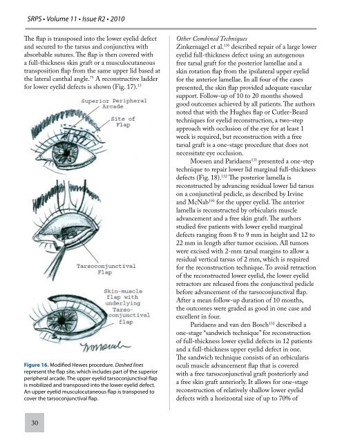
The flap is transposed into the lower eyelid defect<br />
and secured to the tarsus and conjunctiva with<br />
absorbable sutures. The flap is then covered with<br />
a full-thickness skin graft or a musculocutaneous<br />
transposition flap from the same upper lid based at<br />
the lateral canthal angle. 75 A reconstructive ladder<br />
for lower eyelid defects is shown (Fig. 17). 13<br />
Figure 16. Modified Hewes procedure. Dashed lines<br />
represent the flap site, which includes part of the superior<br />
peripheral arcade. The upper eyelid tarsoconjunctival flap<br />
is mobilized and transposed into the lower eyelid defect.<br />
An upper eyelid musculocutaneous flap is transposed to<br />
cover the tarsoconjunctival flap.<br />
30<br />
Other Combined Techniques<br />
Zinkernagel et al. 130 described repair of a large lower<br />
eyelid full-thickness defect using an autogenous<br />
free tarsal graft for the posterior lamellae and a<br />
skin rotation flap from the ipsilateral upper eyelid<br />
for the anterior lamellae. In all four of the cases<br />
presented, the skin flap provided adequate vascular<br />
support. Follow-up of 10 to 20 months showed<br />
good outcomes achieved by all patients. The authors<br />
noted that with the Hughes flap or Cutler-Beard<br />
techniques for eyelid reconstruction, a two-step<br />
approach with occlusion of the eye for at least 1<br />
week is required, but reconstruction with a free<br />
tarsal graft is a one-stage procedure that does not<br />
necessitate eye occlusion.<br />
Moesen and Paridaens 131 presented a one-step<br />
technique to repair lower lid marginal full-thickness<br />
defects (Fig. 18). 132 The posterior lamella is<br />
reconstructed by advancing residual lower lid tarsus<br />
on a conjunctival pedicle, as described by Irvine<br />
and McNab 100 for the upper eyelid. The anterior<br />
lamella is reconstructed by orbicularis muscle<br />
advancement and a free skin graft. The authors<br />
studied five patients with lower eyelid marginal<br />
defects ranging from 8 to 9 mm in height and 12 to<br />
22 mm in length after tumor excision. All tumors<br />
were excised with 2-mm tarsal margins to allow a<br />
residual vertical tarsus of 2 mm, which is required<br />
for the reconstruction technique. To avoid retraction<br />
of the reconstructed lower eyelid, the lower eyelid<br />
retractors are released from the conjunctival pedicle<br />
before advancement of the tarsoconjunctival flap.<br />
After a mean follow-up duration of 10 months,<br />
the outcomes were graded as good in one case and<br />
excellent in four.<br />
Paridaens and van den Bosch 132 described a<br />
one-stage “sandwich technique” for reconstruction<br />
of full-thickness lower eyelid defects in 12 patients<br />
and a full-thickness upper eyelid defect in one.<br />
The sandwich technique consists of an orbicularis<br />
oculi muscle advancement flap that is covered<br />
with a free tarsoconjunctival graft posteriorly and<br />
a free skin graft anteriorly. It allows for one-stage<br />
reconstruction of relatively shallow lower eyelid<br />
defects with a horizontal size of up to 70% of