

Download PDF - The Pancreapedia
Download PDF - The Pancreapedia
Download PDF - The Pancreapedia

You also want an ePaper? Increase the reach of your titles
YUMPU automatically turns print PDFs into web optimized ePapers that Google loves.
of the ducts (i.e., the wider the lumen, the higher<br />
the epithelium). <strong>The</strong> common duct is lined with a<br />
single row (or two) of columnar cells occasionally<br />
with interspersed goblet cells, which are absent in<br />
two-week-old hamsters but occur with moderate<br />
frequency in the entire common duct of adult<br />
animals and become a prominent constituent of<br />
the epithelium after one year 95 . According to<br />
McMinn and Kugler 108 , the luminal surface and<br />
supra-nuclear regions of the common duct cells<br />
contain glycogen and lack alkaline phosphatase<br />
but give a strongly positive reaction for succinic<br />
dehydrogenase. This suggests an active<br />
engagement of cells in transport mechanisms.<br />
<strong>The</strong> common duct has the largest lumen, thickest<br />
periductal connective tissue, and largest epithelial<br />
cells of any other pancreatic duct. Because of its<br />
anatomical and topographical patterns, step<br />
sections of the histological material are necessary<br />
to demonstrate the entire length of the common<br />
duct. A few small pancreatic ducts (peri-biliary<br />
ducts) enter the common bile duct directly. <strong>The</strong><br />
common bile duct merges imperceptibly into the<br />
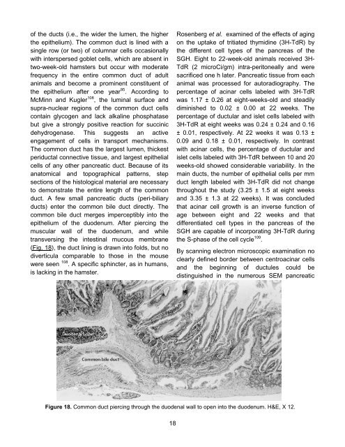
epithelium of the duodenum. After piercing the<br />
muscular wall of the duodenum, and while<br />
transversing the intestinal mucous membrane<br />
(Fig. 18), the duct lining is drawn into folds, but no<br />
diverticula comparable to those in the mouse<br />
were seen 108 . A specific sphincter, as in humans,<br />
is lacking in the hamster.<br />
18<br />
Rosenberg et al. examined of the effects of aging<br />
on the uptake of tritiated thymidine (3H-TdR) by<br />
the different cell types of the pancreas of the<br />
SGH. Eight to 22-week-old animals received 3H-<br />
TdR (2 microCi/gm) intra-peritoneally and were<br />
sacrificed one h later. Pancreatic tissue from each<br />
animal was processed for autoradiography. <strong>The</strong><br />
percentage of acinar cells labeled with 3H-TdR<br />
was 1.17 ± 0.26 at eight-weeks-old and steadily<br />
diminished to 0.02 ± 0.00 at 22 weeks. <strong>The</strong><br />
percentage of ductular and islet cells labeled with<br />
3H-TdR at eight weeks was 0.24 ± 0.24 and 0.16<br />
± 0.01, respectively. At 22 weeks it was 0.13 ±<br />
0.09 and 0.18 ± 0.01, respectively. In contrast<br />
with acinar cells, the percentage of ductular and<br />
islet cells labeled with 3H-TdR between 10 and 20<br />
weeks-old showed considerable variability. In the<br />
main ducts, the number of epithelial cells per mm<br />
duct length labeled with 3H-TdR did not change<br />
throughout the study (3.25 ± 1.5 at eight weeks<br />
and 3.35 ± 1.3 at 22 weeks). It was concluded<br />
that acinar cell growth is an inverse function of<br />
age between eight and 22 weeks and that<br />
differentiated cell types in the pancreas of the<br />
SGH are capable of incorporating 3H-TdR during<br />
the S-phase of the cell cycle 109 .<br />
By scanning electron microscopic examination no<br />
clearly defined border between centroacinar cells<br />
and the beginning of ductules could be<br />
distinguished in the numerous SEM pancreatic<br />
Figure 18. Common duct piercing through the duodenal wall to open into the duodenum. H&E, X 12.


