

Download PDF - The Pancreapedia
Download PDF - The Pancreapedia
Download PDF - The Pancreapedia

Create successful ePaper yourself
Turn your PDF publications into a flip-book with our unique Google optimized e-Paper software.
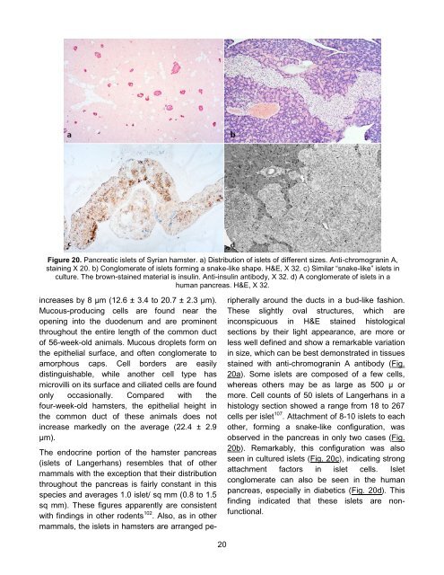
Figure 20. Pancreatic islets of Syrian hamster. a) Distribution of islets of different sizes. Anti-chromogranin A,<br />
staining X 20. b) Conglomerate of islets forming a snake-like shape. H&E, X 32. c) Similar “snake-like” islets in<br />
culture. <strong>The</strong> brown-stained material is insulin. Anti-insulin antibody, X 32. d) A conglomerate of islets in a<br />
human pancreas. H&E, X 32.<br />
increases by 8 μm (12.6 ± 3.4 to 20.7 ± 2.3 μm).<br />
Mucous-producing cells are found near the<br />
opening into the duodenum and are prominent<br />
throughout the entire length of the common duct<br />
of 56-week-old animals. Mucous droplets form on<br />
the epithelial surface, and often conglomerate to<br />
amorphous caps. Cell borders are easily<br />
distinguishable, while another cell type has<br />
microvilli on its surface and ciliated cells are found<br />
only occasionally. Compared with the<br />
four-week-old hamsters, the epithelial height in<br />
the common duct of these animals does not<br />
increase markedly on the average (22.4 ± 2.9<br />
μm).<br />
<strong>The</strong> endocrine portion of the hamster pancreas<br />
(islets of Langerhans) resembles that of other<br />
mammals with the exception that their distribution<br />
throughout the pancreas is fairly constant in this<br />
species and averages 1.0 islet/ sq mm (0.8 to 1.5<br />
sq mm). <strong>The</strong>se figures apparently are consistent<br />
with findings in other rodents 102 . Also, as in other<br />
mammals, the islets in hamsters are arranged pe-<br />
20<br />
ripherally around the ducts in a bud-like fashion.<br />
<strong>The</strong>se slightly oval structures, which are<br />
inconspicuous in H&E stained histological<br />
sections by their light appearance, are more or<br />
less well defined and show a remarkable variation<br />
in size, which can be best demonstrated in tissues<br />
stained with anti-chromogranin A antibody (Fig.<br />
20a). Some islets are composed of a few cells,<br />
whereas others may be as large as 500 μ or<br />
more. Cell counts of 50 islets of Langerhans in a<br />
histology section showed a range from 18 to 267<br />
cells per islet 107 . Attachment of 8-10 islets to each<br />
other, forming a snake-like configuration, was<br />
observed in the pancreas in only two cases (Fig.<br />
20b). Remarkably, this configuration was also<br />
seen in cultured islets (Fig. 20c), indicating strong<br />
attachment factors in islet cells. Islet<br />
conglomerate can also be seen in the human<br />
pancreas, especially in diabetics (Fig. 20d). This<br />
finding indicated that these islets are nonfunctional.


