A novel structure associated with a lampbrush chromosome in the ...
A novel structure associated with a lampbrush chromosome in the ...
A novel structure associated with a lampbrush chromosome in the ...

Create successful ePaper yourself
Turn your PDF publications into a flip-book with our unique Google optimized e-Paper software.
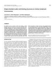
Journal of Cell Science 101, 759-772 (1992)<br />
Pr<strong>in</strong>ted <strong>in</strong> Great Brita<strong>in</strong> © The Company of Biologists Limited 1992<br />
A <strong>novel</strong> <strong>structure</strong> <strong>associated</strong> <strong>with</strong> a <strong>lampbrush</strong> <strong>chromosome</strong> <strong>in</strong> <strong>the</strong> chicken,<br />
Gallus domesticus<br />
IRINA SOLOVEI, ELENA GAGINSKAYA<br />
Biological Research Institute, University of St. Petersburg, Russia<br />
TERRY ALLEN<br />
Paterson Laboratory, Christie Hospital, Manchester, England<br />
and HERBERT MACGREGOR*<br />
Department of Zoology, University of Leicester, Leicester LEI 7RH, England<br />
*Author for correspondence<br />
Summary<br />
At a site near <strong>the</strong> end of <strong>the</strong> short arms of <strong>lampbrush</strong><br />
bivalent 2 <strong>in</strong> <strong>the</strong> chicken {Gallus domesticus) <strong>the</strong>re is<br />
always a marker <strong>structure</strong> that appears <strong>in</strong> <strong>the</strong> phasecontrast<br />
light microscope as a solid object <strong>with</strong> diffuse<br />
edges measur<strong>in</strong>g about 4 //m across. When exam<strong>in</strong>ed by<br />
transmission electron microscopy <strong>in</strong> th<strong>in</strong> section, this<br />
object appears as a loose bundle of fibres. In some<br />
preparations <strong>in</strong>dividual fibres appear 15-16 nm thick,<br />
smooth <strong>in</strong> outl<strong>in</strong>e and solid <strong>in</strong> cross-section. In o<strong>the</strong>r<br />
preparations <strong>the</strong>y are 32-38 nm thick, rougher <strong>in</strong> outl<strong>in</strong>e<br />
and r<strong>in</strong>g-like <strong>in</strong> cross-section. High-resolution scann<strong>in</strong>g<br />
electron micrographs of <strong>the</strong> <strong>chromosome</strong> 2 marker show<br />
it to be a loose bundle of spaghetti-like fibres that is quite<br />
unlike anyth<strong>in</strong>g previously seen on a <strong>lampbrush</strong> <strong>chromosome</strong><br />
of any organism. As <strong>with</strong> <strong>the</strong> sectioned material,<br />
fibres <strong>in</strong> some preparations were smooth and 15-16 nm<br />
<strong>in</strong> diameter, whereas those <strong>in</strong> o<strong>the</strong>rs were more knobbly<br />
and about 35 nm thick. The fibres appear to branch and<br />
Introduction<br />
Lampbrush <strong>chromosome</strong>s are well known as enormously<br />
elongated diplotene bivalents found <strong>in</strong> <strong>the</strong><br />
grow<strong>in</strong>g oocytes of all animals except mammals, some<br />
<strong>in</strong>sects and some reptiles. These <strong>chromosome</strong>s support<br />
a widespread transcription of RNA from many thousands<br />
of promoters distributed throughout <strong>the</strong> lengths<br />
of all <strong>the</strong> <strong>chromosome</strong>s. RNA polymerase transcribes<br />
past <strong>the</strong> end of structural gene sequences and cont<strong>in</strong>ues<br />
on to form contiguous transcripts of whatever sequences<br />
lie downstream (Macgregor and Andrews,<br />
1977; Varley et al., 1980; Diaz et al., 1981; Gall et al.,<br />
1983). The newly formed RNA transcripts rema<strong>in</strong><br />
759<br />
<strong>in</strong> some cases it is clear that <strong>the</strong> daughter strands of a<br />
branch have <strong>the</strong> same dimensions as <strong>the</strong> parent strand.<br />
Free ends are rare. Total length of fibre material present<br />
at one marker locus is estimated to be between 500 and<br />
2000 fan. Similar <strong>structure</strong>s are not present on <strong>the</strong><br />
<strong>lampbrush</strong> <strong>chromosome</strong>s of quail, wood pigeon or<br />
chaff<strong>in</strong>ch.<br />
The nature of this fibrous marker, referred to <strong>in</strong> this<br />
paper as <strong>the</strong> "spaghetti marker", is discussed <strong>in</strong> relation<br />
to <strong>lampbrush</strong> <strong>chromosome</strong> function and to events that<br />
take place dur<strong>in</strong>g <strong>the</strong> <strong>lampbrush</strong> phase of oogenesis <strong>in</strong><br />
chicken. Evidence is discussed <strong>in</strong> relation to <strong>the</strong><br />
possibility that <strong>the</strong> <strong>chromosome</strong> 2 marker represents a<br />
<strong>novel</strong> form of nuclear RNP or <strong>the</strong> specific association of<br />
some structural prote<strong>in</strong> <strong>with</strong> one <strong>chromosome</strong> locus.<br />
Key words: oocyte nucleus, <strong>lampbrush</strong> <strong>chromosome</strong>,<br />
scann<strong>in</strong>g electron microscopy.<br />
<strong>associated</strong> <strong>with</strong> <strong>the</strong> chromosomal DNA template. The<br />
end result of this process is a large number of<br />
exceed<strong>in</strong>gly long transcription units, each of <strong>the</strong>m<br />
polarised <strong>in</strong> <strong>the</strong> sense of carry<strong>in</strong>g RNA transcripts that<br />
are progressively longer from one end of <strong>the</strong> transcription<br />
unit to <strong>the</strong> o<strong>the</strong>r. These transcription units<br />
constitute <strong>the</strong> lateral loops that are <strong>the</strong> basis of <strong>the</strong><br />
<strong>lampbrush</strong> form, and until quite recently RNA transcription<br />
and <strong>the</strong> arrangement and expression of DNA<br />
sequences have been <strong>the</strong> major foci for <strong>lampbrush</strong><br />
<strong>chromosome</strong> research (Callan, 1986; Macgregor, 1987).<br />
This paper is about a study of <strong>the</strong> <strong>lampbrush</strong><br />
<strong>chromosome</strong>s of <strong>the</strong> domestic chicken (Gallus domesticus),<br />
and it focusses specifically on a <strong>novel</strong> <strong>structure</strong>
760 /. Solovei and o<strong>the</strong>rs<br />
such as has never been seen <strong>in</strong> any previous studies of<br />
<strong>lampbrush</strong> <strong>chromosome</strong>s. The earliest studies of <strong>lampbrush</strong><br />
<strong>chromosome</strong>s from birds, carried out <strong>with</strong> <strong>the</strong><br />
techniques <strong>in</strong>troduced by Gall (1954) and Callan and<br />
Lloyd (I960), were those of Koecke and Muller (1965)<br />
and Ahmad (1970). However, methods for prepar<strong>in</strong>g<br />
avian <strong>lampbrush</strong> <strong>chromosome</strong>s for modern molecular<br />
and cytogenetic <strong>in</strong>vestigations were worked out much<br />
later and <strong>in</strong>dependently by Hutchison (Hutchison and<br />
We<strong>in</strong>traub, 1983; Hutchison, 1987) and by Gag<strong>in</strong>skaya<br />
and coworkers (Gag<strong>in</strong>skaya et al., 1984; Kropotova and<br />
Gag<strong>in</strong>skaya, 1984; Chelysheva et al., 1990; Solovei et<br />
al., 1990). These authors have confirmed that avian<br />
<strong>lampbrush</strong> <strong>chromosome</strong>s exhibit all <strong>the</strong> ma<strong>in</strong> features<br />
that have been described for those of amphibians.<br />
At an early stage <strong>in</strong> <strong>the</strong>ir studies of fixed and sta<strong>in</strong>ed<br />
isolated <strong>lampbrush</strong> <strong>chromosome</strong>s from chickens<br />
Chelysheva et al. (1990) noticed that <strong>the</strong> conspicuous<br />
object located near <strong>the</strong> end of <strong>the</strong> short arm of<br />
<strong>chromosome</strong> 2 was different from all o<strong>the</strong>r dist<strong>in</strong>ctive<br />
objects <strong>associated</strong> <strong>with</strong> <strong>the</strong> <strong>chromosome</strong>s <strong>in</strong> <strong>the</strong> sense<br />
that it never <strong>in</strong> any circumstances assumed <strong>the</strong><br />
appearance of a loop. Experimental treatments, such as<br />
reduc<strong>in</strong>g <strong>the</strong> ionic strength of <strong>the</strong> <strong>chromosome</strong> isolation<br />
medium, have <strong>the</strong> effect of remov<strong>in</strong>g <strong>the</strong> RNP from all<br />
loops and often expos<strong>in</strong>g <strong>the</strong> fundamental loop-like<br />
organisation of conspicuous <strong>structure</strong>s that <strong>in</strong> normal<br />
circumstances have a solid, round or irregular appearance<br />
- <strong>the</strong> "lumpy loops" of Callan and Lloyd (1960).<br />
The objects located at <strong>the</strong> Chelysheva et al. locus LL22<br />
never reacted <strong>in</strong> this manner.<br />
Subsequent to <strong>the</strong> studies published <strong>in</strong> 1990<br />
(Chelysheva et al., 1990; Solovei et al., 1990), it was<br />
decided to <strong>in</strong>vestigate certa<strong>in</strong> aspects of <strong>the</strong> f<strong>in</strong>e<br />
<strong>structure</strong> of chicken <strong>lampbrush</strong> <strong>chromosome</strong>s employ<strong>in</strong>g<br />
transmission electron microscopy (TEM) of sectioned<br />
material <strong>with</strong> special regard to <strong>the</strong> marker at<br />
LL22. Preparations of <strong>lampbrush</strong> <strong>chromosome</strong>s were<br />
made accord<strong>in</strong>g to <strong>the</strong> method described by Mott and<br />
Callan (1975). The short arm of <strong>chromosome</strong> 2 was<br />
specifically selected for th<strong>in</strong> section<strong>in</strong>g. Sections<br />
through most regions of <strong>the</strong> <strong>chromosome</strong>s produced no<br />
surprises. In general <strong>the</strong> <strong>structure</strong> of chromomeres and<br />
lateral loop ribonucleoprote<strong>in</strong> appeared more or less<br />
similar to that which had been described <strong>in</strong> electron<br />
micrographs of <strong>lampbrush</strong> <strong>chromosome</strong>s from o<strong>the</strong>r<br />
species (Mott and Callan, 1975; N'Da et al., 1986). The<br />
one outstand<strong>in</strong>g exception was <strong>the</strong> marker at position<br />
LL22. Sections through <strong>the</strong> region of <strong>the</strong> LL22 marker<br />
showed evidence of a loose bundle of strands hav<strong>in</strong>g an<br />
appearance that was quite different from that of<br />
neighbour<strong>in</strong>g loop ribonucleoprote<strong>in</strong>. The area of<br />
section occupied by <strong>the</strong> stranded material was about 4<br />
/an <strong>in</strong> diameter, which was consistent <strong>with</strong> <strong>the</strong> observed<br />
size of <strong>the</strong> LL22 marker as seen <strong>in</strong> unfixed <strong>lampbrush</strong><br />
preparations exam<strong>in</strong>ed <strong>with</strong> a phase-contrast microscope.<br />
In view of <strong>the</strong> extraord<strong>in</strong>ary appearance of <strong>the</strong> LL22<br />
marker <strong>in</strong> light-microscope preparations and TEM<br />
sections, it was decided immediately that this region<br />
required fur<strong>the</strong>r <strong>in</strong>vestigation and, tak<strong>in</strong>g account of<br />
<strong>the</strong> size of <strong>the</strong> whole marker and <strong>the</strong> sizes and pack<strong>in</strong>g<br />
of its <strong>in</strong>dividual strands, high-resolution scann<strong>in</strong>g<br />
electron microscopy (SEM) coupled <strong>with</strong> light microscopy<br />
and cytochemistry seemed <strong>the</strong> most promis<strong>in</strong>g<br />
approaches. In this paper we describe <strong>the</strong> <strong>structure</strong> of<br />
<strong>the</strong> LL22 marker, we present <strong>the</strong> results of some<br />
cytochemical tests and we offer some suggestions as to<br />
<strong>the</strong> nature of this hi<strong>the</strong>rto unknown class of object.<br />
Materials and methods<br />
Oocytes for <strong>lampbrush</strong> studies were obta<strong>in</strong>ed from commercial<br />
l<strong>in</strong>e Zarya 17 and Rhode Island Red crosses of between 21<br />
weeks ("po<strong>in</strong>t of lay") and 31 weeks old. Chickens were killed<br />
by cervical dislocation and <strong>the</strong> whole ovary was removed and<br />
placed <strong>in</strong> a clean dry beaker, covered <strong>with</strong> foil and stored on<br />
ice. The oocytes rema<strong>in</strong> <strong>in</strong> good condition for <strong>lampbrush</strong><br />
studies for a maximum of 12 h after removal from <strong>the</strong> bird.<br />
In <strong>the</strong> chicken, <strong>the</strong> <strong>lampbrush</strong> <strong>chromosome</strong>s are found <strong>in</strong><br />
oocytes rang<strong>in</strong>g from 0.13 mm to 2.5-3 mm <strong>in</strong> diameter<br />
(Gag<strong>in</strong>skaya, 1972a; Hutchison, 1987; Chelysheva et al.,<br />
1990). The best preparations of <strong>lampbrush</strong> <strong>chromosome</strong>s can<br />
be made from oocytes of between 1 mm and 2.5 mm diameter.<br />
The germ<strong>in</strong>al vesicle of a 1.2 mm oocyte measures about 100<br />
/an diameter and that of a 2.5 mm oocyte has a diameter of<br />
about 300 ism. For this study, oocytes of between 0.5 mm to<br />
3.5 mm diameter were ma<strong>in</strong>ly used, although some preparations<br />
were made from post-<strong>lampbrush</strong> oocytes of up to 7<br />
mm diameter. The <strong>chromosome</strong>s were isolated manually<br />
employ<strong>in</strong>g <strong>the</strong> standard <strong>lampbrush</strong> technique as described by<br />
Macgregor and Varley (1988) <strong>with</strong> some m<strong>in</strong>or modifications.<br />
Individual oocytes or small groups of oocytes of <strong>the</strong> required<br />
size were dissected out and transferred to a separate dish<br />
conta<strong>in</strong><strong>in</strong>g "5:1 + phosphate" (Gall et al., 1981; 83 mM KC1,<br />
17 mM NaCl, 6.5 mM Na2HPO4, 3.5 mM KH2PO4, pH 7.2).<br />
Chicken ovary, unlike <strong>the</strong> ovaries of amphibians, is densely<br />
compacted <strong>with</strong> collagen and connective tissue and it is best to<br />
remove most of this material from around <strong>the</strong> oocyte before<br />
try<strong>in</strong>g to isolate <strong>the</strong> germ<strong>in</strong>al vesicle. The germ<strong>in</strong>al vesicle was<br />
removed <strong>in</strong> 5:1 + phosphate by stabb<strong>in</strong>g <strong>the</strong> oocyte <strong>with</strong> a<br />
dissect<strong>in</strong>g needle and <strong>the</strong>n carefully watch<strong>in</strong>g as <strong>the</strong> yolky<br />
cytoplasm streamed out of <strong>the</strong> hole. The germ<strong>in</strong>al vesicle<br />
appears as a small clear <strong>in</strong>terruption <strong>in</strong> <strong>the</strong> flow of yolk as it<br />
emerges from <strong>the</strong> hole. It is perfectly round, turgid,<br />
transparent and glisten<strong>in</strong>g <strong>in</strong> appearance. The germ<strong>in</strong>al<br />
vesicle can be seen <strong>in</strong>side <strong>in</strong>tact oocytes of less than 1.2 mm<br />
diameter if bright substage transmitted illum<strong>in</strong>ation is used on<br />
<strong>the</strong> dissect<strong>in</strong>g b<strong>in</strong>ocular microscope. After isolation, <strong>the</strong><br />
germ<strong>in</strong>al vesicle is picked up immediately <strong>in</strong> a small-bore<br />
Pasteur pipette and transferred to a <strong>lampbrush</strong> observation<br />
chamber (Macgregor and Varley, 1988) conta<strong>in</strong><strong>in</strong>g 3/4<br />
strength 5:1 plus phosphate <strong>with</strong> 0.1% formaldehyde. The<br />
nuclear envelope is removed manually, ei<strong>the</strong>r <strong>with</strong> two pairs<br />
of very f<strong>in</strong>e forceps or <strong>with</strong> tungsten needles, and <strong>the</strong> nuclear<br />
contents are allowed to disperse on <strong>the</strong> bottom of <strong>the</strong><br />
observation chamber.<br />
Some preparations were exam<strong>in</strong>ed directly, <strong>with</strong>out centrifugation<br />
or fixation, by phase-contrast microscopy. In order to<br />
exam<strong>in</strong>e <strong>the</strong> effects of nucleases and proteases, <strong>chromosome</strong>s<br />
were ei<strong>the</strong>r dissected directly <strong>in</strong>to 3/4 strength 5:1 plus<br />
phosphate to which <strong>the</strong> enzyme had been added to a<br />
concentration of 0.1 mg/ml, or <strong>the</strong>y were isolated <strong>in</strong> 3/4<br />
strength 5:1 plus phosphate and <strong>the</strong> enzyme was added later.<br />
With <strong>the</strong> latter technique, <strong>the</strong> observation chamber was<br />
covered <strong>with</strong> a coverslip, <strong>the</strong> <strong>chromosome</strong>s were allowed to
disperse and enzyme solution subsequently added to <strong>the</strong> edge<br />
of <strong>the</strong> coverslip so that it slowly mixed <strong>with</strong> <strong>the</strong> contents of <strong>the</strong><br />
observation chamber. The f<strong>in</strong>al enzyme concentration <strong>in</strong> <strong>the</strong><br />
region of <strong>the</strong> <strong>chromosome</strong>s was estimated to be about 0.01<br />
mg/ml. The effects of deoxyribonuclease (DN-EP, Sigma),<br />
ribonuclease A (Sigma, pre-boiled <strong>in</strong> sodium acetate at pH 5<br />
to elim<strong>in</strong>ate DNase activity), Pronase and tryps<strong>in</strong> were<br />
exam<strong>in</strong>ed <strong>in</strong> this manner and recorded by flash photomicrography<br />
of phase-contrast images. Magnesium chloride (6 x<br />
10~ 4 M) was added to <strong>the</strong> isolation medium for studies of <strong>the</strong><br />
effects of deoxyribonuclease. The effects of several restriction<br />
enzymes, HaeUI, EcoKL and Alul, on <strong>the</strong> <strong>lampbrush</strong><br />
<strong>chromosome</strong>s of chickens and particularly on <strong>the</strong> LL22<br />
marker were also exam<strong>in</strong>ed <strong>in</strong> this study. For all restriction<br />
enzyme studies, <strong>chromosome</strong>s were isolated <strong>in</strong>to Trisbuffered<br />
sal<strong>in</strong>e (TBS) plus magnesium (80 mM KC1, 20 mM<br />
NaCl, 20 mM Tris, 0.6 mM MgCl2, pH 7.8).<br />
Preparations for light-microscopic cytochemistry or for<br />
electron microscopy were centrifuged at 1500 g for 20 m<strong>in</strong> <strong>in</strong> a<br />
Sorvall RT-6000 bench top refrigerated centrifuge (see<br />
Macgregor and Varley, 1988) <strong>in</strong> order to stick <strong>the</strong> <strong>chromosome</strong>s<br />
firmly to <strong>the</strong> base of <strong>the</strong> observation chamber. They<br />
were <strong>the</strong>n fixed <strong>in</strong> 2.5% glutaraldehyde <strong>in</strong> 0.1 M phosphate<br />
buffer, pH 7. Preparations for fluorescence cytochemistry<br />
(Hoechst, Chromomyc<strong>in</strong> A3, DAPI) were fixed <strong>in</strong> 2%<br />
formaldehyde for one hour and <strong>the</strong>n <strong>in</strong> 70% ethanol. They<br />
were <strong>the</strong>n returned to phosphate buffered sal<strong>in</strong>e (PBS: 0.02 M<br />
phosphate buffer, 0.15 M NaCl) and <strong>in</strong>cubated at room<br />
temperature <strong>with</strong> <strong>the</strong> primary and secondary antibodies.<br />
Antibodies to chicken nuclear lam<strong>in</strong>s A and B2 (k<strong>in</strong>dly<br />
provided by Dr. E. A. Nigg) and to mammalian <strong>in</strong>termediate<br />
filaments (k<strong>in</strong>dly provided by Dr. C. Ockleford) were selected<br />
for test<strong>in</strong>g on <strong>the</strong> grounds of <strong>the</strong> f<strong>in</strong>ely fibrous appearance of<br />
<strong>the</strong> LL22 marker. Preparations were pre<strong>in</strong>cubated <strong>in</strong> 10%<br />
horse serum <strong>in</strong> PBS and <strong>the</strong>n <strong>with</strong> <strong>the</strong> first antibody,<br />
employ<strong>in</strong>g ranges of concentrations recommended by Drs.<br />
Nigg and Ockleford. The preparations were <strong>the</strong>n washed <strong>in</strong><br />
10% horse serum, <strong>in</strong>cubated <strong>with</strong> <strong>the</strong> appropriate FTTClabelled<br />
secondary antibody, washed aga<strong>in</strong> <strong>with</strong> 10% horse<br />
serum, mounted and exam<strong>in</strong>ed <strong>with</strong> a Carl Zeiss epifluorescence<br />
microscope.<br />
All preparations for electron microscopy were fixed <strong>in</strong><br />
2.5% glutaraldehyde <strong>in</strong> 0.1 M phosphate buffer, pH 7.<br />
Preparations for subsequent embedd<strong>in</strong>g and th<strong>in</strong>-section<strong>in</strong>g<br />
for transmission electron microscopy and some preparations<br />
for scann<strong>in</strong>g electron microscopy were post-fixed <strong>in</strong> 1% OsO4<br />
<strong>in</strong> 0.1 M phosphate buffer, pH 7, for 30 m<strong>in</strong>utes. Subsequent<br />
treatment of <strong>chromosome</strong>s for th<strong>in</strong> section<strong>in</strong>g was as<br />
described by Mott and Callan (1975). Regions of <strong>the</strong><br />
<strong>chromosome</strong>s that carried dist<strong>in</strong>ctive markers were specifically<br />
selected for th<strong>in</strong> section<strong>in</strong>g. Sections were cut at ei<strong>the</strong>r<br />
100 nm or 150-250 nm thickness and subsequently exam<strong>in</strong>ed<br />
<strong>with</strong> a Tesla BS 500 electron microscope us<strong>in</strong>g accelerat<strong>in</strong>g<br />
voltages of ei<strong>the</strong>r 60 or 90 kV. Preparations for scann<strong>in</strong>g<br />
electron microscopy were dehydrated <strong>in</strong> an ethanol series,<br />
r<strong>in</strong>sed twice <strong>in</strong> amyl acetate and <strong>the</strong>n critical-po<strong>in</strong>t dried from<br />
liquid CO2. They were <strong>the</strong>n coated <strong>with</strong> gold/palladium<br />
(estimated 2-3 nm thickness) or chromium (estimated 1-2 nm<br />
thickness) and exam<strong>in</strong>ed <strong>in</strong> a high-resolution, field emission,<br />
<strong>in</strong>-lens scann<strong>in</strong>g electron microscope (ISI/ABT DS 130F) at a<br />
variety of accelerat<strong>in</strong>g voltages <strong>with</strong><strong>in</strong> <strong>the</strong> range 3-25 kV.<br />
For electron microscopy of negatively sta<strong>in</strong>ed preparations,<br />
<strong>chromosome</strong>s were isolated directly onto a grid <strong>with</strong> a<br />
Formvar support<strong>in</strong>g film situated at <strong>the</strong> bottom of a<br />
<strong>lampbrush</strong> observation chamber. The preparations were<br />
centrifuged, fixed <strong>in</strong> 2.5% glutaraldehyde, washed and air<br />
dried. They were <strong>the</strong>n covered for 30 s <strong>with</strong> a few drops of<br />
Novel <strong>structure</strong> <strong>in</strong> <strong>lampbrush</strong> <strong>chromosome</strong>s 761<br />
saturated uranyl acetate diluted 1:1 <strong>with</strong> water, washed <strong>in</strong><br />
runn<strong>in</strong>g distilled water, air dried and exam<strong>in</strong>ed <strong>in</strong> a JEM-<br />
100CX electron microscope.<br />
Results<br />
Light microscopy<br />
Altoge<strong>the</strong>r, <strong>lampbrush</strong>es from about 90 chickens (commercial<br />
cross Zarya 17) <strong>in</strong> <strong>the</strong> USSR and 15 chickens<br />
(Rhode Island crosses) <strong>in</strong> <strong>the</strong> UK have been exam<strong>in</strong>ed<br />
<strong>in</strong> <strong>the</strong> current programme. All were homozygous for<br />
<strong>the</strong> presence of <strong>the</strong> LL22 marker. The marker has a<br />
dist<strong>in</strong>ctive appearance <strong>in</strong> phase-contrast (Fig. 1A). It<br />
sometimes consists of two components, a small compact<br />
dark <strong>structure</strong> about 1 /an <strong>in</strong> diameter situated <strong>in</strong> l<strong>in</strong>e<br />
<strong>with</strong> <strong>the</strong> <strong>chromosome</strong> axis, and a lighter and more<br />
diffuse area partially or wholly surround<strong>in</strong>g <strong>the</strong> dark<br />
object. The entire <strong>structure</strong> usually measures between 3<br />
and 5 /on across. It is of a similar size <strong>in</strong> all oocytes of all<br />
sizes from a particular bird. LL22 is present <strong>in</strong> oocytes<br />
rang<strong>in</strong>g from 0.4 mm to 3.5 mm diameter, <strong>the</strong> smallest<br />
and largest from which <strong>lampbrush</strong>es can be successfully<br />
isolated. It is absent from post-<strong>lampbrush</strong> <strong>chromosome</strong>s<br />
isolated from oocytes of 7 mm diameter. In<br />
general, its behaviour parallels that of all <strong>the</strong> o<strong>the</strong>r<br />
conspicuous markers <strong>in</strong> <strong>the</strong> <strong>lampbrush</strong> set, <strong>the</strong> majority<br />
of which are typically loop-like <strong>in</strong> organisation.<br />
In some preparations <strong>the</strong> two homologous LL22<br />
markers were fused toge<strong>the</strong>r to form a s<strong>in</strong>gle <strong>structure</strong><br />
that jo<strong>in</strong>ed <strong>the</strong> two half-bivalents at that locus. In all<br />
such cases, <strong>the</strong>re were no chiasmata distal to LL22. In<br />
all of <strong>the</strong> many preparations exam<strong>in</strong>ed <strong>in</strong> which <strong>the</strong>re<br />
was a chiasma distal to LL22 <strong>in</strong> <strong>the</strong> short arm of<br />
<strong>chromosome</strong> 2, <strong>the</strong> LL22 markers were separate from<br />
one ano<strong>the</strong>r.<br />
Simple cytochemical tests <strong>in</strong> which <strong>the</strong> <strong>chromosome</strong>s<br />
were sta<strong>in</strong>ed <strong>with</strong> Hoechst 33258, DAPI, chromomyc<strong>in</strong><br />
A3, gallocyan<strong>in</strong>-chrome alum and Coomassie blue all<br />
<strong>in</strong>dicated that LL22 was not noticeably different from<br />
o<strong>the</strong>r marker <strong>structure</strong>s on <strong>the</strong> <strong>chromosome</strong>s. In DAPI<br />
and Hoechst preparations <strong>the</strong> LL22 marker itself was<br />
unsta<strong>in</strong>ed or very weakly sta<strong>in</strong>ed, but a <strong>structure</strong> that<br />
appeared to be a large chromomere ly<strong>in</strong>g immediately<br />
under or beside LL22 regularly sta<strong>in</strong>ed as brightly as<br />
neighbour<strong>in</strong>g chromomeres <strong>in</strong> <strong>the</strong> <strong>chromosome</strong> axis.<br />
Typical <strong>lampbrush</strong> chromomeres are known to consist<br />
almost entirely of compacted chromat<strong>in</strong> (Callan, 1986).<br />
In studies of <strong>the</strong> effects of nucleases and proteases on<br />
<strong>lampbrush</strong> <strong>chromosome</strong>s two matters are particularly<br />
important. First, conditions must be used that allow <strong>the</strong><br />
enzyme to ga<strong>in</strong> access to freshly isolated and unfixed<br />
<strong>chromosome</strong>s. Secondly, some k<strong>in</strong>d of <strong>in</strong>ternal standard<br />
must be available to provide evidence that <strong>the</strong><br />
enzyme is, <strong>in</strong> fact, do<strong>in</strong>g what is expected of it. In our<br />
experiments, <strong>the</strong> actions of DNase I and of restriction<br />
endonucleases were proved by fragmentation of <strong>lampbrush</strong><br />
loops and breakage of <strong>chromosome</strong> axes. The<br />
action of RNase was proved by <strong>the</strong> destruction of <strong>the</strong><br />
RNP matrix of <strong>lampbrush</strong> loops and <strong>the</strong> consequent<br />
disappearance of <strong>the</strong> loops. These effects were particularly<br />
evident on <strong>the</strong> large landmark loops, TBL21 and
762 /. Solovei and o<strong>the</strong>rs<br />
Fig. 1. Phase-contrast micrograph (A) and accompany<strong>in</strong>g diagrams (B and C) of <strong>lampbrush</strong> bivalent 2 from Gallus<br />
domesticus. The diagrams are redrawn from Chelysheva et al. (1990). (C) Centromeric region of <strong>the</strong> <strong>chromosome</strong>. TBL 21,<br />
<strong>the</strong> "telomeric bow loops" at <strong>the</strong> ends of <strong>the</strong> long arms. LL21, <strong>the</strong> "lumpy loops" near <strong>the</strong> ends of <strong>the</strong> long arms. LL22,<br />
<strong>the</strong> marker <strong>structure</strong>, referred to by Chelysheva et al. as "lumpy loops", that is <strong>the</strong> focus of <strong>the</strong> present study. The<br />
term<strong>in</strong>ology applied to <strong>the</strong>se <strong>structure</strong>s by Chelysheva et al. follows that <strong>in</strong>troduced by Callan and Lloyd (1960). Note that<br />
LL22 on both half-bivalents (arrows on Fig. 1A) appears as a darker blob surrounded by a halo of lighter material <strong>with</strong><br />
<strong>in</strong>dist<strong>in</strong>ct boundaries. Bar <strong>in</strong> A, 20 ftm.
p<br />
Novel <strong>structure</strong> <strong>in</strong> <strong>lampbrush</strong> <strong>chromosome</strong>s 763<br />
Figs 2-7. Phase-contrast micrographs of freshly isolated and unfixed 2nd <strong>lampbrush</strong> bivalents before or after treatment <strong>with</strong><br />
DNase, RNase or tryps<strong>in</strong>. Bar (20 /an) on Fig. 2 applies to all members of this series.<br />
Fig. 2. Before treatment <strong>with</strong> DNase I. Arrow <strong>in</strong>dicates <strong>the</strong> paired LL22 markers.<br />
Fig. 3. The same bivalent as <strong>in</strong> Fig. 2, 2 h 15 m<strong>in</strong> after addition of DNase I at a concentration of 0.1 mg/ml. The bivalent<br />
has been broken <strong>in</strong> several places by <strong>the</strong> enzyme. The LL22 markers (arrows) are reduced <strong>in</strong> size but are more compact<br />
and contrasty than <strong>in</strong> Fig. 2.<br />
Fig. 4. Before treatment <strong>with</strong> RNase A. The LL22 markers <strong>in</strong> this preparation (arrows) were quite widely diffuse and of<br />
correspond<strong>in</strong>gly low phase-contrast.<br />
Fig. 5. The same preparation as <strong>in</strong> Fig. 4, 14 m<strong>in</strong> after addition of RNase A (pre-boiled to elim<strong>in</strong>ate DNase<br />
contam<strong>in</strong>ation). As <strong>with</strong> DNase I, <strong>the</strong> LL22 markers (arrows) are smaller but more compact after RNase treatment. There<br />
was no fur<strong>the</strong>r change <strong>in</strong> <strong>the</strong> appearance of <strong>the</strong> markers over a period of several hours.<br />
Fig. 6. Before treatment <strong>with</strong> tryps<strong>in</strong>. Arrow <strong>in</strong>dicates LL22 markers. The LL21 and TBL markers at <strong>the</strong> o<strong>the</strong>r end of<br />
bivalent 2 are also <strong>in</strong> focus <strong>in</strong> this micrograph.<br />
Fig. 7. The same preparation as <strong>in</strong> Fig. 6, 35 m<strong>in</strong> after addition of tryps<strong>in</strong>. All lateral loop material has disappeared. The<br />
markers at LL21 and TBL have disappeared. LL22 markers are greatly reduced <strong>in</strong> size and contrast.<br />
LL21 (see Fig. 1), at <strong>the</strong> end of <strong>chromosome</strong> 2 opposite<br />
to that bear<strong>in</strong>g LL22. The actions of Pronase and<br />
tryps<strong>in</strong> are characterised by a rapid disappearance of all<br />
loops and landmark <strong>structure</strong>s, leav<strong>in</strong>g bare <strong>chromosome</strong><br />
axes of low phase retractility. DNase I, Haelll,<br />
Alul, EcoRl and RNase A all had <strong>the</strong> expected effects<br />
on loops and <strong>chromosome</strong>s and all had <strong>the</strong> same effect<br />
on <strong>the</strong> LL22 marker. In no case was <strong>the</strong> marker<br />
completely destroyed. After 2 h <strong>in</strong> DNase I several axial<br />
breaks were apparent <strong>in</strong> bivalent 2 and <strong>the</strong> LL22<br />
marker had changed from a relatively large object <strong>with</strong><br />
a loose fluffy appearance to a small, compact and<br />
contrasty granule (Figs 2 and 3). The same effect was<br />
produced by RNase after about 20 m<strong>in</strong>, except that all<br />
loops, <strong>in</strong>clud<strong>in</strong>g <strong>the</strong> large ones at TBL21, disappeared,<br />
and <strong>the</strong>re were no breaks <strong>in</strong> <strong>the</strong> <strong>chromosome</strong>s' axes<br />
(Figs 4 and 5). Tryps<strong>in</strong> and Pronase destroyed all loops,<br />
<strong>in</strong>clud<strong>in</strong>g <strong>the</strong> markers at LL21 and TBL21, and reduced<br />
<strong>the</strong> <strong>chromosome</strong> axis to a fa<strong>in</strong>t and slightly swollen, but<br />
unbroken, strand. LL22 rema<strong>in</strong>ed dist<strong>in</strong>ct even after 50<br />
m<strong>in</strong> <strong>in</strong> tryps<strong>in</strong>, when all o<strong>the</strong>r markers had long s<strong>in</strong>ce<br />
disappeared, although it was much reduced <strong>in</strong> both size<br />
and contrast (Figs 6 and 7).<br />
Imm unofluo rescence<br />
No evidence was found for b<strong>in</strong>d<strong>in</strong>g of ei<strong>the</strong>r chick antilam<strong>in</strong>s<br />
(anti-A and anti-B2) or anti-viment<strong>in</strong> to any<br />
<strong>structure</strong> on chicken <strong>lampbrush</strong> <strong>chromosome</strong>s, <strong>in</strong>clud<strong>in</strong>g<br />
<strong>the</strong> LL22 marker. All three antibodies that were<br />
used were shown by immunofluorescence to b<strong>in</strong>d<br />
specifically to nuclear envelope (anti-lam<strong>in</strong>s) or cytoplasm<br />
(anti-viment<strong>in</strong>) of chicken erythrocytes under<br />
exactly <strong>the</strong> same conditions as used <strong>in</strong> <strong>the</strong> preparation
764 /. Solovei and o<strong>the</strong>rs<br />
NftC<br />
f«<br />
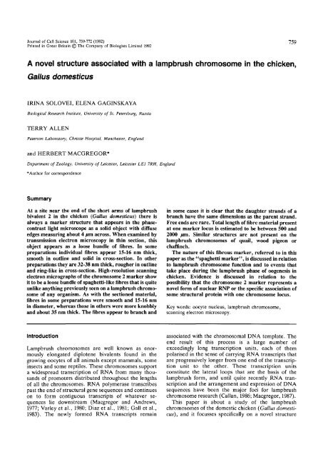
Fig. 8. Transmission electron micrograph (TEM) of a longitud<strong>in</strong>al section through <strong>the</strong> region of <strong>lampbrush</strong> bivalent 2 that<br />
<strong>in</strong>cludes LL22. The small unlabeUed arrows <strong>in</strong>dicate axial chromomeres. ch, position of a chiasma. The material of LL22<br />
lies on ei<strong>the</strong>r side of <strong>the</strong> <strong>chromosome</strong> axis and has a f<strong>in</strong>ely fibrous appearance that is quite different from <strong>the</strong> loop RNP of<br />
<strong>the</strong> rema<strong>in</strong>der of <strong>the</strong> <strong>chromosome</strong>. X7000.<br />
of <strong>lampbrush</strong> <strong>chromosome</strong>s. Anti-lam<strong>in</strong>s also bound<br />
specifically to pieces of oocyte nuclear envelope that lay<br />
alongside <strong>the</strong> <strong>chromosome</strong>s <strong>in</strong> <strong>lampbrush</strong> preparations.<br />
Control preparations, <strong>in</strong> which only <strong>the</strong> second (Fl'l'Clabelled)<br />
antibody was used, were entirely negative.<br />
Transmission electron microscopy of th<strong>in</strong> sections<br />
Electron micrographs have been made from sections <strong>in</strong><br />
two ranges of thickness. The thicker ones, 150-250 nm,<br />
sacrifice some of <strong>the</strong> details of f<strong>in</strong>e <strong>structure</strong> but give an<br />
overview of <strong>the</strong> relative positions of objects along <strong>the</strong><br />
length of a <strong>lampbrush</strong> <strong>chromosome</strong>. Th<strong>in</strong> sections,<br />
about 100 nm, allow <strong>in</strong>terpretation of f<strong>in</strong>e <strong>structure</strong>.<br />
Altoge<strong>the</strong>r 22 <strong>chromosome</strong>s 2 were embedded and<br />
sectioned for transmission electron microscopy.<br />
The marker at LL22 is relatively easy to locate <strong>in</strong><br />
longitud<strong>in</strong>al sections of end-embedded <strong>chromosome</strong>s<br />
on account of <strong>the</strong> dist<strong>in</strong>ctive appearance of its constituent<br />
strands. Sections through <strong>the</strong> region of <strong>the</strong> marker<br />
show it as a loose mass of <strong>in</strong>terwoven fibres ly<strong>in</strong>g<br />
around or to one side of <strong>the</strong> ma<strong>in</strong> <strong>chromosome</strong> axis<br />
(Fig. 8). The fibres are 15-16 nm wide. They appear<br />
straight or curved and of uniform electron contrast<br />
across <strong>the</strong>ir width <strong>in</strong> longitud<strong>in</strong>al and <strong>in</strong> transverse view<br />
(Figs 9 and 10). The entire mass of fibres as seen <strong>in</strong> a<br />
section through <strong>the</strong> widest part of LL22 occupies a<br />
region 4-5 /jm <strong>in</strong> diameter and it usually lies around or<br />
very near to one of <strong>the</strong> chromomeres of <strong>the</strong> <strong>chromosome</strong><br />
axis (Figs 8 and 9). The centre portion of <strong>the</strong> mass<br />
of fibres is much more compact than <strong>the</strong> outer regions<br />
(Fig. 9). Where a section passes through <strong>the</strong> chromomere<br />
it is evident that LL22 fibres are closely <strong>associated</strong><br />
<strong>with</strong> or penetrate right <strong>in</strong>to <strong>the</strong> substance of <strong>the</strong><br />
chromomere (Fig. 10). All of <strong>the</strong>se dimensions and<br />
characteristics were seen <strong>in</strong> <strong>the</strong> great majority of<br />
preparations that we used for th<strong>in</strong>-section<strong>in</strong>g and<br />
transmission electron microscopy.<br />
In an earlier series of sections from which we<br />
obta<strong>in</strong>ed electron micrographs, <strong>the</strong> general organisation<br />
of <strong>the</strong> LL22 marker was exactly as described<br />
above, apart from <strong>the</strong> fact that <strong>the</strong> <strong>in</strong>dividual fibres<br />
were about twice as thick (32 nm), <strong>the</strong>y were much<br />
rougher <strong>in</strong> appearance, sometimes show<strong>in</strong>g evidence of<br />
twist<strong>in</strong>g (Fig. 11), and <strong>in</strong> transverse section <strong>the</strong><br />
<strong>in</strong>dividual fibres showed a less dense central core 3-5 nm<br />
wide (Fig. 12). We suspect that <strong>in</strong> this first set of<br />
preparations some aspect of <strong>the</strong> fixation or dehydration<br />
produced ei<strong>the</strong>r a shorten<strong>in</strong>g and thicken<strong>in</strong>g of <strong>in</strong>dividual<br />
fibres or a coil<strong>in</strong>g of <strong>in</strong>dividual fibres to make<br />
<strong>the</strong>m appear shorter and thicker. Whichever state is<br />
artifactual, we consider it essential that both go on<br />
record, particularly s<strong>in</strong>ce two sets of material prepared<br />
for scann<strong>in</strong>g electron microscopy have also given LL22<br />
fibres <strong>with</strong> correspond<strong>in</strong>gly different properties.<br />
Scann<strong>in</strong>g electron microscopy<br />
The first set of scann<strong>in</strong>g electron micrographs (SEMs)<br />
that we obta<strong>in</strong>ed were from <strong>chromosome</strong>s that had<br />
been lightly centrifuged to flatten <strong>the</strong>m and stick <strong>the</strong>m<br />
to a glass coverslip, fixed <strong>in</strong> glutaraldehyde/osmium,<br />
critical-po<strong>in</strong>t dried and coated <strong>with</strong> chromium to<br />
produce an overall <strong>in</strong>crease <strong>in</strong> thickness of about 2 nm.<br />
In <strong>the</strong>se preparations <strong>the</strong> critical-po<strong>in</strong>t dry<strong>in</strong>g was good<br />
and <strong>in</strong>dividual <strong>chromosome</strong>s and <strong>the</strong>ir marker <strong>structure</strong>s<br />
could be identified <strong>with</strong> confidence. To judge from<br />
our experience <strong>with</strong> SEMs of amphibian <strong>lampbrush</strong>
Novel <strong>structure</strong> <strong>in</strong> <strong>lampbrush</strong> <strong>chromosome</strong>s 765<br />
Fig. 9. TEM of a section through <strong>the</strong> greater part of an LL22 marker show<strong>in</strong>g <strong>the</strong> relatively compact central portion <strong>with</strong><br />
axial chromomeric material (c) close by. x30,000.<br />
Fig. 10. TEM of a section through part of <strong>the</strong> same LL22 marker region as shown <strong>in</strong> Fig. 2. The arrow <strong>in</strong>dicates a close<br />
association between some of <strong>the</strong> LL22 fibres and a chromomere (arrow). This electron micrograph, as well as those shown<br />
<strong>in</strong> Figs 2 and 3, is from <strong>the</strong> second batch of material used for TEM studies <strong>in</strong> which <strong>the</strong> average fibre diameter was <strong>in</strong> <strong>the</strong><br />
region of 15-16 nm, <strong>the</strong> fibres appear<strong>in</strong>g smooth <strong>in</strong> outl<strong>in</strong>e and solid <strong>in</strong> cross-section. x80,000.<br />
Fig. 11. TEM of a section through part of an LL22 marker <strong>in</strong> <strong>the</strong> first batch of material used for TEM studies. Here <strong>the</strong><br />
<strong>in</strong>dividual fibres are thicker, 32 nm, rougher <strong>in</strong> outl<strong>in</strong>e and sometimes show signs of a helical sub<strong>structure</strong> (arrow).<br />
x80,000.<br />
Fig. 12. Same as Fig. 5 but show<strong>in</strong>g <strong>the</strong> r<strong>in</strong>g-like appearance of <strong>the</strong>se larger (32 nm) fibres <strong>in</strong> cross-section. x80,000.<br />
<strong>chromosome</strong>s (HM and TA), <strong>the</strong> chromosomal morphology<br />
and f<strong>in</strong>e <strong>structure</strong> were well preserved. Our<br />
studies were largely conf<strong>in</strong>ed to <strong>chromosome</strong> 2 and<br />
more specifically to <strong>the</strong> LL22 marker that had shown a<br />
dist<strong>in</strong>ctive fibrous <strong>structure</strong> <strong>in</strong> TEM sections. An<br />
example of a typical <strong>chromosome</strong> 2 is shown <strong>in</strong> Fig. 13.<br />
The giant loops at <strong>the</strong> end of <strong>the</strong> long arm of<br />
<strong>chromosome</strong> 2 have reta<strong>in</strong>ed <strong>the</strong>ir form and <strong>the</strong> LL22<br />
marker is conspicuous and loosely fibrous <strong>in</strong> appearance<br />
(Figs 13 and 14). At higher SEM magnifications<br />
(x 18,000 upwards) LL22 varies from a rough and<br />
compact texture <strong>in</strong> one region to a more open loose<br />
network of fibres <strong>in</strong> surround<strong>in</strong>g parts (Figs 14 and 15).<br />
There are few obvious free ends to <strong>the</strong> LL22 fibres.<br />
There is some evidence of branch<strong>in</strong>g of <strong>the</strong> fibres, but it<br />
is usually difficult to determ<strong>in</strong>e whe<strong>the</strong>r a fibre truly<br />
branches <strong>in</strong>to two, or two closely apposed fibres simply<br />
separate <strong>in</strong>to two s<strong>in</strong>gle ones. The fibres are all between<br />
32 and 38 nm wide and <strong>in</strong> many places <strong>the</strong>y show some<br />
evidence of a tightly twisted sub-<strong>structure</strong> (Figs 15 and
766 /. Solovei and o<strong>the</strong>rs<br />
Fig. 13. Low-magnification scann<strong>in</strong>g electron micrograph (SEM) of <strong>the</strong> whole of <strong>lampbrush</strong> bivalent 2 from <strong>the</strong> first set of<br />
material prepared for high-resolution SEM studies. The lateral loops and o<strong>the</strong>r <strong>structure</strong>s <strong>associated</strong> <strong>with</strong> <strong>the</strong> <strong>chromosome</strong>s<br />
are well preserved. The LL22 markers are at <strong>the</strong> left end of <strong>the</strong> bivalent and <strong>the</strong> TBL21 (telomeric bow loops) show at <strong>the</strong><br />
right-hand ends of <strong>the</strong> bivalent. x6,250.<br />
Fig. 14. Higher-magnification view of <strong>the</strong> LL22 regions on <strong>the</strong> bivalent shown <strong>in</strong> Fig. 13. The difference <strong>in</strong> appearances of<br />
<strong>the</strong> normal loop RNP and <strong>the</strong> spaghetti-like strands of <strong>the</strong> LL22 marker is particularly apparent <strong>in</strong> this picture. x6,250.<br />
16). Where <strong>the</strong> fibres are compacted toge<strong>the</strong>r <strong>the</strong>y<br />
frequently have a coarse knobbly appearance that could<br />
be <strong>in</strong>dicative of some supercoil<strong>in</strong>g (Fig. 16). At high<br />
magnification (>x 100,000) <strong>the</strong>se chromium-coated<br />
fibres have a smooth surface <strong>structure</strong> (Fig. 16).<br />
The second set of SEMs that we obta<strong>in</strong>ed were from<br />
<strong>chromosome</strong>s that were isolated, fixed and dried <strong>in</strong><br />
almost <strong>the</strong> same way except for <strong>the</strong> omission of osmium<br />
post-fixation. They were coated <strong>with</strong> gold/palladium to<br />
produce an overall <strong>in</strong>crease <strong>in</strong> thickness of about 5 nm.<br />
In <strong>the</strong>se preparations <strong>the</strong> lateral loops of <strong>the</strong> <strong>chromosome</strong>s<br />
had an appearance that suggested some loss of<br />
material dur<strong>in</strong>g isolation and less than optimal fixation.<br />
Never<strong>the</strong>less, critical-po<strong>in</strong>t dry<strong>in</strong>g was good and <strong>in</strong>di-<br />
vidual <strong>chromosome</strong>s and <strong>the</strong>ir marker <strong>structure</strong>s could<br />
be identified <strong>with</strong> confidence. In <strong>the</strong> first two preparations<br />
that we exam<strong>in</strong>ed <strong>in</strong> this series <strong>the</strong> material at<br />
locus LL22 on both half-bivalents was <strong>in</strong> two forms.<br />
One of <strong>the</strong>se was a smooth, irregularly shaped body<br />
about 1-2 [an across ly<strong>in</strong>g on <strong>the</strong> proximal side of <strong>the</strong><br />
locus and over or alongside <strong>the</strong> ma<strong>in</strong> <strong>chromosome</strong> axis.<br />
The o<strong>the</strong>r material was a complex network of fibres<br />
extend<strong>in</strong>g over an area of several square micrometres<br />
(Fig. 17). The same general organisation was seen <strong>in</strong><br />
several o<strong>the</strong>r preparations, although <strong>the</strong> smooth solid<br />
component was lack<strong>in</strong>g and LL22 consisted entirely of a<br />
meshwork of fibres. Individual fibres <strong>in</strong> LL22 markers<br />
from this second series of preparations were between 15
Novel <strong>structure</strong> <strong>in</strong> <strong>lampbrush</strong> <strong>chromosome</strong>s 767<br />
Fig. 15. Detail at x33,500 magnification of an LL22 marker from <strong>the</strong> first series of preparations. In part of <strong>the</strong> marker, <strong>the</strong><br />
strands are closely packed to give an almost solid <strong>structure</strong>, such as might expla<strong>in</strong> <strong>the</strong> dense region seen <strong>in</strong> phase-contrast<br />
micrographs.<br />
and 16 nm thick. This was best seen <strong>in</strong> preparations<br />
where <strong>the</strong> LL22 marker had been dispersed, probably<br />
mechanically, dur<strong>in</strong>g preparation (Figs 18 and 19).<br />
Fibres often appear to be fused <strong>with</strong> one ano<strong>the</strong>r, two<br />
or more toge<strong>the</strong>r, to produce strands of up to 50 nm<br />
wide. In <strong>the</strong> region of <strong>the</strong> smooth, solid component <strong>the</strong><br />
fibres show extensive fusion to produce strands up to<br />
100 nm thick and <strong>the</strong>y fuse <strong>with</strong> or adhere to <strong>the</strong> surface<br />
of <strong>the</strong> smooth body (Fig. 17). As <strong>in</strong> <strong>the</strong> first series of<br />
preparations, <strong>the</strong>re are few, if any, visible free ends.<br />
High magnification (xl00,000-x200,000) micrographs<br />
show <strong>the</strong> fibres <strong>with</strong> a coarse, granular surface <strong>structure</strong><br />
that we consider to be resolution of <strong>the</strong> gra<strong>in</strong> of <strong>the</strong><br />
gold/palladium coat<strong>in</strong>g (Fig. 19). In both sets of<br />
preparations <strong>the</strong> fibrous component of LL22 has an<br />
appearance that is quite different from that of nearby<br />
lateral loop RNP.<br />
Transmission electron microscopy of negative-sta<strong>in</strong>ed<br />
<strong>chromosome</strong>s<br />
The appearance of LL22 <strong>in</strong> negative-sta<strong>in</strong>ed whole<br />
mounts was exactly as expected on <strong>the</strong> basis of our SEM<br />
studies. Three aspects of <strong>the</strong> fibre <strong>structure</strong> were<br />
particularly clear <strong>in</strong> negative-sta<strong>in</strong>ed preparations (Fig.<br />
20). The fibres are smooth <strong>in</strong> outl<strong>in</strong>e and structurally<br />
homogeneous. The fibre diameter is between 32 and 37<br />
nm. Branch<strong>in</strong>g of <strong>the</strong> fibres is common but, where it<br />
occurs, <strong>the</strong> widths of <strong>the</strong> parent strand and <strong>the</strong> daughter<br />
branches are identical, which would not be expected if<br />
branch<strong>in</strong>g were a consequence of separation of two<br />
closely apposed strands.<br />
Discussion<br />
The phenomenon that we have described <strong>in</strong> this paper<br />
represents someth<strong>in</strong>g entirely new, <strong>in</strong> <strong>the</strong> sense that<br />
noth<strong>in</strong>g remotely comparable has ever been encountered<br />
<strong>in</strong> any previous study of <strong>lampbrush</strong> <strong>chromosome</strong>s.<br />
It has been demonstrated that <strong>in</strong> newt <strong>lampbrush</strong><br />
<strong>chromosome</strong>s all loops, <strong>in</strong>clud<strong>in</strong>g <strong>the</strong> larger "landmark"<br />
<strong>structure</strong>s, no matter what <strong>the</strong>ir appearance <strong>in</strong><br />
<strong>the</strong> light microscope, are made up of <strong>the</strong> same basic 30<br />
nm RNP particles (N'Da et al., 1986; Bonnanfant-Jais<br />
et al., 1991) and all loops exhibit <strong>the</strong> same basic<br />
organisation <strong>with</strong> nascent RNP transcripts attached by<br />
RNA polymerase molecules to <strong>the</strong> DNA loop axis from<br />
which <strong>the</strong>y were transcribed (Scheer et al., 1976). The<br />
two notable exceptions to this rule are <strong>the</strong> "spheres",<br />
such as were characterised by Callan and Lloyd (1960)<br />
on <strong>the</strong> 5th and 8th <strong>chromosome</strong>s of crested newts and<br />
have s<strong>in</strong>ce been shown to be present <strong>in</strong> germ<strong>in</strong>al<br />
vesicles of a wide variety of o<strong>the</strong>r vertebrates (Gall and<br />
Callan, 1989) and <strong>the</strong> "prote<strong>in</strong> bodies" described by<br />
Gag<strong>in</strong>skaya (1972b) and Khut<strong>in</strong>aeva et al. (1989) <strong>in</strong> <strong>the</strong><br />
oocyte nuclei of certa<strong>in</strong> species of bird. The LL22<br />
marker is not made up of 30 nm RNP particles, and<br />
does not resemble a <strong>lampbrush</strong> loop. It seems highly<br />
unlikely that it is related to <strong>the</strong> "spheres" of amphibian
768 /. Solovei and o<strong>the</strong>rs<br />
J6<br />
Fig. 16. High-resolution SEM photographed at an <strong>in</strong>strumental magnification of x 100,000 of part of an LL22 marker from<br />
<strong>the</strong> first series of preparations. Compare <strong>with</strong> Fig. 19. Here <strong>the</strong> fibres have received a high-resolution coat<strong>in</strong>g of chromium<br />
1-2 nm <strong>in</strong> thickness. The average fibre width, allow<strong>in</strong>g a total of 3 nm for coat<strong>in</strong>g thickness, is <strong>in</strong> <strong>the</strong> region of 35 nm.<br />
They have a smooth surface <strong>structure</strong> but an overall knobbly appearance, perhaps suggest<strong>in</strong>g some degree of supercoil<strong>in</strong>g.<br />
x192,000.<br />
oocytes and <strong>in</strong>deed it does not sta<strong>in</strong> by <strong>in</strong>direct<br />
immunofluorescence <strong>with</strong> antibodies that are known to<br />
react <strong>with</strong> sphere prote<strong>in</strong>s <strong>in</strong> Triturus, Notophthalmus<br />
and Xenopus (H. Macgregor, unpublished observations).<br />
It has to be admitted immediately that even after<br />
quite a wide-rang<strong>in</strong>g structural study and a range of<br />
experimental approaches we still do not know what<br />
LL22 is made of. Our objective at this stage must<br />
<strong>the</strong>refore be simply to publish <strong>the</strong> f<strong>in</strong>d<strong>in</strong>gs, <strong>in</strong>tegrate<br />
<strong>the</strong>m <strong>in</strong>to some reasonable hypo<strong>the</strong>ses and <strong>the</strong>n <strong>in</strong>vite<br />
o<strong>the</strong>r <strong>in</strong>vestigators to help <strong>in</strong> discover<strong>in</strong>g <strong>the</strong> nature and<br />
significance of this curious <strong>structure</strong>.<br />
There are three ma<strong>in</strong> possibilities. First, <strong>the</strong> material<br />
at LL22 could represent a <strong>novel</strong> form of RNP <strong>with</strong> a<br />
l<strong>in</strong>ear super<strong>structure</strong> ra<strong>the</strong>r than a particulate one;<br />
secondly, LL22 could be a <strong>novel</strong> form of chroma t<strong>in</strong>; and<br />
thirdly, LL22 could represent an accumulation of large<br />
amounts of a specific prote<strong>in</strong> or structural macromolecule<br />
at a s<strong>in</strong>gle chromosomal locus. Whichever of<br />
<strong>the</strong>se possibilities is nearest to <strong>the</strong> truth, one observation<br />
would seem to be of paramount importance.<br />
LL22 is a constant marker <strong>structure</strong> <strong>associated</strong> <strong>with</strong> a<br />
specific chromosomal locus, and it is present at <strong>the</strong> same<br />
position on both half-bivalents. Moreover, none of <strong>the</strong><br />
many chickens that we have exam<strong>in</strong>ed has been<br />
heterozygous for <strong>the</strong> presence of LL22. It is <strong>the</strong>refore<br />
difficult to avoid <strong>the</strong> conclusion that LL22 reflects some<br />
property of <strong>the</strong> chromosomal DNA at that locus.<br />
Undoubtedly <strong>the</strong> simplest and most conservative<br />
hypo<strong>the</strong>sis would be one <strong>in</strong>volv<strong>in</strong>g a <strong>novel</strong> form of<br />
transcription and packag<strong>in</strong>g of RNP. In this regard <strong>the</strong><br />
follow<strong>in</strong>g po<strong>in</strong>ts are significant. Few free ends are<br />
evident. They might, of course, be obscured by fusion<br />
or just general tangl<strong>in</strong>g of <strong>the</strong> material, but <strong>the</strong>y are<br />
probably not common. Careful modell<strong>in</strong>g of <strong>the</strong><br />
<strong>structure</strong>s shown <strong>in</strong> Figs 14 and 17, <strong>with</strong> close attention<br />
to scale, has shown that <strong>the</strong> total strand length would be<br />
of <strong>the</strong> order of 500 to 2000 pcm. Allow<strong>in</strong>g a factor of 5:1<br />
for normal foreshorten<strong>in</strong>g of RNP transcripts on<br />
<strong>lampbrush</strong> loops (Hill and Macgregor, 1980), <strong>the</strong>se<br />
lengths rise to 2.5 to 10 mm. If we allow ano<strong>the</strong>r factor<br />
of 5:1 for tight coil<strong>in</strong>g of <strong>the</strong> 15 nm fibre such as can be<br />
shown, aga<strong>in</strong> by scale modell<strong>in</strong>g, to produce <strong>the</strong> 35 nm<br />
fibres shown <strong>in</strong> Fig. 16, <strong>the</strong>n that particular marker<br />
might be estimated to conta<strong>in</strong> up to 50 mm of primary
Novel <strong>structure</strong> <strong>in</strong> lamp brush <strong>chromosome</strong>s 769<br />
Fig. 17. SEM view of an LL22 marker from <strong>the</strong> second set of preparations made for scann<strong>in</strong>g electron microscopy. The<br />
marker appears as a tangled mass of relatively smooth spaghetti-like fibres (s) ly<strong>in</strong>g alongside a smooth-surfaced solid mass<br />
of material. Note how <strong>the</strong> fibres become thicker <strong>in</strong> <strong>the</strong> region of <strong>the</strong> smooth solid <strong>structure</strong> and merge <strong>with</strong> its surface<br />
where <strong>the</strong>y touch (arrowhead). Small spherical bodies <strong>with</strong> <strong>the</strong> same general appearance as <strong>the</strong> large solid <strong>structure</strong> lie<br />
amongst <strong>the</strong> spaghetti fibres (arrows). x34,000.<br />
RNA transcript. That would seem to be an <strong>in</strong>ord<strong>in</strong>ately<br />
large amount of RNA to accumulate at one <strong>lampbrush</strong><br />
locus, but it is actually comparable to <strong>the</strong> amount that is<br />
<strong>associated</strong> <strong>with</strong> <strong>the</strong> axes of some of <strong>the</strong> large marker<br />
loops that regularly form on <strong>the</strong> <strong>lampbrush</strong> <strong>chromosome</strong>s<br />
of urodele amphibians. Could it be that LL22<br />
represents transcription of a highly repeated DNA<br />
sequence at a level that, for purely physical reasons<br />
<strong>with</strong><strong>in</strong> <strong>the</strong> environment of <strong>the</strong> small chicken germ<strong>in</strong>al<br />
vesicle, requires a system of packag<strong>in</strong>g and process<strong>in</strong>g<br />
that is different from that which operates on <strong>the</strong> normal<br />
<strong>lampbrush</strong> transcription unit? None of <strong>the</strong> evidence<br />
that is available conflicts <strong>with</strong> such a hypo<strong>the</strong>sis. The<br />
marker is characteristic of <strong>the</strong> <strong>lampbrush</strong> phase. Like<br />
most conspicuous marker <strong>structure</strong>s, it persists after <strong>the</strong><br />
normal loops have retracted and <strong>the</strong>n disappears right<br />
at <strong>the</strong> end of <strong>the</strong> <strong>lampbrush</strong> phase. The <strong>structure</strong> is<br />
reduced <strong>in</strong> size by both ribonuclease and proteases. The<br />
substance of LL22 has been reported to b<strong>in</strong>d total DNA<br />
from chicken under conditions that would favour<br />
hybridisation of DNA to nascent loop RNP (Hutchison,<br />
1987). However, recent experiments carried out <strong>in</strong> <strong>the</strong><br />
Leicester laboratory and <strong>in</strong> Dr. Hutchison's laboratory<br />
<strong>in</strong> <strong>the</strong> USA lead us to <strong>the</strong> conclusion that <strong>the</strong> b<strong>in</strong>d<strong>in</strong>g of<br />
labelled DNA to LL22 does not represent <strong>the</strong> formation<br />
of a hybrid nucleic acid complex. The details of <strong>the</strong>se<br />
experiments will be reported elsewhere <strong>in</strong> <strong>the</strong> context<br />
of a study of <strong>the</strong> arrangement of DNA sequences on<br />
<strong>lampbrush</strong> <strong>chromosome</strong>s of chickens.<br />
In a second hypo<strong>the</strong>sis, <strong>the</strong> material at LL22 would<br />
consist of chromosomal DNA and <strong>associated</strong> prote<strong>in</strong>s,<br />
essentially a modified form of chromat<strong>in</strong>. The evidence<br />
<strong>in</strong> support of this idea is m<strong>in</strong>imal. The LL22 fibres seem<br />
to be closely <strong>associated</strong> <strong>with</strong> or perhaps cont<strong>in</strong>uous <strong>with</strong><br />
<strong>the</strong> conventional chromat<strong>in</strong> of <strong>the</strong> <strong>chromosome</strong> axis.<br />
The general histochemistry of LL22 is not wholly<br />
<strong>in</strong>consistent <strong>with</strong> loosely organised DNP, and <strong>the</strong><br />
<strong>structure</strong> becomes smaller and more contrasty when<br />
exposed to DNase. Never<strong>the</strong>less, on all accounts we<br />
consider <strong>the</strong> modified chromat<strong>in</strong> hypo<strong>the</strong>sis to be<br />
unlikely and we will not consider it fur<strong>the</strong>r.<br />
A third hypo<strong>the</strong>sis would present LL22 as <strong>the</strong> result<br />
of a specific <strong>in</strong>teraction between a chromomeric DNA<br />
sequence and a specific macromolecule that was<br />
abundant <strong>in</strong> <strong>the</strong> germ<strong>in</strong>al vesicle. That hypo<strong>the</strong>sis starts<br />
<strong>with</strong> <strong>the</strong> premise that LL22 does not consist of<br />
nucleoprote<strong>in</strong> and has noth<strong>in</strong>g to do <strong>with</strong> transcription,<br />
and it takes special account of <strong>the</strong> fact that <strong>the</strong> strands<br />
of LL22 seem to cluster around what appears to be an<br />
axial chromomere and <strong>in</strong> some cases penetrate right<br />
<strong>in</strong>to <strong>the</strong> substance of <strong>the</strong> chromomere. The chromomere<br />
itself is relatively large. Identification of <strong>the</strong> ma<strong>in</strong>
770 /. Solovei and o<strong>the</strong>rs<br />
j<br />
Fig. 18. SEM detail (x58,500) of LL22 fibres <strong>in</strong> a preparation where <strong>the</strong>y were more dispersed, probably as a consequence<br />
of mechanical disturbance dur<strong>in</strong>g manual isolation of <strong>the</strong> <strong>chromosome</strong>s. Fusion of fibres produces strands of up to 100 nm<br />
<strong>in</strong> thickness. Round objects <strong>with</strong> <strong>the</strong> same general surface properties and appearance as <strong>the</strong> material of <strong>the</strong> fibres give <strong>the</strong><br />
impression of swell<strong>in</strong>gs along <strong>the</strong> lengths of some fibres.<br />
Fig. 19. High-resolution SEM, photographed at x 100,000 <strong>in</strong>strumental magnification, <strong>in</strong> which <strong>the</strong> gold/palladium coat<strong>in</strong>g<br />
on <strong>the</strong> surface of <strong>the</strong> fibres has been resolved. The average s<strong>in</strong>gle fibre width <strong>in</strong> micrographs such as this was estimated to<br />
be <strong>in</strong> <strong>the</strong> region of 15-16 nm. x 195,000. Compare <strong>with</strong> Fig. 16.<br />
molecular components of LL22 strands is, of course, of<br />
paramount importance. So far this has proved difficult.<br />
Nucleases have effects that could be <strong>in</strong>terpreted as<br />
<strong>in</strong>dicat<strong>in</strong>g <strong>the</strong> presence of ei<strong>the</strong>r DNA or RNA, and<br />
proteases fail to destroy <strong>the</strong> <strong>structure</strong> completely,<br />
although <strong>the</strong>y do remove a substantial part of its<br />
substance. Immunofluorescence experiments <strong>with</strong> antilam<strong>in</strong>s,<br />
anti-viment<strong>in</strong> and anti-histone (H. Macgregor,<br />
unpublished) antibodies have all proved <strong>in</strong>conclusive or<br />
negative, although only <strong>the</strong> anti-lam<strong>in</strong> immunofluorescence<br />
tests employed antibodies to prote<strong>in</strong>s from<br />
chickens.<br />
In <strong>the</strong> search for clues to <strong>the</strong> molecular nature of<br />
LL22 several quite simple observations have to be kept<br />
<strong>in</strong> m<strong>in</strong>d. The fibres are homogeneous and smooth, <strong>the</strong>y<br />
are capable of branch<strong>in</strong>g, and two different forms of<br />
<strong>the</strong>m (15 nm and 35 nm) have both been identified<br />
<strong>in</strong>dependently <strong>in</strong> two separate laboratories and employ<strong>in</strong>g<br />
different techniques, th<strong>in</strong> section<strong>in</strong>g and SEM.<br />
Structurally, <strong>the</strong> nearest resemblance is between LL22<br />
strands and <strong>the</strong> 'membranous tubules' identified <strong>in</strong><br />
association <strong>with</strong> nucleoli and <strong>the</strong> nuclear envelope <strong>in</strong><br />
germ<strong>in</strong>al vesicles of amphibians (Leon et al., 1991). The<br />
latter have a diameter of about 30 nm (accord<strong>in</strong>g to our<br />
measurements of Leon et al.'s micrographs). Leon et al.<br />
speculate that <strong>the</strong>se "membranous tubules" may represent<br />
a store of prefabricated nuclear lam<strong>in</strong>a components<br />
assembled <strong>in</strong> <strong>the</strong> germ<strong>in</strong>al vesicle for later use<br />
<strong>in</strong> oogenesis. Our own experiments <strong>with</strong> monoclonal<br />
antibodies to chicken lam<strong>in</strong>s would seem to rule out <strong>the</strong><br />
lam<strong>in</strong> hypo<strong>the</strong>sis, but we never<strong>the</strong>less consider that <strong>the</strong><br />
structural resemblance between LL22 fibres and Leon
Novel <strong>structure</strong> <strong>in</strong> <strong>lampbrush</strong> <strong>chromosome</strong>s 11\<br />
20 •;<br />
Fig. 20. Transmission electron micrograph of isolated LL22 fibres sta<strong>in</strong>ed <strong>with</strong> uranyl acetate for negative contrast. Fibre<br />
thickness here is estimated to be <strong>in</strong> <strong>the</strong> region of 35 nm. Note <strong>the</strong> apparent branch<strong>in</strong>g of fibres on <strong>the</strong> left of <strong>the</strong> picture,<br />
<strong>with</strong> all three arms of <strong>the</strong> branch be<strong>in</strong>g of <strong>the</strong> same thickness. x210,000.<br />
et al.'s "membranous tubules" and <strong>the</strong> fact that both<br />
occur <strong>in</strong> <strong>the</strong> same cell type and at correspond<strong>in</strong>g stages<br />
are likely to be significant.<br />
A search has been made for LL22-like material<br />
<strong>associated</strong> <strong>with</strong> <strong>the</strong> <strong>lampbrush</strong> <strong>chromosome</strong>s of o<strong>the</strong>r<br />
bird species, Japanese quail (Coturnix coturnix), wood<br />
pigeon (Columba palumbus) and chaff<strong>in</strong>ch (Fr<strong>in</strong>gilla<br />
coelebs). The first of <strong>the</strong>se species belongs to <strong>the</strong> same<br />
family, <strong>the</strong> Phasianidae, as <strong>the</strong> chicken. Noth<strong>in</strong>g<br />
resembl<strong>in</strong>g <strong>the</strong> LL22 marker could be found <strong>in</strong><br />
Japanese quail. Likewise, noth<strong>in</strong>g could be found <strong>in</strong><br />
wood pigeon, although one of <strong>the</strong> longer <strong>chromosome</strong>s<br />
of this species does carry an extremely large and loosely<br />
organised <strong>lampbrush</strong> marker such as might represent a<br />
counterpart for LL22 <strong>in</strong> chicken. The marker to which<br />
we refer is not related <strong>in</strong> any way to <strong>the</strong> many<br />
conspicuous "prote<strong>in</strong> bodies" described by Gag<strong>in</strong>skaya<br />
(1972b) and Khut<strong>in</strong>aeva et al. (1989) <strong>in</strong> pigeon<br />
{Columba livia) germ<strong>in</strong>al vesicles. It should be added<br />
that <strong>the</strong> wood pigeon marker has a f<strong>in</strong>e <strong>structure</strong>, as<br />
seen <strong>in</strong> scann<strong>in</strong>g electron micrographs, that is quite<br />
different from that of LL22 and from anyth<strong>in</strong>g<br />
previously seen on <strong>lampbrush</strong> <strong>chromosome</strong>s of birds or<br />
amphibians (Allen and Macgregor, unpublished observations).<br />
The Russian authors of this paper report that,<br />
on <strong>the</strong> basis of transmission EM studies of th<strong>in</strong> sections<br />
of oocyte nuclei, <strong>the</strong>re is noth<strong>in</strong>g comparable <strong>with</strong> <strong>the</strong><br />
LL22 marker on chaff<strong>in</strong>ch <strong>lampbrush</strong> <strong>chromosome</strong>s,<br />
although an exhaustive search by high-resolution<br />
scann<strong>in</strong>g electron microscopy has not been made.<br />
Whatever we may eventually discover about <strong>the</strong><br />
LL22 marker one th<strong>in</strong>g is certa<strong>in</strong>: it is an entirely <strong>novel</strong><br />
<strong>structure</strong> <strong>the</strong> like of which has never been seen before <strong>in</strong><br />
association <strong>with</strong> a <strong>lampbrush</strong> <strong>chromosome</strong>. It is a<br />
<strong>structure</strong> that can only be seen <strong>in</strong> th<strong>in</strong> sections<br />
exam<strong>in</strong>ed <strong>with</strong> a transmission electron microscope or <strong>in</strong><br />
whole mounts exam<strong>in</strong>ed <strong>with</strong> a high-resolution scann<strong>in</strong>g<br />
electron microscope. We th<strong>in</strong>k it may offer opportunities<br />
for some <strong>in</strong>terest<strong>in</strong>g new <strong>in</strong>sights <strong>in</strong>to <strong>the</strong> role of <strong>the</strong><br />
<strong>chromosome</strong>s and <strong>the</strong> germ<strong>in</strong>al vesicle <strong>in</strong> oogenesis.<br />
Throughout this paper we have referred to <strong>the</strong> object<br />
of study as LL22. The name was <strong>in</strong>troduced by<br />
Chelysheva et al. (1990) on account of <strong>the</strong> superficial<br />
resemblance of <strong>the</strong> object to <strong>the</strong> "lumpy loops"<br />
described by Callan and Lloyd (1960) on <strong>the</strong> <strong>lampbrush</strong><br />
<strong>chromosome</strong>s of crested newts. We now know that<br />
LL22 is not a <strong>lampbrush</strong> loop but has a peculiar identity<br />
of its own that has yet to be expla<strong>in</strong>ed. S<strong>in</strong>ce <strong>the</strong> very<br />
start of our SEM study we have referred to Chelysheva<br />
et al.'s (1990) LL22 as <strong>the</strong> "spaghetti marker", and we<br />
th<strong>in</strong>k it may be useful to reta<strong>in</strong> this morphologically<br />
descriptive name, just as Callan and Lloyd (1960) did<br />
<strong>with</strong> <strong>the</strong>ir "lumpy loops" "spheres" and "currant buns"<br />
on <strong>the</strong> <strong>lampbrush</strong> <strong>chromosome</strong>s of Triturus, until it is<br />
possible to def<strong>in</strong>e <strong>the</strong> molecular and functional nature<br />
of this bizarre <strong>structure</strong>.
772 /. Solovei and o<strong>the</strong>rs<br />
We thank The British Council and The Wellcome Trust for<br />
<strong>the</strong>ir help and for fund<strong>in</strong>g visits to <strong>the</strong> UK by <strong>the</strong> two Russian<br />
authors of this paper, so mak<strong>in</strong>g possible <strong>the</strong> collaborative<br />
effort that was needed to accomplish this research. We thank<br />
Lesley Barnett and Evie Roberts for <strong>the</strong>ir valuable technical<br />
assistance.<br />
References<br />
Ahmad, M. S. (1970). Development, <strong>structure</strong> and composition of<br />
<strong>lampbrush</strong> <strong>chromosome</strong>s <strong>in</strong> domestic fowl. Can. J. Genet. Cytol.<br />
12, 728-737.<br />
Bonnanfant-Jais, M. L., Penrad-Mobayed, M. and Angelier, N.<br />
(1991). Ultrastructural similarity <strong>in</strong> landmark loops of amphibian<br />
<strong>lampbrush</strong> <strong>chromosome</strong>s. Biol. Cell 71, 105-114.<br />
Callan, H. G. (1986). Lampbrush Chromosomes. Spr<strong>in</strong>ger Verlag,<br />
Berl<strong>in</strong>, Heidelberg, New York, Tokyo.<br />
Callan, H. G. and Lloyd, L. (1960). Lampbrush <strong>chromosome</strong>s of<br />
crested newts Triturus cristatus (Laurenti). Phil. Trans. Roy. Soc.<br />
Lond. B, 243, 135-219.<br />
Chelysheva, L. A., Solovei, I. V., Rodlonov, A. V., Yakovlev, A. F.<br />
and Gag<strong>in</strong>skaya, E. R. (1990). The <strong>lampbrush</strong> <strong>chromosome</strong>s of <strong>the</strong><br />
chicken: <strong>the</strong> cytological maps of macrobivalents. Cytology (Russ.)<br />
32, 303-316.<br />
Diaz, M. O., Barsacchl-Pilone, G., Mahon, K. A. and Gall, J. G.<br />
(1981). Transcripts from both strands of a satellite DNA occur on<br />
<strong>lampbrush</strong> <strong>chromosome</strong> loops of <strong>the</strong> newt Notophthalmus. Cell 24,<br />
649-659.<br />
Gag<strong>in</strong>skaya, E. R. (1972a). The nuclear <strong>structure</strong>s of oocytes <strong>in</strong> adult<br />
birds: <strong>chromosome</strong> behaviour dur<strong>in</strong>g <strong>the</strong> oocyte growth. Cytology<br />
(Russ.) 14, 426-432.<br />
Gag<strong>in</strong>skaya, E. R. (1972b). The nuclear <strong>structure</strong>s of oocytes <strong>in</strong> adult<br />
birds: prote<strong>in</strong> bodies and karyosphere. Cytology (Russ.) 14, 568-<br />
578.<br />
Gag<strong>in</strong>skaya, E. R., Kropotova, E. V., Tsvetkov, A. G. and Vengerov,<br />
Yu. Yu. (1984). Morphology of transcription <strong>in</strong> <strong>lampbrush</strong><br />
<strong>chromosome</strong>s from avian oocytes. Cytology (Russ.) 26, 1064-1065.<br />
Gall, J. G. (1954). Lampbrush <strong>chromosome</strong>s from oocyte nuclei of<br />
<strong>the</strong> newt. J. Morphol. 94, 283-352.<br />
Gall, J. G. and Callan, H. G. (1989). The sphere organelle conta<strong>in</strong>s<br />
small nuclear ribonucleoprote<strong>in</strong>s. Proc. Natl Acad. Set. U.S.A. 86,<br />
6635-6639.<br />
Gall, J. G., Diaz, M. O., Stephenson, E. C. and Mahon, J. A. (1983).<br />
The transcription unit of <strong>lampbrush</strong> <strong>chromosome</strong>s. In Gene<br />
Structure and Regulation <strong>in</strong> Development (41st Symposium of <strong>the</strong><br />
Society for Developmental Biology), pp. 137-146. Alan R. Liss,<br />
Inc., New York.<br />
Gall, J. G., Stephenson, E. C, Erba, H. P., Diaz, M. O. and<br />
Barsacchi-Pilone, G. (1981). Histone genes are located at <strong>the</strong><br />
sphere loci of newt <strong>lampbrush</strong> <strong>chromosome</strong>s. Chromosoma 84,<br />
159-171.<br />
Hill, R. S. and Macgregor, H. C. (1980). The development of<br />
<strong>lampbrush</strong> <strong>chromosome</strong>-type transcription <strong>in</strong> <strong>the</strong> early diplotene<br />
oocytes olXenopus laevis: an electron microscope analysis. /. Cell<br />
Sci. 44, 87-101.<br />
Hutchison, N. J. (1987). Lampbrush <strong>chromosome</strong>s of <strong>the</strong> chicken,<br />
Gallus domesticus. J. Cell Biol. 105, 1493-1500.<br />
Hutchison, N. J. and We<strong>in</strong>traub, H. (1983). Lampbrush <strong>chromosome</strong><br />
<strong>structure</strong> and expression <strong>in</strong> chicken oocytes. J. Cell Biol. 97, 388a.<br />
Khut<strong>in</strong>aeva, M. A., Kropotova, E. V. and Gag<strong>in</strong>skaya, E. R. (1989).<br />
Morphofunctional organisation peculiarities of <strong>the</strong> <strong>lampbrush</strong><br />
<strong>chromosome</strong>s of oocytes <strong>in</strong> <strong>the</strong> pigeon, Columbia livia. Cytology<br />
(Russ.) 31, 1185-1193.<br />
Koecke, H. U. and Muller, M. (1965). Formweschsel und Anzahl der<br />
Chromosomen bei Huhn und Ente. Naturwissenschaften 52, 483.<br />
Kropotova, E. V. and Gag<strong>in</strong>skaya, E. R. (1984). Lampbrush<br />
<strong>chromosome</strong>s from <strong>the</strong> Japanese quail oocytes: light and electron<br />
microscopic study. Cytology (Russ.) 26, 1008-1015.<br />
Leon, P., Kezer, J. and Schabtach, E. (1991). Fur<strong>the</strong>r observations on<br />
nucleolar tails <strong>in</strong> amphibian oocytes. J. Cell Sci. 99, 515-522.<br />
Macgregor, H. C. (1987). Lampbrush <strong>chromosome</strong>s. J. Cell Sci. 88, 7-<br />
9.<br />
Macgregor, H. C. and Andrews, C. (1977). The arrangement and<br />
transcription of "middle repetitive" DNA sequences on <strong>lampbrush</strong><br />
<strong>chromosome</strong>s of Triturus. Chromosoma 63, 109-126.<br />
Macgregor, H. C. and Varley, J. M. (1988). Work<strong>in</strong>g <strong>with</strong> Animal<br />
Chromosomes (second edition). John Wiley and Sons, Chichester<br />
and New York.<br />
Mott, M. R. and Callan, H. G. (1975). An electron microscope study<br />
of <strong>the</strong> <strong>lampbrush</strong> <strong>chromosome</strong>s of <strong>the</strong> newt Triturus cristatus. J.<br />
Cell Sci. 17, 241-261.<br />
N'Da, E., Bonnanfant-Jais, M. L., Penrad-Mobayed, M. and<br />
Angelier, N. (1986). Size uniformity of ribonucleoprote<strong>in</strong> matrix<br />
particles <strong>in</strong> loops of Pleurodeles waltlii <strong>lampbrush</strong> <strong>chromosome</strong>s<br />
visualized by electron microscopy. J. Cell Sci. 81, 17-27.<br />
Scheer, U., Frankc, M. F., Trendelenburg, M. F. and Spr<strong>in</strong>g, H.<br />
(1976). Classification of loops of <strong>lampbrush</strong> <strong>chromosome</strong>s<br />
accord<strong>in</strong>g to <strong>the</strong> arrangement of transcriptional complexes. J. Cell<br />
Sci. 22, 503-519.<br />
Solovei, I. V., Khut<strong>in</strong>aeva, M. A., Chelysheva, L. A., Rodionov, A. V.<br />
and Gag<strong>in</strong>skaya, E. R. (1990). Lampbrush sex bivalent of birds:<br />
identification and <strong>structure</strong>. Cytology (Russ.) 32, 817-825.<br />
Varley, J. M., Macgregor, H. C, Nardi, I., Andrews, C. and Erba, H.<br />
P. (1980). Cytological evidence of transcription of highly repeated<br />
DNA sequences dur<strong>in</strong>g <strong>the</strong> <strong>lampbrush</strong> stage <strong>in</strong> Triturus cristatus<br />
carnifex. Chromosoma 80, 289-307.<br />
(Received 23 August 1991 - Accepted, <strong>in</strong> revised form,<br />
9 January 1992)