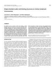
A novel structure associated with a lampbrush chromosome in the ...
A novel structure associated with a lampbrush chromosome in the ...
A novel structure associated with a lampbrush chromosome in the ...

Create successful ePaper yourself
Turn your PDF publications into a flip-book with our unique Google optimized e-Paper software.
760 /. Solovei and o<strong>the</strong>rs<br />
such as has never been seen <strong>in</strong> any previous studies of<br />
<strong>lampbrush</strong> <strong>chromosome</strong>s. The earliest studies of <strong>lampbrush</strong><br />
<strong>chromosome</strong>s from birds, carried out <strong>with</strong> <strong>the</strong><br />
techniques <strong>in</strong>troduced by Gall (1954) and Callan and<br />
Lloyd (I960), were those of Koecke and Muller (1965)<br />
and Ahmad (1970). However, methods for prepar<strong>in</strong>g<br />
avian <strong>lampbrush</strong> <strong>chromosome</strong>s for modern molecular<br />
and cytogenetic <strong>in</strong>vestigations were worked out much<br />
later and <strong>in</strong>dependently by Hutchison (Hutchison and<br />
We<strong>in</strong>traub, 1983; Hutchison, 1987) and by Gag<strong>in</strong>skaya<br />
and coworkers (Gag<strong>in</strong>skaya et al., 1984; Kropotova and<br />
Gag<strong>in</strong>skaya, 1984; Chelysheva et al., 1990; Solovei et<br />
al., 1990). These authors have confirmed that avian<br />
<strong>lampbrush</strong> <strong>chromosome</strong>s exhibit all <strong>the</strong> ma<strong>in</strong> features<br />
that have been described for those of amphibians.<br />
At an early stage <strong>in</strong> <strong>the</strong>ir studies of fixed and sta<strong>in</strong>ed<br />
isolated <strong>lampbrush</strong> <strong>chromosome</strong>s from chickens<br />
Chelysheva et al. (1990) noticed that <strong>the</strong> conspicuous<br />
object located near <strong>the</strong> end of <strong>the</strong> short arm of<br />
<strong>chromosome</strong> 2 was different from all o<strong>the</strong>r dist<strong>in</strong>ctive<br />
objects <strong>associated</strong> <strong>with</strong> <strong>the</strong> <strong>chromosome</strong>s <strong>in</strong> <strong>the</strong> sense<br />
that it never <strong>in</strong> any circumstances assumed <strong>the</strong><br />
appearance of a loop. Experimental treatments, such as<br />
reduc<strong>in</strong>g <strong>the</strong> ionic strength of <strong>the</strong> <strong>chromosome</strong> isolation<br />
medium, have <strong>the</strong> effect of remov<strong>in</strong>g <strong>the</strong> RNP from all<br />
loops and often expos<strong>in</strong>g <strong>the</strong> fundamental loop-like<br />
organisation of conspicuous <strong>structure</strong>s that <strong>in</strong> normal<br />
circumstances have a solid, round or irregular appearance<br />
- <strong>the</strong> "lumpy loops" of Callan and Lloyd (1960).<br />
The objects located at <strong>the</strong> Chelysheva et al. locus LL22<br />
never reacted <strong>in</strong> this manner.<br />
Subsequent to <strong>the</strong> studies published <strong>in</strong> 1990<br />
(Chelysheva et al., 1990; Solovei et al., 1990), it was<br />
decided to <strong>in</strong>vestigate certa<strong>in</strong> aspects of <strong>the</strong> f<strong>in</strong>e<br />
<strong>structure</strong> of chicken <strong>lampbrush</strong> <strong>chromosome</strong>s employ<strong>in</strong>g<br />
transmission electron microscopy (TEM) of sectioned<br />
material <strong>with</strong> special regard to <strong>the</strong> marker at<br />
LL22. Preparations of <strong>lampbrush</strong> <strong>chromosome</strong>s were<br />
made accord<strong>in</strong>g to <strong>the</strong> method described by Mott and<br />
Callan (1975). The short arm of <strong>chromosome</strong> 2 was<br />
specifically selected for th<strong>in</strong> section<strong>in</strong>g. Sections<br />
through most regions of <strong>the</strong> <strong>chromosome</strong>s produced no<br />
surprises. In general <strong>the</strong> <strong>structure</strong> of chromomeres and<br />
lateral loop ribonucleoprote<strong>in</strong> appeared more or less<br />
similar to that which had been described <strong>in</strong> electron<br />
micrographs of <strong>lampbrush</strong> <strong>chromosome</strong>s from o<strong>the</strong>r<br />
species (Mott and Callan, 1975; N'Da et al., 1986). The<br />
one outstand<strong>in</strong>g exception was <strong>the</strong> marker at position<br />
LL22. Sections through <strong>the</strong> region of <strong>the</strong> LL22 marker<br />
showed evidence of a loose bundle of strands hav<strong>in</strong>g an<br />
appearance that was quite different from that of<br />
neighbour<strong>in</strong>g loop ribonucleoprote<strong>in</strong>. The area of<br />
section occupied by <strong>the</strong> stranded material was about 4<br />
/an <strong>in</strong> diameter, which was consistent <strong>with</strong> <strong>the</strong> observed<br />
size of <strong>the</strong> LL22 marker as seen <strong>in</strong> unfixed <strong>lampbrush</strong><br />
preparations exam<strong>in</strong>ed <strong>with</strong> a phase-contrast microscope.<br />
In view of <strong>the</strong> extraord<strong>in</strong>ary appearance of <strong>the</strong> LL22<br />
marker <strong>in</strong> light-microscope preparations and TEM<br />
sections, it was decided immediately that this region<br />
required fur<strong>the</strong>r <strong>in</strong>vestigation and, tak<strong>in</strong>g account of<br />
<strong>the</strong> size of <strong>the</strong> whole marker and <strong>the</strong> sizes and pack<strong>in</strong>g<br />
of its <strong>in</strong>dividual strands, high-resolution scann<strong>in</strong>g<br />
electron microscopy (SEM) coupled <strong>with</strong> light microscopy<br />
and cytochemistry seemed <strong>the</strong> most promis<strong>in</strong>g<br />
approaches. In this paper we describe <strong>the</strong> <strong>structure</strong> of<br />
<strong>the</strong> LL22 marker, we present <strong>the</strong> results of some<br />
cytochemical tests and we offer some suggestions as to<br />
<strong>the</strong> nature of this hi<strong>the</strong>rto unknown class of object.<br />
Materials and methods<br />
Oocytes for <strong>lampbrush</strong> studies were obta<strong>in</strong>ed from commercial<br />
l<strong>in</strong>e Zarya 17 and Rhode Island Red crosses of between 21<br />
weeks ("po<strong>in</strong>t of lay") and 31 weeks old. Chickens were killed<br />
by cervical dislocation and <strong>the</strong> whole ovary was removed and<br />
placed <strong>in</strong> a clean dry beaker, covered <strong>with</strong> foil and stored on<br />
ice. The oocytes rema<strong>in</strong> <strong>in</strong> good condition for <strong>lampbrush</strong><br />
studies for a maximum of 12 h after removal from <strong>the</strong> bird.<br />
In <strong>the</strong> chicken, <strong>the</strong> <strong>lampbrush</strong> <strong>chromosome</strong>s are found <strong>in</strong><br />
oocytes rang<strong>in</strong>g from 0.13 mm to 2.5-3 mm <strong>in</strong> diameter<br />
(Gag<strong>in</strong>skaya, 1972a; Hutchison, 1987; Chelysheva et al.,<br />
1990). The best preparations of <strong>lampbrush</strong> <strong>chromosome</strong>s can<br />
be made from oocytes of between 1 mm and 2.5 mm diameter.<br />
The germ<strong>in</strong>al vesicle of a 1.2 mm oocyte measures about 100<br />
/an diameter and that of a 2.5 mm oocyte has a diameter of<br />
about 300 ism. For this study, oocytes of between 0.5 mm to<br />
3.5 mm diameter were ma<strong>in</strong>ly used, although some preparations<br />
were made from post-<strong>lampbrush</strong> oocytes of up to 7<br />
mm diameter. The <strong>chromosome</strong>s were isolated manually<br />
employ<strong>in</strong>g <strong>the</strong> standard <strong>lampbrush</strong> technique as described by<br />
Macgregor and Varley (1988) <strong>with</strong> some m<strong>in</strong>or modifications.<br />
Individual oocytes or small groups of oocytes of <strong>the</strong> required<br />
size were dissected out and transferred to a separate dish<br />
conta<strong>in</strong><strong>in</strong>g "5:1 + phosphate" (Gall et al., 1981; 83 mM KC1,<br />
17 mM NaCl, 6.5 mM Na2HPO4, 3.5 mM KH2PO4, pH 7.2).<br />
Chicken ovary, unlike <strong>the</strong> ovaries of amphibians, is densely<br />
compacted <strong>with</strong> collagen and connective tissue and it is best to<br />
remove most of this material from around <strong>the</strong> oocyte before<br />
try<strong>in</strong>g to isolate <strong>the</strong> germ<strong>in</strong>al vesicle. The germ<strong>in</strong>al vesicle was<br />
removed <strong>in</strong> 5:1 + phosphate by stabb<strong>in</strong>g <strong>the</strong> oocyte <strong>with</strong> a<br />
dissect<strong>in</strong>g needle and <strong>the</strong>n carefully watch<strong>in</strong>g as <strong>the</strong> yolky<br />
cytoplasm streamed out of <strong>the</strong> hole. The germ<strong>in</strong>al vesicle<br />
appears as a small clear <strong>in</strong>terruption <strong>in</strong> <strong>the</strong> flow of yolk as it<br />
emerges from <strong>the</strong> hole. It is perfectly round, turgid,<br />
transparent and glisten<strong>in</strong>g <strong>in</strong> appearance. The germ<strong>in</strong>al<br />
vesicle can be seen <strong>in</strong>side <strong>in</strong>tact oocytes of less than 1.2 mm<br />
diameter if bright substage transmitted illum<strong>in</strong>ation is used on<br />
<strong>the</strong> dissect<strong>in</strong>g b<strong>in</strong>ocular microscope. After isolation, <strong>the</strong><br />
germ<strong>in</strong>al vesicle is picked up immediately <strong>in</strong> a small-bore<br />
Pasteur pipette and transferred to a <strong>lampbrush</strong> observation<br />
chamber (Macgregor and Varley, 1988) conta<strong>in</strong><strong>in</strong>g 3/4<br />
strength 5:1 plus phosphate <strong>with</strong> 0.1% formaldehyde. The<br />
nuclear envelope is removed manually, ei<strong>the</strong>r <strong>with</strong> two pairs<br />
of very f<strong>in</strong>e forceps or <strong>with</strong> tungsten needles, and <strong>the</strong> nuclear<br />
contents are allowed to disperse on <strong>the</strong> bottom of <strong>the</strong><br />
observation chamber.<br />
Some preparations were exam<strong>in</strong>ed directly, <strong>with</strong>out centrifugation<br />
or fixation, by phase-contrast microscopy. In order to<br />
exam<strong>in</strong>e <strong>the</strong> effects of nucleases and proteases, <strong>chromosome</strong>s<br />
were ei<strong>the</strong>r dissected directly <strong>in</strong>to 3/4 strength 5:1 plus<br />
phosphate to which <strong>the</strong> enzyme had been added to a<br />
concentration of 0.1 mg/ml, or <strong>the</strong>y were isolated <strong>in</strong> 3/4<br />
strength 5:1 plus phosphate and <strong>the</strong> enzyme was added later.<br />
With <strong>the</strong> latter technique, <strong>the</strong> observation chamber was<br />
covered <strong>with</strong> a coverslip, <strong>the</strong> <strong>chromosome</strong>s were allowed to