A novel structure associated with a lampbrush chromosome in the ...
A novel structure associated with a lampbrush chromosome in the ...
A novel structure associated with a lampbrush chromosome in the ...

You also want an ePaper? Increase the reach of your titles
YUMPU automatically turns print PDFs into web optimized ePapers that Google loves.
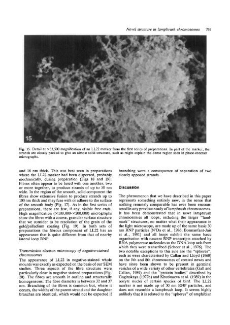
Novel <strong>structure</strong> <strong>in</strong> <strong>lampbrush</strong> <strong>chromosome</strong>s 767<br />
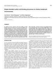
Fig. 15. Detail at x33,500 magnification of an LL22 marker from <strong>the</strong> first series of preparations. In part of <strong>the</strong> marker, <strong>the</strong><br />
strands are closely packed to give an almost solid <strong>structure</strong>, such as might expla<strong>in</strong> <strong>the</strong> dense region seen <strong>in</strong> phase-contrast<br />
micrographs.<br />
and 16 nm thick. This was best seen <strong>in</strong> preparations<br />
where <strong>the</strong> LL22 marker had been dispersed, probably<br />
mechanically, dur<strong>in</strong>g preparation (Figs 18 and 19).<br />
Fibres often appear to be fused <strong>with</strong> one ano<strong>the</strong>r, two<br />
or more toge<strong>the</strong>r, to produce strands of up to 50 nm<br />
wide. In <strong>the</strong> region of <strong>the</strong> smooth, solid component <strong>the</strong><br />
fibres show extensive fusion to produce strands up to<br />
100 nm thick and <strong>the</strong>y fuse <strong>with</strong> or adhere to <strong>the</strong> surface<br />
of <strong>the</strong> smooth body (Fig. 17). As <strong>in</strong> <strong>the</strong> first series of<br />
preparations, <strong>the</strong>re are few, if any, visible free ends.<br />
High magnification (xl00,000-x200,000) micrographs<br />
show <strong>the</strong> fibres <strong>with</strong> a coarse, granular surface <strong>structure</strong><br />
that we consider to be resolution of <strong>the</strong> gra<strong>in</strong> of <strong>the</strong><br />
gold/palladium coat<strong>in</strong>g (Fig. 19). In both sets of<br />
preparations <strong>the</strong> fibrous component of LL22 has an<br />
appearance that is quite different from that of nearby<br />
lateral loop RNP.<br />
Transmission electron microscopy of negative-sta<strong>in</strong>ed<br />
<strong>chromosome</strong>s<br />
The appearance of LL22 <strong>in</strong> negative-sta<strong>in</strong>ed whole<br />
mounts was exactly as expected on <strong>the</strong> basis of our SEM<br />
studies. Three aspects of <strong>the</strong> fibre <strong>structure</strong> were<br />
particularly clear <strong>in</strong> negative-sta<strong>in</strong>ed preparations (Fig.<br />
20). The fibres are smooth <strong>in</strong> outl<strong>in</strong>e and structurally<br />
homogeneous. The fibre diameter is between 32 and 37<br />
nm. Branch<strong>in</strong>g of <strong>the</strong> fibres is common but, where it<br />
occurs, <strong>the</strong> widths of <strong>the</strong> parent strand and <strong>the</strong> daughter<br />
branches are identical, which would not be expected if<br />
branch<strong>in</strong>g were a consequence of separation of two<br />
closely apposed strands.<br />
Discussion<br />
The phenomenon that we have described <strong>in</strong> this paper<br />
represents someth<strong>in</strong>g entirely new, <strong>in</strong> <strong>the</strong> sense that<br />
noth<strong>in</strong>g remotely comparable has ever been encountered<br />
<strong>in</strong> any previous study of <strong>lampbrush</strong> <strong>chromosome</strong>s.<br />
It has been demonstrated that <strong>in</strong> newt <strong>lampbrush</strong><br />
<strong>chromosome</strong>s all loops, <strong>in</strong>clud<strong>in</strong>g <strong>the</strong> larger "landmark"<br />
<strong>structure</strong>s, no matter what <strong>the</strong>ir appearance <strong>in</strong><br />
<strong>the</strong> light microscope, are made up of <strong>the</strong> same basic 30<br />
nm RNP particles (N'Da et al., 1986; Bonnanfant-Jais<br />
et al., 1991) and all loops exhibit <strong>the</strong> same basic<br />
organisation <strong>with</strong> nascent RNP transcripts attached by<br />
RNA polymerase molecules to <strong>the</strong> DNA loop axis from<br />
which <strong>the</strong>y were transcribed (Scheer et al., 1976). The<br />
two notable exceptions to this rule are <strong>the</strong> "spheres",<br />
such as were characterised by Callan and Lloyd (1960)<br />
on <strong>the</strong> 5th and 8th <strong>chromosome</strong>s of crested newts and<br />
have s<strong>in</strong>ce been shown to be present <strong>in</strong> germ<strong>in</strong>al<br />
vesicles of a wide variety of o<strong>the</strong>r vertebrates (Gall and<br />
Callan, 1989) and <strong>the</strong> "prote<strong>in</strong> bodies" described by<br />
Gag<strong>in</strong>skaya (1972b) and Khut<strong>in</strong>aeva et al. (1989) <strong>in</strong> <strong>the</strong><br />
oocyte nuclei of certa<strong>in</strong> species of bird. The LL22<br />
marker is not made up of 30 nm RNP particles, and<br />
does not resemble a <strong>lampbrush</strong> loop. It seems highly<br />
unlikely that it is related to <strong>the</strong> "spheres" of amphibian