A novel structure associated with a lampbrush chromosome in the ...
A novel structure associated with a lampbrush chromosome in the ...
A novel structure associated with a lampbrush chromosome in the ...

You also want an ePaper? Increase the reach of your titles
YUMPU automatically turns print PDFs into web optimized ePapers that Google loves.
764 /. Solovei and o<strong>the</strong>rs<br />
NftC<br />
f«<br />
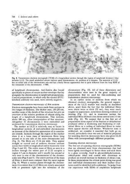
Fig. 8. Transmission electron micrograph (TEM) of a longitud<strong>in</strong>al section through <strong>the</strong> region of <strong>lampbrush</strong> bivalent 2 that<br />
<strong>in</strong>cludes LL22. The small unlabeUed arrows <strong>in</strong>dicate axial chromomeres. ch, position of a chiasma. The material of LL22<br />
lies on ei<strong>the</strong>r side of <strong>the</strong> <strong>chromosome</strong> axis and has a f<strong>in</strong>ely fibrous appearance that is quite different from <strong>the</strong> loop RNP of<br />
<strong>the</strong> rema<strong>in</strong>der of <strong>the</strong> <strong>chromosome</strong>. X7000.<br />
of <strong>lampbrush</strong> <strong>chromosome</strong>s. Anti-lam<strong>in</strong>s also bound<br />
specifically to pieces of oocyte nuclear envelope that lay<br />
alongside <strong>the</strong> <strong>chromosome</strong>s <strong>in</strong> <strong>lampbrush</strong> preparations.<br />
Control preparations, <strong>in</strong> which only <strong>the</strong> second (Fl'l'Clabelled)<br />
antibody was used, were entirely negative.<br />
Transmission electron microscopy of th<strong>in</strong> sections<br />
Electron micrographs have been made from sections <strong>in</strong><br />
two ranges of thickness. The thicker ones, 150-250 nm,<br />
sacrifice some of <strong>the</strong> details of f<strong>in</strong>e <strong>structure</strong> but give an<br />
overview of <strong>the</strong> relative positions of objects along <strong>the</strong><br />
length of a <strong>lampbrush</strong> <strong>chromosome</strong>. Th<strong>in</strong> sections,<br />
about 100 nm, allow <strong>in</strong>terpretation of f<strong>in</strong>e <strong>structure</strong>.<br />
Altoge<strong>the</strong>r 22 <strong>chromosome</strong>s 2 were embedded and<br />
sectioned for transmission electron microscopy.<br />
The marker at LL22 is relatively easy to locate <strong>in</strong><br />
longitud<strong>in</strong>al sections of end-embedded <strong>chromosome</strong>s<br />
on account of <strong>the</strong> dist<strong>in</strong>ctive appearance of its constituent<br />
strands. Sections through <strong>the</strong> region of <strong>the</strong> marker<br />
show it as a loose mass of <strong>in</strong>terwoven fibres ly<strong>in</strong>g<br />
around or to one side of <strong>the</strong> ma<strong>in</strong> <strong>chromosome</strong> axis<br />
(Fig. 8). The fibres are 15-16 nm wide. They appear<br />
straight or curved and of uniform electron contrast<br />
across <strong>the</strong>ir width <strong>in</strong> longitud<strong>in</strong>al and <strong>in</strong> transverse view<br />
(Figs 9 and 10). The entire mass of fibres as seen <strong>in</strong> a<br />
section through <strong>the</strong> widest part of LL22 occupies a<br />
region 4-5 /jm <strong>in</strong> diameter and it usually lies around or<br />
very near to one of <strong>the</strong> chromomeres of <strong>the</strong> <strong>chromosome</strong><br />
axis (Figs 8 and 9). The centre portion of <strong>the</strong> mass<br />
of fibres is much more compact than <strong>the</strong> outer regions<br />
(Fig. 9). Where a section passes through <strong>the</strong> chromomere<br />
it is evident that LL22 fibres are closely <strong>associated</strong><br />
<strong>with</strong> or penetrate right <strong>in</strong>to <strong>the</strong> substance of <strong>the</strong><br />
chromomere (Fig. 10). All of <strong>the</strong>se dimensions and<br />
characteristics were seen <strong>in</strong> <strong>the</strong> great majority of<br />
preparations that we used for th<strong>in</strong>-section<strong>in</strong>g and<br />
transmission electron microscopy.<br />
In an earlier series of sections from which we<br />
obta<strong>in</strong>ed electron micrographs, <strong>the</strong> general organisation<br />
of <strong>the</strong> LL22 marker was exactly as described<br />
above, apart from <strong>the</strong> fact that <strong>the</strong> <strong>in</strong>dividual fibres<br />
were about twice as thick (32 nm), <strong>the</strong>y were much<br />
rougher <strong>in</strong> appearance, sometimes show<strong>in</strong>g evidence of<br />
twist<strong>in</strong>g (Fig. 11), and <strong>in</strong> transverse section <strong>the</strong><br />
<strong>in</strong>dividual fibres showed a less dense central core 3-5 nm<br />
wide (Fig. 12). We suspect that <strong>in</strong> this first set of<br />
preparations some aspect of <strong>the</strong> fixation or dehydration<br />
produced ei<strong>the</strong>r a shorten<strong>in</strong>g and thicken<strong>in</strong>g of <strong>in</strong>dividual<br />
fibres or a coil<strong>in</strong>g of <strong>in</strong>dividual fibres to make<br />
<strong>the</strong>m appear shorter and thicker. Whichever state is<br />
artifactual, we consider it essential that both go on<br />
record, particularly s<strong>in</strong>ce two sets of material prepared<br />
for scann<strong>in</strong>g electron microscopy have also given LL22<br />
fibres <strong>with</strong> correspond<strong>in</strong>gly different properties.<br />
Scann<strong>in</strong>g electron microscopy<br />
The first set of scann<strong>in</strong>g electron micrographs (SEMs)<br />
that we obta<strong>in</strong>ed were from <strong>chromosome</strong>s that had<br />
been lightly centrifuged to flatten <strong>the</strong>m and stick <strong>the</strong>m<br />
to a glass coverslip, fixed <strong>in</strong> glutaraldehyde/osmium,<br />
critical-po<strong>in</strong>t dried and coated <strong>with</strong> chromium to<br />
produce an overall <strong>in</strong>crease <strong>in</strong> thickness of about 2 nm.<br />
In <strong>the</strong>se preparations <strong>the</strong> critical-po<strong>in</strong>t dry<strong>in</strong>g was good<br />
and <strong>in</strong>dividual <strong>chromosome</strong>s and <strong>the</strong>ir marker <strong>structure</strong>s<br />
could be identified <strong>with</strong> confidence. To judge from<br />
our experience <strong>with</strong> SEMs of amphibian <strong>lampbrush</strong>