A novel structure associated with a lampbrush chromosome in the ...
A novel structure associated with a lampbrush chromosome in the ...
A novel structure associated with a lampbrush chromosome in the ...

Create successful ePaper yourself
Turn your PDF publications into a flip-book with our unique Google optimized e-Paper software.
p<br />
Novel <strong>structure</strong> <strong>in</strong> <strong>lampbrush</strong> <strong>chromosome</strong>s 763<br />
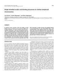
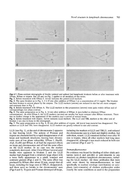
Figs 2-7. Phase-contrast micrographs of freshly isolated and unfixed 2nd <strong>lampbrush</strong> bivalents before or after treatment <strong>with</strong><br />
DNase, RNase or tryps<strong>in</strong>. Bar (20 /an) on Fig. 2 applies to all members of this series.<br />
Fig. 2. Before treatment <strong>with</strong> DNase I. Arrow <strong>in</strong>dicates <strong>the</strong> paired LL22 markers.<br />
Fig. 3. The same bivalent as <strong>in</strong> Fig. 2, 2 h 15 m<strong>in</strong> after addition of DNase I at a concentration of 0.1 mg/ml. The bivalent<br />
has been broken <strong>in</strong> several places by <strong>the</strong> enzyme. The LL22 markers (arrows) are reduced <strong>in</strong> size but are more compact<br />
and contrasty than <strong>in</strong> Fig. 2.<br />
Fig. 4. Before treatment <strong>with</strong> RNase A. The LL22 markers <strong>in</strong> this preparation (arrows) were quite widely diffuse and of<br />
correspond<strong>in</strong>gly low phase-contrast.<br />
Fig. 5. The same preparation as <strong>in</strong> Fig. 4, 14 m<strong>in</strong> after addition of RNase A (pre-boiled to elim<strong>in</strong>ate DNase<br />
contam<strong>in</strong>ation). As <strong>with</strong> DNase I, <strong>the</strong> LL22 markers (arrows) are smaller but more compact after RNase treatment. There<br />
was no fur<strong>the</strong>r change <strong>in</strong> <strong>the</strong> appearance of <strong>the</strong> markers over a period of several hours.<br />
Fig. 6. Before treatment <strong>with</strong> tryps<strong>in</strong>. Arrow <strong>in</strong>dicates LL22 markers. The LL21 and TBL markers at <strong>the</strong> o<strong>the</strong>r end of<br />
bivalent 2 are also <strong>in</strong> focus <strong>in</strong> this micrograph.<br />
Fig. 7. The same preparation as <strong>in</strong> Fig. 6, 35 m<strong>in</strong> after addition of tryps<strong>in</strong>. All lateral loop material has disappeared. The<br />
markers at LL21 and TBL have disappeared. LL22 markers are greatly reduced <strong>in</strong> size and contrast.<br />
LL21 (see Fig. 1), at <strong>the</strong> end of <strong>chromosome</strong> 2 opposite<br />
to that bear<strong>in</strong>g LL22. The actions of Pronase and<br />
tryps<strong>in</strong> are characterised by a rapid disappearance of all<br />
loops and landmark <strong>structure</strong>s, leav<strong>in</strong>g bare <strong>chromosome</strong><br />
axes of low phase retractility. DNase I, Haelll,<br />
Alul, EcoRl and RNase A all had <strong>the</strong> expected effects<br />
on loops and <strong>chromosome</strong>s and all had <strong>the</strong> same effect<br />
on <strong>the</strong> LL22 marker. In no case was <strong>the</strong> marker<br />
completely destroyed. After 2 h <strong>in</strong> DNase I several axial<br />
breaks were apparent <strong>in</strong> bivalent 2 and <strong>the</strong> LL22<br />
marker had changed from a relatively large object <strong>with</strong><br />
a loose fluffy appearance to a small, compact and<br />
contrasty granule (Figs 2 and 3). The same effect was<br />
produced by RNase after about 20 m<strong>in</strong>, except that all<br />
loops, <strong>in</strong>clud<strong>in</strong>g <strong>the</strong> large ones at TBL21, disappeared,<br />
and <strong>the</strong>re were no breaks <strong>in</strong> <strong>the</strong> <strong>chromosome</strong>s' axes<br />
(Figs 4 and 5). Tryps<strong>in</strong> and Pronase destroyed all loops,<br />
<strong>in</strong>clud<strong>in</strong>g <strong>the</strong> markers at LL21 and TBL21, and reduced<br />
<strong>the</strong> <strong>chromosome</strong> axis to a fa<strong>in</strong>t and slightly swollen, but<br />
unbroken, strand. LL22 rema<strong>in</strong>ed dist<strong>in</strong>ct even after 50<br />
m<strong>in</strong> <strong>in</strong> tryps<strong>in</strong>, when all o<strong>the</strong>r markers had long s<strong>in</strong>ce<br />
disappeared, although it was much reduced <strong>in</strong> both size<br />
and contrast (Figs 6 and 7).<br />
Imm unofluo rescence<br />
No evidence was found for b<strong>in</strong>d<strong>in</strong>g of ei<strong>the</strong>r chick antilam<strong>in</strong>s<br />
(anti-A and anti-B2) or anti-viment<strong>in</strong> to any<br />
<strong>structure</strong> on chicken <strong>lampbrush</strong> <strong>chromosome</strong>s, <strong>in</strong>clud<strong>in</strong>g<br />
<strong>the</strong> LL22 marker. All three antibodies that were<br />
used were shown by immunofluorescence to b<strong>in</strong>d<br />
specifically to nuclear envelope (anti-lam<strong>in</strong>s) or cytoplasm<br />
(anti-viment<strong>in</strong>) of chicken erythrocytes under<br />
exactly <strong>the</strong> same conditions as used <strong>in</strong> <strong>the</strong> preparation