Gorlin syndrome: the PTCH gene links ocular developmental defects ...
Gorlin syndrome: the PTCH gene links ocular developmental defects ...
Gorlin syndrome: the PTCH gene links ocular developmental defects ...

You also want an ePaper? Increase the reach of your titles
YUMPU automatically turns print PDFs into web optimized ePapers that Google loves.
Downloaded from<br />
bjo.bmj.com on July 1, 2013 - Published by group.bmj.com<br />
<strong>Gorlin</strong> <strong>syndrome</strong> 989<br />
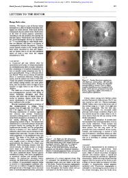
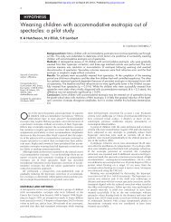
Figure 2 Sagittal section of MRI scan of patient demonstrating<br />
enhancing mass in <strong>the</strong> posterior fossa (long arrow), and a fur<strong>the</strong>r<br />
enhancing lesion in <strong>the</strong> pineal region (short arrow).<br />
number of neuroectodermal markers (MB84, CD 56, NSE),<br />
and <strong>the</strong> proliferation marker, Ki67, showed a high cell<br />
turnover in <strong>the</strong> cellular areas. In summary <strong>the</strong> features were<br />
consistent with a desmoplastic medulloblastoma.<br />
After surgery, MRI of <strong>the</strong> brain also showed <strong>the</strong> previously<br />
noted hetero<strong>gene</strong>ously enhancing partly cystic pineal mass<br />
but no evidence of residual posterior fossa tumour.<br />
Cerebrospinal fluid cytology did not reveal <strong>the</strong> presence of<br />
malignant cells. A Hickman line was inserted for delivery of<br />
treatment and bone marrow was harvested. Treatment was<br />
with <strong>the</strong> Infant PNET (primitive neuroectodermal tumour)<br />
protocol, which consists of six courses of dose intensive<br />
‘‘induction’’ chemo<strong>the</strong>rapy with growth factor support. This<br />
is usually followed by focal radio<strong>the</strong>rapy and consolidation<br />
chemo<strong>the</strong>rapy. Following induction chemo<strong>the</strong>rapy <strong>the</strong>re was<br />
no evidence of residual or recurrent tumour. The parents<br />
decided against radio<strong>the</strong>rapy after careful discussion with <strong>the</strong><br />
treating team and with a second oncologist, because of <strong>the</strong><br />
possible neuropsychological late effects. Follow up included<br />
3 monthly MRI spine and brain scans. The last was<br />
30 months after surgery with no recurrence demonstrated.<br />
She had <strong>the</strong> orbital cyst excised at <strong>the</strong> age of 4 years<br />
8 months.<br />
The unusual association of early onset medulloblastoma<br />
with microphthalmia suggested this may be <strong>Gorlin</strong> <strong>syndrome</strong><br />
and we undertook mutational analysis of <strong>the</strong> <strong>PTCH</strong> <strong>gene</strong>.<br />
Methodology<br />
Exons of <strong>the</strong> <strong>PTCH</strong> <strong>gene</strong> were screened by combined SSCP<br />
and heteroduplex analysis. 15 Exons that showed a variant<br />
band pattern were sequenced to confirm <strong>the</strong> presence of a<br />
mutation.<br />
RESULTS<br />
A deletion of G at nucleotide position 1402 within exon 10 of<br />
<strong>the</strong> <strong>PTCH</strong> <strong>gene</strong> was detected by sequencing. This frameshift<br />
mutation results in <strong>the</strong> introduction of a stop codon within<br />
exon 10 and is predicted to result in <strong>the</strong> translation of a<br />
truncated <strong>PTCH</strong> protein (fig 3). Nei<strong>the</strong>r parent had this<br />
mutation on sequencing of lymphocyte DNA.<br />
DISCUSSION<br />
Although <strong>ocular</strong> phenotypes listed for <strong>Gorlin</strong> <strong>syndrome</strong><br />
include microphthalmia and coloboma, <strong>the</strong>se are relatively<br />
rare signs and no case of microphthalmia with cyst has been<br />
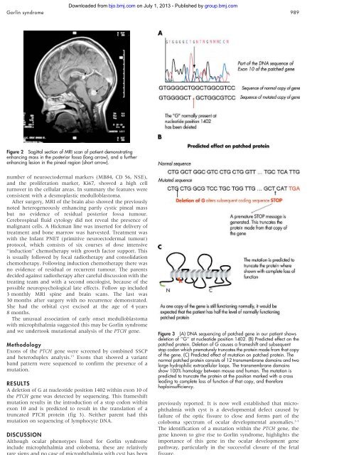
Figure 3 (A) DNA sequencing of patched <strong>gene</strong> in our patient shows<br />
deletion of ‘‘G’’ at nucleotide position 1402. (B) Predicted effect on <strong>the</strong><br />
patched protein. Deletion of G causes a frameshift and subsequent<br />
stop codon which prematurely truncates <strong>the</strong> protein made from that copy<br />
of <strong>the</strong> <strong>gene</strong>. (C) Predicted effect of mutation on patched protein. The<br />
normal patched protein consists of 12 transmembrane domains and two<br />
large hydrophilic extracellular loops. The transmembrane domains<br />
show 100% homology between mouse and human. The mutation is<br />
predicted to truncate <strong>the</strong> protein at <strong>the</strong> position marked with a cross<br />
leading to complete loss of function of that copy, and <strong>the</strong>refore<br />
haploinsufficiency.<br />
previously reported. It is now well established that microphthalmia<br />
with cyst is a <strong>developmental</strong> defect caused by<br />
failure of <strong>the</strong> optic fissure to close and forms part of <strong>the</strong><br />
coloboma spectrum of <strong>ocular</strong> <strong>developmental</strong> anomalies. 1–3<br />
The identification of a mutation within <strong>the</strong> <strong>PTCH</strong> <strong>gene</strong>, <strong>the</strong><br />
<strong>gene</strong> known to give rise to <strong>Gorlin</strong> <strong>syndrome</strong>, highlights <strong>the</strong><br />
importance of this <strong>gene</strong> in <strong>the</strong> <strong>ocular</strong> development <strong>gene</strong><br />
pathway, particularly in <strong>the</strong> successful closure of <strong>the</strong> fetal<br />
fissure.<br />
www.bjophthalmol.com