Gorlin syndrome: the PTCH gene links ocular developmental defects ...
Gorlin syndrome: the PTCH gene links ocular developmental defects ...
Gorlin syndrome: the PTCH gene links ocular developmental defects ...

Create successful ePaper yourself
Turn your PDF publications into a flip-book with our unique Google optimized e-Paper software.
988<br />
SCIENTIFIC REPORT<br />
<strong>Gorlin</strong> <strong>syndrome</strong>: <strong>the</strong> <strong>PTCH</strong> <strong>gene</strong> <strong>links</strong> <strong>ocular</strong> <strong>developmental</strong><br />
<strong>defects</strong> and tumour formation<br />
N K Ragge, A Salt, J R O Collin, A Michalski, P A Farndon<br />
...............................................................................................................................<br />
Aim: To identify a <strong>gene</strong> linking microphthalmia with cyst with<br />
early onset medulloblastoma.<br />
Methods: Mutation analysis of <strong>the</strong> <strong>PTCH</strong> <strong>gene</strong>.<br />
Results: A mutation in exon 10 of <strong>the</strong> <strong>PTCH</strong> <strong>gene</strong> was<br />
identified, confirming a diagnosis of <strong>Gorlin</strong> <strong>syndrome</strong>.<br />
Conclusions: This is <strong>the</strong> first <strong>gene</strong>tically identified mutation<br />
giving rise to microphthalmia with cyst and provides a<br />
valuable link in <strong>the</strong> eye <strong>developmental</strong> <strong>gene</strong> pathway.<br />
M icrophthalmia<br />
with cyst is a relatively rare <strong>ocular</strong><br />
<strong>developmental</strong> defect that is caused by failure of optic<br />
fissure closure, thus forming part of <strong>the</strong> spectrum of<br />
colobomatous abnormalities. 1–3 Although microphthalmia<br />
with cyst is usually an isolated phenomenon, it can occur<br />
in association with systemic abnormalities notably clefting<br />
<strong>syndrome</strong>s, oculo-cerebro-cutaneous <strong>syndrome</strong>, branchiooculo-facial<br />
<strong>syndrome</strong>, and cardiovascular anomalies including<br />
coarctation of <strong>the</strong> aorta and atrial septal <strong>defects</strong>. 4<br />
However, any described conditions and <strong>gene</strong>s (for example<br />
SHH (sonic hedgehog), or SIX3) associated with coloboma<br />
formation could in <strong>the</strong>ory also be associated with microphthalmia<br />
or anophthalmia with cyst formation. 5 6 Isolated<br />
microphthalmia with cyst is usually sporadic, with occasional<br />
familial occurrence described, 7 including a family with<br />
autosomal recessive inheritance. 8 All inheritance patterns<br />
have been described for microphthalmia with coloboma, and<br />
in <strong>the</strong>ory this could also be seen in microphthalmia with cyst<br />
1 9 10<br />
formation.<br />
Microphthalmia, with or without coloboma, is one of<br />
around 100 potential features seen in <strong>the</strong> naevoid basal cell<br />
carcinoma (BCC) <strong>syndrome</strong> or <strong>Gorlin</strong> <strong>syndrome</strong>, an autosomal<br />
dominant condition first described by <strong>Gorlin</strong> and<br />
Goltz. 11 12 The o<strong>the</strong>r <strong>ocular</strong> features include anterior segment<br />
dys<strong>gene</strong>sis with cataract and Peter’s anomaly, and vitreoretinal<br />
anomalies including epiretinal membrane formation,<br />
myelinated nerve fibres, and persistent fetal hyaloid anomalies.<br />
13 The main systemic features include <strong>developmental</strong><br />
<strong>defects</strong>, such as skeletal abnormalities (bifid ribs and<br />
kyphoscoliosis), palmar and plantar pits, odontogenic keratocysts,<br />
falx calcification, as well as a predisposition to<br />
tumour development, including multiple basal cell carcinomas,<br />
medulloblastoma, and rhabdomyosarcoma. The <strong>gene</strong> for<br />
<strong>Gorlin</strong> <strong>syndrome</strong> has been identified as <strong>the</strong> human homologue<br />
of <strong>the</strong> Drosophila segment polarity <strong>gene</strong>, ‘‘patched,’’ 14<br />
known as <strong>the</strong> <strong>PTCH</strong> <strong>gene</strong>. We present an unusual case of<br />
microphthalmia with cyst associated with early medulloblastoma<br />
formation where a mutation within <strong>the</strong> <strong>PTCH</strong> <strong>gene</strong><br />
has been identified, confirming a diagnosis of <strong>Gorlin</strong><br />
<strong>syndrome</strong>. We discuss <strong>the</strong> implications of <strong>developmental</strong><br />
<strong>ocular</strong> <strong>gene</strong> <strong>defects</strong> as indicators of tumour suppressor <strong>gene</strong><br />
<strong>syndrome</strong>s.<br />
www.bjophthalmol.com<br />
Downloaded from<br />
bjo.bmj.com on July 1, 2013 - Published by group.bmj.com<br />
Br J Ophthalmol 2005;89:988–991. doi: 10.1136/bjo.2004.061390<br />
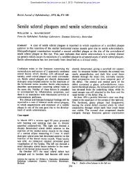
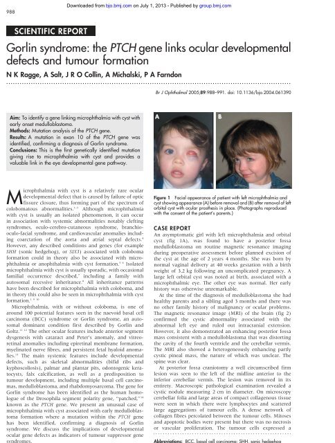
Figure 1 Facial appearance of patient with left microphthalmia and<br />
cyst showing appearance (A) before removal and (B) after removal of left<br />
orbital cyst with <strong>ocular</strong> pros<strong>the</strong>sis in place. (Photographs reproduced<br />
with <strong>the</strong> consent of <strong>the</strong> patient’s parents.)<br />
CASE REPORT<br />
An asymptomatic girl with left microphthalmia and orbital<br />
cyst (fig 1A), was found to have a posterior fossa<br />
medulloblastoma on routine magnetic resonance imaging<br />
during preoperative assessment before planned excision of<br />
<strong>the</strong> cyst at <strong>the</strong> age of 2 years 4 months. She was born by<br />
normal vaginal delivery at 40 weeks gestation with a birth<br />
weight of 3.2 kg following an uncomplicated pregnancy. A<br />
large left orbital cyst was noted at birth, associated with a<br />
microphthalmic eye. The o<strong>the</strong>r eye was normal. Her early<br />
history was o<strong>the</strong>rwise unremarkable.<br />
At <strong>the</strong> time of <strong>the</strong> diagnosis of medulloblastoma she had<br />
healthy parents and a sibling aged 3 months and <strong>the</strong>re was<br />
no o<strong>the</strong>r family history of malignancy or <strong>ocular</strong> problems.<br />
The magnetic resonance image (MRI) of <strong>the</strong> brain (fig 2)<br />
confirmed <strong>the</strong> cystic abnormality associated with <strong>the</strong><br />
abnormal left eye and ruled out intracranial extension.<br />
However, it also demonstrated an enhancing posterior fossa<br />
mass consistent with a medulloblastoma that was distorting<br />
<strong>the</strong> cavity of <strong>the</strong> fourth ventricle and <strong>the</strong> cerebellar vermis.<br />
The MRI also showed a hetero<strong>gene</strong>ously enhancing partly<br />
cystic pineal mass, <strong>the</strong> nature of which was unclear. The<br />
spine was clear.<br />
At posterior fossa craniotomy a well circumscribed firm<br />
lesion was seen to <strong>the</strong> left of <strong>the</strong> midline anterior to <strong>the</strong><br />
inferior cerebellar vermis. The lesion was removed in its<br />
entirety. Macroscopic pathological examination revealed a<br />
cystic nodule measuring 2 cm in diameter. On microscopy<br />
cerebellar folia and large areas of compact collagenous tissue<br />
were seen in which <strong>the</strong>re were lymphocytes and scattered<br />
large aggregations of tumour cells. A dense network of<br />
collagen fibres percolated between <strong>the</strong> tumour cells. Mitoses<br />
and apoptotic bodies were present but <strong>the</strong>re was no necrosis<br />
or vascular proliferation. The tumour cells expressed a<br />
Abbreviations: BCC, basal cell carcinoma; SHH, sonic hedgehog
Downloaded from<br />
bjo.bmj.com on July 1, 2013 - Published by group.bmj.com<br />
<strong>Gorlin</strong> <strong>syndrome</strong> 989<br />
Figure 2 Sagittal section of MRI scan of patient demonstrating<br />
enhancing mass in <strong>the</strong> posterior fossa (long arrow), and a fur<strong>the</strong>r<br />
enhancing lesion in <strong>the</strong> pineal region (short arrow).<br />
number of neuroectodermal markers (MB84, CD 56, NSE),<br />
and <strong>the</strong> proliferation marker, Ki67, showed a high cell<br />
turnover in <strong>the</strong> cellular areas. In summary <strong>the</strong> features were<br />
consistent with a desmoplastic medulloblastoma.<br />
After surgery, MRI of <strong>the</strong> brain also showed <strong>the</strong> previously<br />
noted hetero<strong>gene</strong>ously enhancing partly cystic pineal mass<br />
but no evidence of residual posterior fossa tumour.<br />
Cerebrospinal fluid cytology did not reveal <strong>the</strong> presence of<br />
malignant cells. A Hickman line was inserted for delivery of<br />
treatment and bone marrow was harvested. Treatment was<br />
with <strong>the</strong> Infant PNET (primitive neuroectodermal tumour)<br />
protocol, which consists of six courses of dose intensive<br />
‘‘induction’’ chemo<strong>the</strong>rapy with growth factor support. This<br />
is usually followed by focal radio<strong>the</strong>rapy and consolidation<br />
chemo<strong>the</strong>rapy. Following induction chemo<strong>the</strong>rapy <strong>the</strong>re was<br />
no evidence of residual or recurrent tumour. The parents<br />
decided against radio<strong>the</strong>rapy after careful discussion with <strong>the</strong><br />
treating team and with a second oncologist, because of <strong>the</strong><br />
possible neuropsychological late effects. Follow up included<br />
3 monthly MRI spine and brain scans. The last was<br />
30 months after surgery with no recurrence demonstrated.<br />
She had <strong>the</strong> orbital cyst excised at <strong>the</strong> age of 4 years<br />
8 months.<br />
The unusual association of early onset medulloblastoma<br />
with microphthalmia suggested this may be <strong>Gorlin</strong> <strong>syndrome</strong><br />
and we undertook mutational analysis of <strong>the</strong> <strong>PTCH</strong> <strong>gene</strong>.<br />
Methodology<br />
Exons of <strong>the</strong> <strong>PTCH</strong> <strong>gene</strong> were screened by combined SSCP<br />
and heteroduplex analysis. 15 Exons that showed a variant<br />
band pattern were sequenced to confirm <strong>the</strong> presence of a<br />
mutation.<br />
RESULTS<br />
A deletion of G at nucleotide position 1402 within exon 10 of<br />
<strong>the</strong> <strong>PTCH</strong> <strong>gene</strong> was detected by sequencing. This frameshift<br />
mutation results in <strong>the</strong> introduction of a stop codon within<br />
exon 10 and is predicted to result in <strong>the</strong> translation of a<br />
truncated <strong>PTCH</strong> protein (fig 3). Nei<strong>the</strong>r parent had this<br />
mutation on sequencing of lymphocyte DNA.<br />
DISCUSSION<br />
Although <strong>ocular</strong> phenotypes listed for <strong>Gorlin</strong> <strong>syndrome</strong><br />
include microphthalmia and coloboma, <strong>the</strong>se are relatively<br />
rare signs and no case of microphthalmia with cyst has been<br />
Figure 3 (A) DNA sequencing of patched <strong>gene</strong> in our patient shows<br />
deletion of ‘‘G’’ at nucleotide position 1402. (B) Predicted effect on <strong>the</strong><br />
patched protein. Deletion of G causes a frameshift and subsequent<br />
stop codon which prematurely truncates <strong>the</strong> protein made from that copy<br />
of <strong>the</strong> <strong>gene</strong>. (C) Predicted effect of mutation on patched protein. The<br />
normal patched protein consists of 12 transmembrane domains and two<br />
large hydrophilic extracellular loops. The transmembrane domains<br />
show 100% homology between mouse and human. The mutation is<br />
predicted to truncate <strong>the</strong> protein at <strong>the</strong> position marked with a cross<br />
leading to complete loss of function of that copy, and <strong>the</strong>refore<br />
haploinsufficiency.<br />
previously reported. It is now well established that microphthalmia<br />
with cyst is a <strong>developmental</strong> defect caused by<br />
failure of <strong>the</strong> optic fissure to close and forms part of <strong>the</strong><br />
coloboma spectrum of <strong>ocular</strong> <strong>developmental</strong> anomalies. 1–3<br />
The identification of a mutation within <strong>the</strong> <strong>PTCH</strong> <strong>gene</strong>, <strong>the</strong><br />
<strong>gene</strong> known to give rise to <strong>Gorlin</strong> <strong>syndrome</strong>, highlights <strong>the</strong><br />
importance of this <strong>gene</strong> in <strong>the</strong> <strong>ocular</strong> development <strong>gene</strong><br />
pathway, particularly in <strong>the</strong> successful closure of <strong>the</strong> fetal<br />
fissure.<br />
www.bjophthalmol.com
Downloaded from<br />
bjo.bmj.com on July 1, 2013 - Published by group.bmj.com<br />
990 Ragge, Salt, Collin, et al<br />
The eye develops by a precisely orchestrated sequence of<br />
interdependent morpho<strong>gene</strong>tic programmes allowing inductive<br />
interactions between tissues of different embryonic<br />
origin. All <strong>the</strong>se <strong>ocular</strong> <strong>developmental</strong> processes are controlled<br />
by a complex network of regulatory <strong>gene</strong>s which are<br />
co-expressed in precise temporal and spatial patterns to form<br />
various eye compartments. These <strong>gene</strong>s are highly conserved<br />
across species. These same <strong>developmental</strong> <strong>gene</strong>s are also<br />
expressed in o<strong>the</strong>r parts of <strong>the</strong> developing embryo and are<br />
important in organo<strong>gene</strong>sis of o<strong>the</strong>r systems. Such observations<br />
provide an explanation for <strong>the</strong> plethora of associated<br />
phenotypes seen in cases of anophthalmia-microphthalmia.<br />
The <strong>gene</strong> <strong>PTCH</strong> is an important <strong>developmental</strong> regulator<br />
and tumour suppressor <strong>gene</strong>. The <strong>PTCH</strong> protein is a binding<br />
protein for SHH—which is crucial to many aspects of<br />
<strong>developmental</strong> patterning in <strong>the</strong> vertebrate embryo. <strong>PTCH</strong><br />
functions with SHH as part of a dosage sensitive pathway<br />
resulting in activation of downstream target <strong>gene</strong>s, including<br />
Smoo<strong>the</strong>ned and GLI. 16 During central nervous system<br />
development, SHH is required for ventral specification along<br />
<strong>the</strong> entire neural axis. Heterozygous mutations within <strong>the</strong><br />
SHH <strong>gene</strong> in humans can lead to holoprosencephaly, cyclops,<br />
and microphthalmia with coloboma. 5 6 17–19 Mice homozygous<br />
for mutations in <strong>the</strong> Ptch <strong>gene</strong> die as embryos and have<br />
massive neural tube <strong>defects</strong>. 20 Heterozygote Ptch+/2 mice are<br />
larger than wild type mice, and have hindlimb <strong>defects</strong> (for<br />
example, polydactyly or syndactyly), medulloblastomas, and<br />
soft tissue tumours. 21 Of interest, mutations in Ptch in mice<br />
also cause vitreoretinal abnormalities similar to those seen in<br />
<strong>the</strong> human. 13<br />
The <strong>PTCH</strong> <strong>gene</strong> is an especially interesting <strong>gene</strong> since it acts<br />
both as a <strong>developmental</strong> <strong>gene</strong> and as a tumour suppressor<br />
<strong>gene</strong> akin to <strong>the</strong> retinoblastoma <strong>gene</strong>. 22 The congenital<br />
anomalies are associated with a mutation in one copy of<br />
<strong>the</strong> <strong>PTCH</strong> <strong>gene</strong> and are predicted to be caused by <strong>gene</strong> dosage<br />
alterations in <strong>the</strong> hedgehog pathway during development.<br />
Our patient had a truncating <strong>PTCH</strong> mutation in one allele<br />
reducing <strong>the</strong> predicted amount of <strong>PTCH</strong> protein by 50%. As<br />
her structural eye malformation was unilateral (<strong>the</strong> most<br />
usual pattern in <strong>Gorlin</strong> <strong>syndrome</strong> 13 ) <strong>PTCH</strong> haploinsufficiency<br />
must interact with o<strong>the</strong>r localised effects such as <strong>the</strong> o<strong>the</strong>r<br />
<strong>gene</strong>s expressed in <strong>the</strong> immediate vicinity, critical timing<br />
periods, and external factors. Loss of function of <strong>the</strong> second<br />
<strong>PTCH</strong> allele leads to <strong>the</strong> formation of jaw cysts, 23 basal cell<br />
carcinomas, 24 and in medulloblastoma, 25 26 as presumably in<br />
this case.<br />
The identification of <strong>the</strong> mutation within <strong>the</strong> <strong>PTCH</strong> <strong>gene</strong>,<br />
and <strong>the</strong>refore a firm diagnosis of <strong>Gorlin</strong> <strong>syndrome</strong>, is<br />
important for several reasons. Firstly, it provides an<br />
explanation to <strong>the</strong> family for an apparently rare coincidence<br />
of a serious <strong>ocular</strong> <strong>developmental</strong> defect and early onset<br />
potentially life threatening medulloblastoma. Secondly, an<br />
understanding of <strong>the</strong> more benign natural history of early<br />
medulloblastoma in <strong>Gorlin</strong> <strong>syndrome</strong> provides reassurance to<br />
<strong>the</strong> family and clinicians and supports <strong>the</strong> serendipitous<br />
decision to withhold radio<strong>the</strong>rapy because of <strong>the</strong> risk of<br />
27 28<br />
developing hundreds of BCCs in <strong>the</strong> treatment zone.<br />
Indeed, in future, analysis of <strong>the</strong> <strong>gene</strong> expression patterns<br />
within medulloblastomas may assist in predicting <strong>the</strong> likely<br />
response to various chemo<strong>the</strong>rapeutic agents. 26 Thirdly, a<br />
knowledge of o<strong>the</strong>r potential features of <strong>Gorlin</strong> <strong>syndrome</strong> can<br />
lead to appropriate screening—for example, for odontogenic<br />
cysts, and advice about sun avoidance and use of sunscreen<br />
that may result in a reduction in <strong>the</strong> development of BCCs.<br />
Finally, <strong>gene</strong>tic testing can be offered to <strong>the</strong> family to identify<br />
o<strong>the</strong>r members at risk so that surveillance for complications<br />
can be instituted.<br />
Although often helpful in confirming <strong>the</strong> diagnosis, 29 skull,<br />
chest, and spine radiographs were not undertaken in our<br />
www.bjophthalmol.com<br />
patient’s case because a pathogenic mutation had been<br />
identified on DNA analysis. Her continuing management for<br />
<strong>Gorlin</strong> <strong>syndrome</strong> 30 will include orthopantograms for jaw cysts<br />
from about 8 years of age and detailed skin examination<br />
from puberty for BCCs.<br />
As result of this study fur<strong>the</strong>r work is in progress to screen<br />
o<strong>the</strong>r patients with microphthalmia-anophthalmia and o<strong>the</strong>r<br />
clinical abnormalities—for example, cleft lip-palate, midline<br />
abnormalities to establish <strong>the</strong> prevalence of <strong>PTCH</strong> mutations,<br />
which may not result in full <strong>Gorlin</strong> <strong>syndrome</strong>. We would like<br />
to draw attention to <strong>the</strong> association between <strong>developmental</strong><br />
eye anomalies and tumour suppressor <strong>gene</strong>s <strong>syndrome</strong>s and<br />
to alert clinicians to potential future tumour development in<br />
those patients with apparently isolated <strong>developmental</strong> eye<br />
anomalies.<br />
ACKNOWLEDGEMENTS<br />
We would like to thank <strong>the</strong> family for <strong>the</strong>ir participation in our study<br />
and West Midlands Regional Genetics Laboratory, Birmingham<br />
Women’s Hospital, Edgbaston, Birmingham for performing <strong>the</strong><br />
molecular analysis. Nicola Ragge is a senior surgical scientist<br />
supported by <strong>the</strong> Academy of Medical Sciences/The Health<br />
Foundation.<br />
.....................<br />
Authors’ affiliations<br />
N K Ragge, A Salt, J R O Collin, Adnexal Service, Moorfields Eye<br />
Hospital, London, UK<br />
N K Ragge, Department of Human Anatomy and Genetics, University of<br />
Oxford, Oxford, and Department of Ophthalmology, Birmingham<br />
Children’s Hospital, Birmingham, UK<br />
A Salt, A Michalski, Great Ormond Street Hospital for Children, London,<br />
UK<br />
P A Farndon, Clinical Genetics Unit, Birmingham Women’s Hospital,<br />
Birmingham, UK<br />
Ethical approval: This patient and family were part of a research study<br />
that was carried out with full ethical approval obtained from <strong>the</strong> ethics<br />
committee, Moorfields Eye Hospital, London, UK.<br />
Correspondence to: Nicola K Ragge, MD, FRCPCH, FRCOphth,<br />
Department of Human Anatomy and Genetics, South Parks Road,<br />
Oxford OX1 3QX, UK; nicky.ragge@anat.ox.ac.uk<br />
Accepted for publication 9 December 2004<br />
REFERENCES<br />
1 Ravine D, Ragge NK, Stephens D, et al. Dominant coloboma-microphthalmos<br />
<strong>syndrome</strong> associated with sensorineural hearing loss, hematuria, and cleft-lip/<br />
palate. Am J Med Genet 1997;72:227–36.<br />
2 Ragge NK, Ravine D, Wilkie AOM. Dominant inheritance of optic pits.<br />
Am J Ophthalmol 1998;125:124.<br />
3 McLean CJ, Ragge NK, Jones RB, et al. The management of orbital cysts<br />
associated with congenital microphthalmos and anophthalmos.<br />
Br J Ophthalmol 2003;87:860–3.<br />
4 McLean JR, Boswell R, O’Donnell J. Cloning and molecular characterization of<br />
a metabolic <strong>gene</strong> with development functions in Drosophila. I. Analysis of <strong>the</strong><br />
head function of Punch. Genetics 1990;126:1007–19.<br />
5 Schimmenti LA, de la Cruz J, Lewis RA, et al. Novel mutation in sonic<br />
hedgehog in non-syndromic colobomatous microphthalmia. Am J Med Genet<br />
2003;116A:215–21.<br />
6 Dubourg C, Lazaro L, Pasquier L, et al. Molecular screening of SHH, ZIC2,<br />
SIX3, and TGIF <strong>gene</strong>s in patients with features of holoprosencephaly spectrum:<br />
mutation review and genotype-phenotype correlations. Hum Mutat<br />
2004;24:43–51.<br />
7 Lea<strong>the</strong>rbarrow B, Kwartz J, Noble JL. Microphthalmos with cyst in<br />
monozygous twins. J Pediatr Ophthalmol Strabismus 1990;27:294–8.<br />
8 Porges Y, Gershoni-Baruch R, Leibu R, et al. Hereditary microphthalmia with<br />
colobomatous cyst. Am J Ophthalmol 1992;114:30–4.<br />
9 Lehman DM, Sponsel WE, Stratton RF, et al. Genetic mapping of a novel Xlinked<br />
recessive colobomatous microphthalmia. Am J Med Genet<br />
2001;101:114–19.<br />
10 Bar-Yosef U, Abuelaish I, Harel T, et al. CHX10 mutations cause nonsyndromic<br />
microphthalmia/anophthalmia in Arab and Jewish kindreds. Hum<br />
Genet 2004.<br />
11 <strong>Gorlin</strong> R, Goltz R. Multiple nevoid basal cell epi<strong>the</strong>lioma, jaw cysts and bifid<br />
rib: a <strong>syndrome</strong>. N Engl J Med 1960;262:908.<br />
12 Manners RM, Morris RJ, Francis PJ, et al. Microphthalmos in association with<br />
<strong>Gorlin</strong>’s <strong>syndrome</strong>. Br J Ophthalmol 1996;80:378.
Downloaded from<br />
bjo.bmj.com on July 1, 2013 - Published by group.bmj.com<br />
<strong>Gorlin</strong> <strong>syndrome</strong> 991<br />
13 Black GC, Mazerolle CJ, Wang Y, et al. Abnormalities of <strong>the</strong> vitreoretinal<br />
interface caused by dysregulated Hedgehog signaling during retinal<br />
development. Hum Mol Genet 2003;12:3269–76.<br />
14 Wicking C, Shanley S, Smyth I, et al. Most germ-line mutations in <strong>the</strong> nevoid<br />
basal cell carcinoma <strong>syndrome</strong> lead to a premature termination of <strong>the</strong><br />
PATCHED protein, and no genotype-phenotype correlations are evident.<br />
Am J Hum Genet 1997;60:21–6.<br />
15 Hahn H, Wicking C, Zaphiropoulous PG, et al. Mutations of <strong>the</strong> human<br />
homolog of Drosophila patched in <strong>the</strong> nevoid basal cell carcinoma <strong>syndrome</strong>.<br />
Cell 1996;85:841–51.<br />
16 Villavicencio EH, Walterhouse DO, Iannaccone PM. The sonic hedgehogpatched-gli<br />
pathway in human development and disease. Am J Hum Genet<br />
2000;67:1047–54.<br />
17 Belloni E, Muenke M, Roessler E, et al. Identification of Sonic hedgehog as a<br />
candidate <strong>gene</strong> responsible for holoprosencephaly. Nat Genet<br />
1996;14:353–6.<br />
18 Roessler E, Belloni E, Gaudenz K, et al. Mutations in <strong>the</strong> human Sonic<br />
hedgehog <strong>gene</strong> cause holoprosencephaly. Nat Genet 1996;14:357–60.<br />
19 Roessler E, Belloni E, Gaudenz K, et al. Mutations in <strong>the</strong> C-terminal domain of<br />
Sonic Hedgehog cause holoprosencephaly. Hum Mol Genet<br />
1997;6:1847–53.<br />
20 Goodrich LV, Milenkovic L, Higgins KM, et al. Altered neural cell fates and<br />
medulloblastoma in mouse patched mutants. Science 1997;277:1109–13.<br />
21 Milenkovic L, Goodrich LV, Higgins KM, et al. Mouse patched1 controls body<br />
size determination and limb patterning. Development 1999;126:4431–40.<br />
22 Knudson AGJ. Mutation and Cancer: statistical study of retinoblastoma. Proc<br />
Natl Acad Sci 1971;68:820–3.<br />
23 Levanat S, <strong>Gorlin</strong> RJ, Fallet S, et al. A two-hit model for <strong>developmental</strong> <strong>defects</strong><br />
in <strong>Gorlin</strong> <strong>syndrome</strong>. Nat Genet 1996;12:85–7.<br />
24 Bonifas JM, Bare JW, Kerschmann RL, et al. Parental origin of chromosome<br />
9q22.3–q31 lost in basal cell carcinomas from basal cell nevus <strong>syndrome</strong><br />
patients. Hum Mol Genet 1994;3:447–8.<br />
25 Cowan R, Hoban P, Kelsey A, et al. The <strong>gene</strong> for <strong>the</strong> naevoid basal cell<br />
carcinoma <strong>syndrome</strong> acts as a tumour-suppressor <strong>gene</strong> in medulloblastoma.<br />
Br J Cancer 1997;76:141–5.<br />
26 Pomeroy SL, Tamayo P, Gaasenbeek M, et al. Prediction of central nervous<br />
system embryonal tumour outcome based on <strong>gene</strong> expression. Nature<br />
2002;415:436–42.<br />
27 O’Malley S, Weitman D, Olding M, et al. Multiple neoplasms following<br />
craniospinal irradiation for medulloblastoma in a patient with nevoid<br />
basal cell carcinoma <strong>syndrome</strong>. Case report. J Neurosurg<br />
1997;86:286–8.<br />
28 Atahan IL, Yildiz F, Ozyar E, et al. Basal cell carcinomas developing in a case<br />
of medulloblastoma associated with <strong>Gorlin</strong>’s <strong>syndrome</strong>. Pediatr Hematol<br />
Oncol 1998;15:187–91.<br />
29 Ratcliffe JF, Shanley S, Chenevix-Trench G. The prevalence of cervical and<br />
thoracic congenital skeletal abnormalities in basal cell naevus <strong>syndrome</strong>; a<br />
review of cervical and chest radiographs in 80 patients with BCNS. Br J Radiol<br />
1995;68:596–9.<br />
30 Evans DG, Farndon PA. Nevoid basal cell carcinoma <strong>syndrome</strong>. In:<br />
Washington U, ed. GeneReviews at <strong>gene</strong> tests: Medical Genetics Information<br />
Resource [database online]. Available at www.<strong>gene</strong>tests.org. Seattle, 1997–<br />
2004 (updated October 2004).<br />
www.bjophthalmol.com
References<br />
Email alerting<br />
service<br />
Notes<br />
<strong>Gorlin</strong> <strong>syndrome</strong>: <strong>the</strong> <strong>PTCH</strong> <strong>gene</strong> <strong>links</strong> <strong>ocular</strong><br />
<strong>developmental</strong> <strong>defects</strong> and tumour formation<br />
N K Ragge, A Salt, J R O Collin, et al.<br />
Br J Ophthalmol 2005 89: 988-991<br />
doi: 10.1136/bjo.2004.061390<br />
Updated information and services can be found at:<br />
http://bjo.bmj.com/content/89/8/988.full.html<br />
These include:<br />
To request permissions go to:<br />
http://group.bmj.com/group/rights-licensing/permissions<br />
To order reprints go to:<br />
http://journals.bmj.com/cgi/reprintform<br />
To subscribe to BMJ go to:<br />
http://group.bmj.com/subscribe/<br />
Downloaded from<br />
bjo.bmj.com on July 1, 2013 - Published by group.bmj.com<br />
This article cites 26 articles, 9 of which can be accessed free at:<br />
http://bjo.bmj.com/content/89/8/988.full.html#ref-list-1<br />
Article cited in:<br />
http://bjo.bmj.com/content/89/8/988.full.html#related-urls<br />
Receive free email alerts when new articles cite this article. Sign up in <strong>the</strong><br />
box at <strong>the</strong> top right corner of <strong>the</strong> online article.