Methylcellulose and lens implantation - British Journal of ...
Methylcellulose and lens implantation - British Journal of ...
Methylcellulose and lens implantation - British Journal of ...
You also want an ePaper? Increase the reach of your titles
YUMPU automatically turns print PDFs into web optimized ePapers that Google loves.
Downloaded from bjo.bmj.com on February 4, 2014 - Published by group.bmj.com<br />
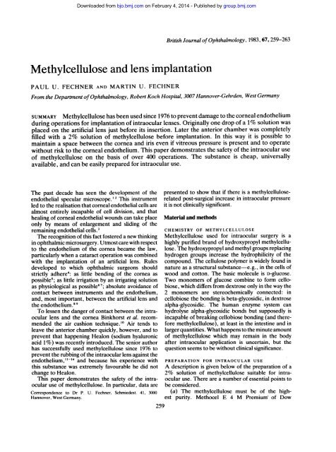
<strong>British</strong> <strong>Journal</strong> <strong>of</strong> Ophthalmology, 1983, 67, 259-263<br />
<strong>Methylcellulose</strong> <strong>and</strong> <strong>lens</strong> <strong>implantation</strong><br />
PAUL U. FECHNER AND MARTIN U. FECHNER<br />
From the Department <strong>of</strong> Ophthalmology, Robert Koch Hospital, 3007 Hannover-Gehrden, West Germany<br />
SUMMARY <strong>Methylcellulose</strong> has been used since 1976 to prevent damage to the corneal endothelium<br />
during operations for <strong>implantation</strong> <strong>of</strong> intraocular <strong>lens</strong>es. Originally one drop <strong>of</strong> a 1% solution was<br />
placed on the artificial <strong>lens</strong> just before its insertion. Later the anterior chamber was completely<br />
filled with a 2% solution <strong>of</strong> methylcellulose before <strong>implantation</strong>. In this way it is possible to<br />
maintain a space between the comea <strong>and</strong> iris even if vitreous pressure is present <strong>and</strong> to operate<br />
without risk to the comeal endothelium. This paper demonstrates the safety <strong>of</strong> the intraocular use<br />
<strong>of</strong> methylcellulose on the basis <strong>of</strong> over 400 operations. The substance is cheap, universally<br />
available, <strong>and</strong> can be easily prepared for intraocular use.<br />
The past decade has seen the development <strong>of</strong> the<br />
endothelial specular microscope.'2 This instrument<br />
led to the realisation that corneal endothelial cells are<br />
almost entirely incapable <strong>of</strong> cell division, <strong>and</strong> that<br />
healing <strong>of</strong> corneal endothelial wounds can take place<br />
only by means <strong>of</strong> enlargement <strong>and</strong> sliding <strong>of</strong> the<br />
remaining endothelial cells.3<br />
The recognition <strong>of</strong> this fact fostered a new thinking<br />
in ophthalmic microsurgery. Utmost care with respect<br />
to the endothelium <strong>of</strong> the cornea became the law,<br />
particularly when a cataract operation was combined<br />
with the <strong>implantation</strong> <strong>of</strong> an artificial <strong>lens</strong>. Rules<br />
developed to which ophthalmic surgeons should<br />
strictly adhere4: as little bending <strong>of</strong> the cornea as<br />
possible5; as little irrigation by an irrigating solution<br />
as physiological as possible67; absolute avoidance <strong>of</strong><br />
contact between instruments <strong>and</strong> the endothelium,<br />
<strong>and</strong>, most important, between the artificial <strong>lens</strong> <strong>and</strong><br />
the endothelium.89<br />
To lessen the danger <strong>of</strong> contact between the intraocular<br />
<strong>lens</strong> <strong>and</strong> the cornea Binkhorst et al. recommended<br />
the air cushion technique.'0 Air tends to<br />
leave the anterior chamber quickly, however, <strong>and</strong> to<br />
prevent that happening Healon (sodium hyaluronic<br />
acid 1%) was recently introduced. The senior author<br />
has successfully used methylcellulose since<br />
1976 to<br />
prevent the rubbing <strong>of</strong> the intraocular <strong>lens</strong> against the<br />
endothelium,' -'' <strong>and</strong> because his experience with<br />
this substance was extremely favourable he did not<br />
change to Healon.<br />
This paper demonstrates the safety <strong>of</strong> the intraocular<br />
use <strong>of</strong> methylcellulose. In particular, data are<br />
Correspondence to Dr P. U. Fechner, Schmiedest. 41, 3000<br />
Hannover, West Gernany.<br />
presented to show that if there is a methylcelluloserelated<br />
post-surgical increase in intraocular pressure<br />
it is not clinically significant.<br />
Material <strong>and</strong> methods<br />
CHEMISTRY OF METHYLCELLULOSE<br />
<strong>Methylcellulose</strong> used for intraocular surgery is a<br />
highly purified br<strong>and</strong> <strong>of</strong> hydroxypropyl methylcellulose.<br />
The hydroxypropyl <strong>and</strong> methyl groups replacing<br />
hydrogen groups increase the hydrophilicity <strong>of</strong> the<br />
compound. The cellulose polymer is widely found in<br />
nature as a structural substance-e.g., in the cells <strong>of</strong><br />
wood <strong>and</strong> cotton. The basic molecule is D-glucose.<br />
Two monomers <strong>of</strong> glucose combine to form cellobiose,<br />
which differs from dextrose only in the way the<br />
2 monomers are stereochemically connected: in<br />
cellobiose the bonding is beta-glycosidic, in dextrose<br />
alpha-glycosidic. The human enzyme system can<br />
hydrolyse alpha-glycosidic bonds but supposedly is<br />
incapable <strong>of</strong> breaking cellobiose bonding (<strong>and</strong> therefore<br />
methylcellulose), at least in the intestine <strong>and</strong> in<br />
larger quantities. What happens to the minute amount<br />
<strong>of</strong> methylcellulose which may remain in the body<br />
after intraocular application is uncertain, but the<br />
question seems to be without clinical significance.<br />
PREPARATION<br />
FOR INTRAOCULAR USE<br />
A description is given below <strong>of</strong> the preparation <strong>of</strong> a<br />
2% solution <strong>of</strong> methylcellulose suitable for intraocular<br />
use. There are a number <strong>of</strong> essential points to<br />
be considered.<br />
(a) The methylcellulose must be <strong>of</strong> the highest<br />
purity. Methocel E 4 M Premium' <strong>of</strong> Dow<br />
259
Downloaded from bjo.bmj.com on February 4, 2014 - Published by group.bmj.com<br />
260<br />
Chemical Corporation is recommended for use.<br />
(b) A physiological solvent should be used. The<br />
following solvents are suitable: Balanced salt solution<br />
(BSS) (Alcon), Ringer's solution, Ringer's lactate<br />
solution, or equivalents.<br />
(c) Antiseptic compounds like benzalkonium,<br />
chlorbutanol, thimerosal, <strong>and</strong> others are damaging to<br />
the endothelium <strong>and</strong> must not be used. The solution<br />
therefore has to be sterilised by autoclaving. Since the<br />
internal temperature <strong>of</strong> a viscous solution rises slowly,<br />
autoclaving should last longer than the usual 20<br />
minutes, i.e., 30-40 minutes.<br />
(d) Some stains which in the past have been used<br />
as 'vital stains' were recently shown to be carcinogenic,<br />
e.g., trypan blue.'5 Though this may be <strong>of</strong><br />
negligible importance in our context, we now recommend<br />
'patent blue' V (sulphan blue), which is known<br />
not to be carcinogenic. 15<br />
(e) A freshly prepared solution <strong>of</strong> methylcellulose<br />
contains crystalline complexes which form corpuscular<br />
elements. Filtration, therefore, is an essential part<br />
<strong>of</strong> the preparation <strong>of</strong> a solution destined for intraocular<br />
use.<br />
Preparation <strong>of</strong> 100 ml <strong>of</strong> a 2% solution <strong>of</strong> methylcellulose<br />
is as follows: Dissolve 2 g in 30 ml <strong>of</strong> boiling<br />
solvent (BSS, Ringer's lactate solution, Ringer's<br />
solution). Allow to cool. Add 70 ml <strong>of</strong> cold or icy<br />
solvent. The solvent may contain 5 mg <strong>of</strong> patent blue<br />
V (sulphan blue).'5 Store in a refrigerator overnight<br />
at -10 to 0°C.<br />
Next day the solution can be filtered. The pore size<br />
should be between 16 /Am <strong>and</strong> 05 ,um. One may use<br />
suction by means <strong>of</strong> a water-stream pump through a<br />
G 3-Fritte, Jenaer Glass (pore size approximately 16<br />
,um) or more conveniently pressure (nitrogen). The<br />
solution is filtered through a Sparkler Submicron<br />
Filter Tube, cartridge CS-0-5 with a pore size <strong>of</strong> 0-5<br />
um (Sparkler Inc., Conroe, Texas, USA).<br />
After the filtered solution has been divided into<br />
small bottles, these are sterilised by autoclaving at<br />
120°C for 30-40 minutes.<br />
OPERATIVE TECHNIQUES<br />
All 417 operations on which this paper is based were<br />
performed by the senior author or in his presence by a<br />
junior colleague. The <strong>lens</strong>es used were mostly<br />
Medallion, but Binkhorst-four-loop, Shearing,<br />
Sinskey, or Sinskey-Kratz <strong>lens</strong>es were also implanted.<br />
Since the type <strong>of</strong> <strong>lens</strong> has no bearing on the postoperative<br />
intraocular pressure, it will not be considered<br />
further in this paper.<br />
The technique <strong>of</strong> operation was st<strong>and</strong>ardised as<br />
follows. All wounds were corneoscleral under a<br />
limbus-based flap reaching from 9.30 to 2.30. All<br />
intracapsular operations were performed with the<br />
help <strong>of</strong> alpha-chymotrypsine 1: 10000 for21/2 minutes.<br />
Paul U. Fechner <strong>and</strong> Martin U. Fechner<br />
After <strong>implantation</strong> <strong>of</strong> the <strong>lens</strong> the wound was closed<br />
with approximately 12 8-0 silk interrupted sutures.<br />
During the operation methylcellulose was applied in 2<br />
different ways. From 1976 to 1979 one drop <strong>of</strong> a 1%<br />
solution was applied to the artificial <strong>lens</strong> immediately<br />
previous to its insertion. Since January 1980 the<br />
anterior chamber has been filled with a 2% methylcellulose<br />
solution prior to implanting the <strong>lens</strong>. After<br />
the <strong>lens</strong> was in place, sutured to the iris, <strong>and</strong> some<br />
corneoscleral sutures had been inserted, most <strong>of</strong> the<br />
methylcellulose was removed from the eye by one <strong>of</strong><br />
the following methods: (a) diluting it <strong>and</strong> rinsing out<br />
with a McIntyre-type coaxial infusion <strong>and</strong> aspiration<br />
cannula; (b) forcing it out by inflating the anterior<br />
chamber with air; (c) a combination <strong>of</strong> these 2<br />
methods, either simultaneously or consecutively.<br />
For the past few months a methylcellulose solution<br />
has been used which is stained slightly blue to help<br />
observe its disappearance. No attempt was made to<br />
remove all the methylcellulose, since the blue specks<br />
which remained at the end <strong>of</strong> the procedure had<br />
disappeared on the first postoperative day.<br />
POSTOPERATIVE MEASURES<br />
All patients received topical applications <strong>of</strong> dexamethasone<br />
0 -1% q.i.d. from the last preoperative day<br />
for several weeks. This might have contributed to the<br />
fact that there was a slight rise <strong>of</strong> pressure during the<br />
early postoperative period. Therefore all patients<br />
received acetazolamide in sustained release form<br />
(Diamox Sequels) 500 mg daily, from the day <strong>of</strong> the<br />
operation to the third postoperative day. Diamox<br />
Sequels was given irrespective <strong>of</strong> whether the<br />
operation was an intracapsular (with alphachymotrypsin)<br />
or extracapsular (without alphachymotrypsin)<br />
procedure. The postoperative intraocular<br />
pressures <strong>of</strong> days 1 to 3 were under the<br />
influence <strong>of</strong> acetazolamide; at day 4 there may have<br />
been a residual effect.<br />
Applanation tonometry was performed at the first,<br />
third, <strong>and</strong> fifth postoperative days. A pressurelowering<br />
treatment was reinstituted in about 5% <strong>of</strong><br />
the cases on the fifth postoperative day. This did not<br />
influence the results presented below.<br />
Results<br />
OPERATION<br />
The use <strong>of</strong> methylcellulose greatly facilitated the<br />
operation. If despite ocular massage the anterior<br />
chamber remained shallow, the methylcellulose<br />
always increased the depth <strong>of</strong> the chamber to allow<br />
for an easy <strong>and</strong> safe <strong>implantation</strong> (even <strong>of</strong> 3-plane<br />
<strong>lens</strong>es with the 'open-sky' route) without a Sheets<br />
glide or posterior vitrectomy. The <strong>implantation</strong> never<br />
had to be ab<strong>and</strong>oned, in contrast to its ab<strong>and</strong>onment
Downloaded from bjo.bmj.com on February 4, 2014 - Published by group.bmj.com<br />
<strong>Methylcellulose</strong> <strong>and</strong> <strong>lens</strong> <strong>implantation</strong><br />
261<br />
50*<br />
50<br />
40'<br />
30'<br />
mm Hg<br />
20<br />
10'<br />
40*<br />
30<br />
mmHg<br />
20<br />
10<br />
n=81<br />
1 i<br />
Acetazolamide 500mg per day<br />
Op 1st 2nd 3rd 4th 5th<br />
Postop day<br />
Fig. 1 Postoperative pressures after 324 planned<br />
intracapsular operations with 1% or 2% methylcellulose as<br />
an adjunct. Alpha-chymotrypsin 1/10 000 was used for21/2<br />
minutes in all cases. Acetazolamide (Diamox) 500 mg per<br />
day was given from the day <strong>of</strong> the operation to the third<br />
postoperative day.<br />
in about 10% <strong>of</strong> cases prior to the use <strong>of</strong> methylcellulose.<br />
All the types <strong>of</strong> intraocular <strong>lens</strong> used by us could be<br />
implanted without touching the cornea <strong>and</strong> could, as<br />
was generally done, be sutured to the iris without<br />
harming the endothelium. Moreover, it was easier to<br />
observe the tying <strong>of</strong> the iris suture knot through the<br />
cornea if the anterior chamber was filled with methylcellulose<br />
rather than with air.<br />
POSTOPERATIVE INTRAOCULAR PRESSURE<br />
When the pressures after use <strong>of</strong> 1% <strong>and</strong> <strong>of</strong> 2%<br />
methylcellulose were compared it was evident that<br />
they were no higher after 2% methylcellulose. (The<br />
pressures were slightly lower in fact after both intracapsular<br />
<strong>and</strong> extracapsular procedures.)Therefore the<br />
groups <strong>of</strong> patients receiving 1% <strong>and</strong> 2% methylcellulose<br />
were combined in the figures presented below.<br />
Fig. 1 shows the postoperative intraocular pressure<br />
after planned intracapsular operations (i.e., with<br />
application <strong>of</strong> alpha-chymotrypsin 1/10000 for 21/2<br />
minutes). It should be remembered that the intraocular<br />
pressures from day 1 through day 3 were<br />
influenced by the application <strong>of</strong> acetazolamide.<br />
Fig. 2 shows the intraocular pressure after planned<br />
extracapsular operations (alpha-chymotrypsin was<br />
not used in these eyes, but the eyes did receive<br />
acetazolamide).<br />
Fig. 3 shows the intraocular pressure in a small<br />
group <strong>of</strong> secondary <strong>implantation</strong>s <strong>of</strong>medallion <strong>lens</strong>es.<br />
These operations differ from those shown in Figs. 1<br />
<strong>and</strong> 2 in that they were less traumatic. Acetazolamide<br />
was given in these cases also.<br />
Acetazolamide<br />
500mg per day<br />
Op 1st 2nd 3rd 4th 5th<br />
Postop day<br />
Fig. 2 Postoperative pressures after 81 planned<br />
extracapsular operations. <strong>Methylcellulose</strong> 1% <strong>and</strong> 2% was<br />
used as an adjunct in all cases. No operation was performed<br />
with enzymatic zonulolysis. Acetazolamide (Diamox) 500<br />
mg per day was given from the day <strong>of</strong> the operation to the<br />
third postoperative day.<br />
The 3 figures demonstrate clearly that the increases<br />
in intraocular pressure after a cataract operation with<br />
<strong>lens</strong> <strong>implantation</strong> (or after a secondary <strong>lens</strong> <strong>implantation</strong>),<br />
during which methylcellulose has been applied,<br />
are not a complication <strong>of</strong> clinical significance.<br />
POSTOPERATIVE INFLAMMATION<br />
No postoperative infections occurred. Possibly<br />
because <strong>of</strong> steroid treatment postoperative intraocular<br />
inflammation was minimal. Only slight flare<br />
50'<br />
40-<br />
30-<br />
mm Hg<br />
20'<br />
10'<br />
TT<br />
I<br />
n=12<br />
Acetazolarride 500mg per day<br />
__<br />
Op 1St 2nd 3rd 4th<br />
Postop day<br />
Fig. 3 Postoperative pressures after 12 secondary <strong>lens</strong><br />
<strong>implantation</strong>s <strong>of</strong>medallion <strong>lens</strong>es with 1% or2%<br />
methylcellulose used as an adjunct. Acetazolamide<br />
(Diamox) 500 mg per day was given from the day <strong>of</strong> the<br />
operation to the third postoperative day.<br />
5th
Downloaded from bjo.bmj.com on February 4, 2014 - Published by group.bmj.com<br />
262<br />
<strong>and</strong> a few cells were observed for a few days. No<br />
hypopyon occurred. (All <strong>lens</strong>es had been wetsterilised<br />
by Medical Workshop.) Only a few eyes<br />
with a more pronounced exudation were observed.<br />
This could not have been the result <strong>of</strong> the methylcellulose,<br />
because the methylcellulose was always<br />
withdrawn from one bottle for several <strong>implantation</strong>s,<br />
while the postoperative cases <strong>of</strong> iritis occurred only<br />
sporadically, never epidemically.<br />
POSTOPERATIVE STATE OF THE CORNEA<br />
Each operation was graded according to a subjective<br />
impression <strong>of</strong> quality, <strong>and</strong> later compared with the<br />
postoperative appearance <strong>of</strong> the cornea. In the cases<br />
with the highest grade (most eyes), the corneae were<br />
nearly clear at the first postoperative day, <strong>and</strong><br />
completely clear by the third day.<br />
The endothelium was observed postoperatively in<br />
about 100 cases by noncontact specular microscopy<br />
<strong>and</strong> with the help <strong>of</strong> a McIntyre grid <strong>and</strong> compared<br />
with the nonoperated eye. In most <strong>of</strong> the operated<br />
eyes there were about 2000 endothelial cells per<br />
square mm in both eyes. To us this indicated a lack <strong>of</strong><br />
any toxic effect <strong>of</strong> methylcellulose on the endothelium.<br />
Therefore as a clinical impression on the basis <strong>of</strong><br />
approximately 2,000 postoperative slit-lamp<br />
examinations on 417 eyes it can be stated that<br />
methylcellulose did not harm the endothelium. Since<br />
the mechanical advantage <strong>of</strong> this adjunct to the<br />
operation is so evident, the overall effect on the<br />
cornea must obviously be beneficial.<br />
Discussion<br />
Healon was recently considered 'the most important<br />
substance to come along in cataract surgery since<br />
alpha-chymotrypsin'. 6 This statement, however,<br />
must be qualified. What matters is not Healon but<br />
appropriate viscous material in the eye. Healon,<br />
despite its value, has its disadvantages: it is very<br />
expensive, not universally available, <strong>and</strong> it is difficult<br />
to dilute. For the last reason a relatively large amount<br />
<strong>of</strong> the material may remain in the eye <strong>and</strong> cause a<br />
dangerous rise <strong>of</strong> pressure postoperatively. 17<br />
We prefer methylcellulose 2% dissolved in BSS.<br />
Fleming et al.'8 have shown that methylcellulose<br />
inside the eye is harmless. <strong>Methylcellulose</strong> has many<br />
advantages. As an adjunct to <strong>lens</strong> <strong>implantation</strong> it is<br />
perfect. It coats the <strong>lens</strong> <strong>and</strong> deepens a shallow<br />
anterior chamber. It is universally available <strong>and</strong><br />
inexpensive. The solution is comparatively easy to<br />
prepare-at least for a moderately equipped<br />
pharmacy. This is advantageous in underdeveloped<br />
<strong>and</strong> developing countries. The solution can be<br />
resterilised, thereby further decreasing the cost<br />
Paul U. Fechner <strong>and</strong> Martin U. Fechner<br />
factor. It can be stained blue for easy recognition<br />
inside the anterior chamber. It is-<strong>and</strong> this is a very<br />
important point-easily diluted <strong>and</strong> can be largely<br />
removed from the anterior chamber without difficulty.<br />
If postoperative rises <strong>of</strong> intraocular pressure<br />
occur owing to its use, they are easily manageable <strong>and</strong><br />
not clinically significant.<br />
Because methylcellulose is a nonphysiological <strong>and</strong><br />
possibly nonmetabolic substance consisting <strong>of</strong> large<br />
polymers, it could be argued that the residue which<br />
remains in the eye at the end <strong>of</strong> the operation might<br />
clog the trabecular meshwork <strong>and</strong> cause dangerous<br />
bouts <strong>of</strong> glaucoma, as has been reported after the use<br />
<strong>of</strong> Healon"' Our results show that this is definitely<br />
not so. As can be seen from Figs. 1 <strong>and</strong> 2, the intraocular<br />
pressure at the first postoperative day was 33-1<br />
mmHg after intracapsular operation, <strong>and</strong> 30-2 mmHg<br />
after extracapsular operation (with acetazolamide).<br />
Obviously the difference between the pressures must<br />
be due to the increased tension following the<br />
enzymatic zonulolysis. But even after extracapsular<br />
operation pressure rises occur which seem in the<br />
first place to be due to the trauma <strong>of</strong> the operation<br />
<strong>and</strong> <strong>lens</strong> material remaining in the anterior chamber.<br />
It is unclear whether remnants <strong>of</strong> methylcellulose in<br />
the anterior chamber in addition increase the postoperative<br />
intraocular pressure. Whether this is so<strong>and</strong><br />
if so, to what extent-could not be clarified.<br />
What was shown, however, was the unequivocally<br />
benign behaviour <strong>of</strong> the methylcellulose inside the<br />
eye with respect to the postoperative pressure.<br />
As regards the argument that methylcellulose<br />
cannot be metabolised, one must consider the<br />
quantity under discussion. If 20% <strong>of</strong> what was<br />
injected into the eye-say 0-5 ml-stays in the<br />
anterior chamber, this amounts to only 2 mg <strong>of</strong> the<br />
dry substance <strong>of</strong> methylcellulose, a substance<br />
considered to be inert <strong>and</strong> confirmed by our<br />
experience to be so. None <strong>of</strong> the patients showed any<br />
ill effect either systemically or locally which could be<br />
related to methylcellulose.<br />
With respect to the cornea we did not prove that<br />
methylcellulose was more beneficial than Healon or<br />
air or no cushioning substance at all. But since most<br />
eyes which where examined by noncontact specular<br />
microscopy did show a very high endothelial cell<br />
count (practically equal to that <strong>of</strong> the nonoperated<br />
eyes) we feel justified in the assumption that methylcellulose<br />
is at least not toxic to the endothelium. That<br />
it is <strong>of</strong> benefit is a belief based on our experience that<br />
<strong>implantation</strong> in so many cases is much facilitated by<br />
its use.<br />
In summary, our experience <strong>of</strong> over 6 years with<br />
methylcellulose leads us to believe that this substance<br />
is most valuable in intraocular surgery whenever a<br />
cushioning substance other than air is needed in the
Downloaded from bjo.bmj.com on February 4, 2014 - Published by group.bmj.com<br />
<strong>Methylcellulose</strong> <strong>and</strong> <strong>lens</strong> <strong>implantation</strong><br />
anterior chamber. Admittedly this statement is based<br />
on clinical experience. We hope that this paper may<br />
stimulate further investigations to obtain confirmation.<br />
We obtained the solution <strong>of</strong> methylcellulose from Raths-Apotheke,<br />
Oster-St 51, 3250 Hameln, West Germany.<br />
References<br />
I Laing R A, S<strong>and</strong>strom M M, Leibowitz H M. In vivo photomicrography<br />
<strong>of</strong> the corneal endothelium. Arch Ophthalmol 1975;<br />
93:143-5.<br />
2 Bourne W M, Kaufman H E. Specular microscopy <strong>of</strong> human<br />
corneal endothelium in vivo. Am JOphthalmol 1976; 81: 319-23.<br />
3 H<strong>of</strong>fer K J, Philippi G. A cell membrane theory <strong>of</strong> endothelial<br />
repair <strong>and</strong> vertical cell loss after cataract surgery. Am Intra-ocular<br />
Implant Soc J 1978; 4: 18-25.<br />
4 Fechner P U. Intraocular <strong>lens</strong>es. Stuttgart: Enke, 1980: 66.<br />
5 Norn M S. Vital staining <strong>of</strong> corneal endothelium in cataract<br />
extraction. Acta Ophthalmol (Kbh) 1971; 49: 725-33.<br />
6 Edelhauser H F, van Horn D L, Schultz R 0, Hyndiuk R A.<br />
Comparative toxicity <strong>of</strong> intraocular irrigating solutions on the<br />
corneal endothelium. Am J Ophthalmol 1976; 81: 473-81.<br />
7 Edelhauser H F, Connering R, van Horn D L. Intraocular<br />
irrigating solutions. Arch Ophthalmol 1978; 96: 516-20.<br />
263<br />
8 Kaufman H E, Katz J. Endothelial damage from intraocular <strong>lens</strong><br />
insertion. Invest Ophthalmol Visual Sci 1976; 15: 996-1000.<br />
9 Kaufman H E, Katz J. Endothelial damage from intraocular <strong>lens</strong><br />
insertion. Invest Ophthalmol Visual Sci 1977; 16: 265.<br />
10 Binkhorst C D, Loones, L H, Nygaard P. Die Bedeutung der<br />
Spiegelmikroskopie und -graphie des Hornhautendothels fur die<br />
Einpflanzung kunstlicher Augenlinsen. Beschreibung einer<br />
Tiefkammertechnik fur Einpflanzung. Ber Dtsch OphthalmolGes<br />
1978; 75:93-103.<br />
11 Fechner P U. <strong>Methylcellulose</strong> in <strong>lens</strong> <strong>implantation</strong>. Am Intra-<br />
Ocular Implant Soc J 1977; 3: 180-1.<br />
12 Fechner P U. Erfahrungen mit der Implantation einer kunstlichen<br />
Augenlinse. Klin Monatsbl Augenheilkd 1979; 172: 852-62.<br />
13 Fechner P U. <strong>Methylcellulose</strong> als Gleitsubstanz fur die Implantation<br />
kunstlicher Augenlinsen. Klin Monatsbl Augenheilkd 1979;<br />
174:136-18.<br />
14 Fechner P U. Use <strong>of</strong> methylcellulose during <strong>lens</strong> <strong>implantation</strong>.<br />
Am Intra-Ocular Implant Soc J 1980; 6: 368-9.<br />
15 Martindale: the extra pharmacopoeia. 27th ed. London: Pharmaceutical<br />
Press, 1977: 475, 615.<br />
16 Drews R C. The highlights on intraocular <strong>lens</strong>es, merits <strong>of</strong> extracapsulars<br />
versus intracapsulars. In: Boyd B, ed. Highlights <strong>of</strong><br />
ophthalmology. 25th ed. Panama, Highlights <strong>of</strong> Ophthalmology:<br />
1981; 2: 904.<br />
17 Binkhorst C D. Inflammation <strong>and</strong> intraocular pressure after the<br />
use <strong>of</strong> Healon in intraocular <strong>lens</strong> surgery. Am Intra-Ocular<br />
Implant Soc J 1980; 6: 340-1.<br />
18 Fleming M A, Merrill D L, Girard L J. Studies <strong>of</strong> the irritating<br />
action <strong>of</strong> methylcellulose. Arch Ophthalmol 1959; 61: 565-7.
Downloaded from bjo.bmj.com on February 4, 2014 - Published by group.bmj.com<br />
<strong>Methylcellulose</strong> <strong>and</strong> <strong>lens</strong><br />
<strong>implantation</strong>.<br />
P. U. Fechner <strong>and</strong> M. U. Fechner<br />
Br J Ophthalmol 1983 67: 259-263<br />
doi: 10.1136/bjo.67.4.259<br />
Updated information <strong>and</strong> services can be found at:<br />
http://bjo.bmj.com/content/67/4/259<br />
References<br />
Email alerting<br />
service<br />
These include:<br />
Article cited in:<br />
http://bjo.bmj.com/content/67/4/259#related-urls<br />
Receive free email alerts when new articles cite this article.<br />
Sign up in the box at the top right corner <strong>of</strong> the online article.<br />
Notes<br />
To request permissions go to:<br />
http://group.bmj.com/group/rights-licensing/permissions<br />
To order reprints go to:<br />
http://journals.bmj.com/cgi/reprintform<br />
To subscribe to BMJ go to:<br />
http://group.bmj.com/subscribe/


