Confocal microscopy in cornea guttata and Fuchs' endothelial ...
Confocal microscopy in cornea guttata and Fuchs' endothelial ...
Confocal microscopy in cornea guttata and Fuchs' endothelial ...

You also want an ePaper? Increase the reach of your titles
YUMPU automatically turns print PDFs into web optimized ePapers that Google loves.
Br J Ophthalmol 1999;83:185–189 185<br />
Louisiana State<br />
University Eye Center,<br />
New Orleans, LA, USA<br />
A G-Y Chiou<br />
S C Kaufman<br />
R W Beuerman<br />
T Ohta<br />
H Soliman<br />
H E Kaufman<br />
Correspondence to:<br />
Auguste G-Y Chiou, MD,<br />
LSU Eye Center, 2020<br />
Gravier Street, Suite B, New<br />
Orleans, LA 70112-2234,<br />
USA.<br />
Accepted for publication<br />
26 August 1998<br />
Downloaded from<br />
bjo.bmj.com on April 9, 2013 - Published by group.bmj.com<br />
<strong>Confocal</strong> <strong>microscopy</strong> <strong>in</strong> <strong>cornea</strong> <strong>guttata</strong> <strong>and</strong> Fuchs’<br />
<strong>endothelial</strong> dystrophy<br />
Auguste G-Y Chiou, Stephen C Kaufman, Roger W Beuerman, Toshihiko Ohta,<br />
Hisham Soliman, Herbert E Kaufman<br />
Abstract<br />
Aims—To report the appearances of <strong>cornea</strong><br />
<strong>guttata</strong> <strong>and</strong> Fuchs’ <strong>endothelial</strong> dystrophy<br />
from white light confocal <strong>microscopy</strong>.<br />
Methods—Seven eyes of four consecutive<br />
patients with <strong>cornea</strong> <strong>guttata</strong> were prospectively<br />
exam<strong>in</strong>ed. Of the seven eyes,<br />
three also had <strong>cornea</strong>l oedema (Fuchs’<br />
dystrophy). In vivo white light t<strong>and</strong>em<br />
scann<strong>in</strong>g confocal <strong>microscopy</strong> was performed<br />
<strong>in</strong> all eyes. Results were compared<br />
with non-contact specular <strong>microscopy</strong>.<br />
Results—Specular <strong>microscopy</strong> was precluded<br />
by <strong>cornea</strong>l oedema <strong>in</strong> one eye. In<br />
the rema<strong>in</strong><strong>in</strong>g six eyes, it demonstrated<br />
typical changes <strong>in</strong>clud<strong>in</strong>g pleomorphism,<br />
polymegathism, <strong>and</strong> the presence of guttae<br />
appear<strong>in</strong>g as dark bodies, some with a<br />
central bright reflex. In all seven eyes,<br />
confocal <strong>microscopy</strong> revealed the presence<br />
of round hyporeflective images with<br />
an occasional central highlight at the level<br />
of the endothelium. Changes <strong>in</strong> cell morphology<br />
<strong>and</strong> size were readily appreciated.<br />
Conclusion—By comparison with specular<br />
<strong>microscopy</strong>, the hyporeflective images<br />
with an occasional central highlight seen<br />
on confocal <strong>microscopy</strong> are consistent<br />
with the presence of guttae. <strong>Confocal</strong><br />
<strong>microscopy</strong> may confirm the diagnosis of<br />
<strong>cornea</strong> <strong>guttata</strong> <strong>and</strong> Fuchs’ <strong>endothelial</strong><br />
dystrophy by demonstrat<strong>in</strong>g the presence<br />
of guttae. This technique is especially<br />
valuable <strong>in</strong> cases of <strong>cornea</strong>l oedema,<br />
where specular <strong>microscopy</strong> may fail to<br />
visualise the endothelium. However,<br />
specular <strong>microscopy</strong> should rema<strong>in</strong> the<br />
method of choice to evaluate the endothelium,<br />
pr<strong>in</strong>cipally because it is easier to<br />
use.<br />
(Br J Ophthalmol 1999;83:185–189)<br />
Corneal guttae consist of focal accumulation of<br />
collagen at the posterior surface of Descemet’s<br />
membrane. 1 They are probably secreted by<br />
abnormal <strong>endothelial</strong> cells <strong>and</strong> may appear as a<br />
result of ag<strong>in</strong>g. Guttae are characteristic for<br />
Fuchs’ <strong>endothelial</strong> dystrophy, which is a familial<br />
dom<strong>in</strong>antly <strong>in</strong>herited disorder, also characterised<br />
by <strong>cornea</strong>l oedema. 2 Usually the condition<br />
is bilateral, although asymmetric<br />
presentation is not uncommon. When <strong>cornea</strong>l<br />
oedema is absent <strong>and</strong> only guttae are present,<br />
the condition is called <strong>cornea</strong> <strong>guttata</strong>.<br />
The endothelium is usually best exam<strong>in</strong>ed<br />
by specular <strong>microscopy</strong>. However, confocal<br />
<strong>microscopy</strong> allows superior image contrast <strong>and</strong><br />
vertical <strong>and</strong> lateral resolution, compared with<br />
conventional imag<strong>in</strong>g methods. 3–13 Because of<br />
its ability to focus the light source <strong>and</strong> the<br />
image on the same focal plane, it allows real<br />
time <strong>in</strong> vivo assessment of the diVerent layers<br />
of the <strong>cornea</strong>, <strong>in</strong>clud<strong>in</strong>g the <strong>endothelial</strong> layer.<br />
Therefore, it may be an alternative method <strong>in</strong><br />
evaluat<strong>in</strong>g <strong>cornea</strong> <strong>guttata</strong> or Fuchs’ <strong>endothelial</strong><br />
dystrophy.<br />
In the current study, we analysed the<br />
appearances of <strong>cornea</strong> <strong>guttata</strong> <strong>and</strong> Fuchs’ dystrophy<br />
from confocal <strong>microscopy</strong> <strong>and</strong> compare<br />
the technique with non-contact specular <strong>microscopy</strong>.<br />
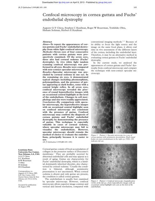
Figure 1 Patient 1. Specular <strong>microscopy</strong> <strong>in</strong> a case of<br />
<strong>cornea</strong> <strong>guttata</strong> with asymmetric presentation. Dark round<br />
bodies are more predom<strong>in</strong>ant <strong>in</strong> the right eye (OD). OS<br />
<strong>in</strong>dicates left eye.<br />
Figure 2 Patient 1. <strong>Confocal</strong> <strong>microscopy</strong> (magnification<br />
×210) of the left eye. A few hyporeflective images with an<br />
occasional central highlight were seen at the level of the<br />
endothelium.
Downloaded from<br />
bjo.bmj.com on April 9, 2013 - Published by group.bmj.com<br />
186 Chiou, Kaufman, Beuerman, et al<br />
Figure 3 Patient 1. <strong>Confocal</strong> <strong>microscopy</strong> (magnification<br />
×210) of the right eye. Compared with the left eye,<br />
significantly more hyporeflective images were seen.<br />
Hyperreflective <strong>endothelial</strong> cells were found between the<br />
lesions.<br />
Figure 4 Patient 2. Specular <strong>microscopy</strong> <strong>in</strong> the right<br />
(OD) <strong>and</strong> left (OS) eyes <strong>in</strong> a case of bilateral <strong>cornea</strong>l<br />
guttae <strong>and</strong> oedema (Fuchs’ dystrophy).<br />
Figure 5 Patient 2. <strong>Confocal</strong> <strong>microscopy</strong> (magnification ×210) of the right eye.<br />
Hyporeflective images with a central highlight were seen <strong>in</strong> the endothelium.<br />
Materials <strong>and</strong> methods<br />
Seven eyes of four consecutive patients with<br />
cl<strong>in</strong>ical signs of <strong>cornea</strong> <strong>guttata</strong> or Fuchs’<br />
dystrophy were prospectively studied. Specular<br />
<strong>microscopy</strong> us<strong>in</strong>g a non-contact microscope<br />
(Topcon SP 2000 P, Tokyo, Japan) was<br />
performed. Patients then underwent <strong>in</strong> vivo<br />
confocal microscopic exam<strong>in</strong>ation of their<br />
eyes. We used a prototype white light t<strong>and</strong>em<br />
scann<strong>in</strong>g confocal microscope (LSU Eye<br />
Center, New Orleans, LA, USA). The <strong>in</strong>strument<br />
utilised a 24×/0.60 contact objective <strong>and</strong><br />
allowed optical section<strong>in</strong>g of the <strong>cornea</strong> with a<br />
depth of field of 12 µm. The images were captured<br />
us<strong>in</strong>g a video camera (CCD 200 E,<br />
Videoscope International, Wash<strong>in</strong>gton DC,<br />
USA) <strong>and</strong> stored on S-VHS video tapes. <strong>Confocal</strong><br />
<strong>and</strong> specular microscopic f<strong>in</strong>d<strong>in</strong>gs were<br />
analysed <strong>and</strong> compared.<br />
Results<br />
CASE 1<br />
A 41 year old black male patient with a history<br />
of bilateral <strong>cornea</strong> <strong>guttata</strong> was exam<strong>in</strong>ed.<br />
Familial <strong>and</strong> general medical history was unremarkable.<br />
The patient had been treated with<br />
hypertonic sal<strong>in</strong>e drops <strong>in</strong> the right eye. Best<br />
corrected visual acuity was 20/30 <strong>in</strong> the right<br />
eye, <strong>and</strong> 20/20 <strong>in</strong> the left eye. Slit lamp exam<strong>in</strong>ation<br />
of the right eye disclosed advanced<br />
beaten metal appearance of the <strong>cornea</strong>l endothelium.<br />
In the left eye the endothelium<br />
showed moderate beaten metal appearance.<br />
Intraocular pressure was 16 mm Hg <strong>in</strong> the<br />
right eye <strong>and</strong> 18 mm Hg <strong>in</strong> the left eye. The<br />
rema<strong>in</strong>der of the ocular exam<strong>in</strong>ation was<br />
normal. Corneal thickness was measured by<br />
ultrasonic pachymetry. The right <strong>cornea</strong><br />
measured 594 µm versus 553 µm <strong>in</strong> the left eye.<br />
Specular microscopic exam<strong>in</strong>ation (Fig 1) of<br />
the right <strong>cornea</strong> demonstrated large numbers<br />
of dark bodies. Between the dark bodies, the<br />
<strong>endothelial</strong> cells appeared hyperreflective <strong>and</strong><br />
their boundaries could not be identified. In the<br />
left eye the same alterations were demonstrated,<br />
however, to a much lesser extent.<br />
There were fewer dark bodies <strong>and</strong> the <strong>endothelial</strong><br />
cells were well <strong>in</strong>dividualised. They presented<br />
polymegathism <strong>and</strong> pleomorphism.<br />
In the left eye (Fig 2), confocal <strong>microscopy</strong><br />
demonstrated <strong>endothelial</strong> cells with polymegathism,<br />
pleomorphism, <strong>and</strong> few hyporeflective<br />
round images conta<strong>in</strong><strong>in</strong>g occasionally a central<br />
highlight. The right eye (Fig 3) presented more<br />
significant alterations. At the level of the<br />
endothelium, hyporeflective round images<br />
were found <strong>in</strong> larger size <strong>and</strong> number. Between<br />
them, the <strong>endothelial</strong> cells appeared hyperreflective<br />
<strong>and</strong> could not be identified <strong>in</strong>dividually.<br />
CASE 2<br />
A 72 year old white woman with progressive<br />
decrease of vision <strong>in</strong> both eyes was referred for<br />
evaluation. She had a long term history of <strong>cornea</strong><br />
<strong>guttata</strong>. However, <strong>cornea</strong>l oedema was<br />
never documented. Besides a medically well<br />
controlled hypothyroidism, no other abnormalities<br />
were reported. She mentioned a sister<br />
diagnosed with Fuchs’ <strong>endothelial</strong> dystrophy.
Downloaded from<br />
bjo.bmj.com on April 9, 2013 - Published by group.bmj.com<br />
<strong>Confocal</strong> <strong>microscopy</strong> <strong>in</strong> <strong>cornea</strong> <strong>guttata</strong> <strong>and</strong> Fuchs’ <strong>endothelial</strong> dystrophy 187<br />
Figure 6 Patient 2. <strong>Confocal</strong> <strong>microscopy</strong> (magnification ×210) of the left eye. Confluent<br />
hyporeflective images with a sporadic central highlight were demonstrated.<br />
Best corrected vision was 20/60 <strong>and</strong> 20/80 <strong>in</strong><br />
the right <strong>and</strong> left eye, respectively. In both eyes,<br />
<strong>cornea</strong>l thickness appeared <strong>in</strong>creased on slit<br />
lamp exam<strong>in</strong>ation, <strong>and</strong> mild epithelial oedema<br />
was noted <strong>in</strong> the left eye. Both eyes presented<br />
significant beaten metal appearance at the level<br />
of the endothelium <strong>and</strong> moderate nuclear cataract.<br />
Intraocular pressure was 14 mm Hg <strong>in</strong><br />
both eyes. The rema<strong>in</strong>der of the exam<strong>in</strong>ations<br />
was with<strong>in</strong> normal ranges. Corneal thickness<br />
measured by ultrasonic pachymetry was 632<br />
µm <strong>and</strong> 643 µm <strong>in</strong> the right <strong>and</strong> left eye,<br />
respectively.<br />
Specular <strong>microscopy</strong> demonstrated <strong>in</strong> both<br />
eyes large numbers of dark bodies <strong>in</strong> the<br />
<strong>cornea</strong>l endothelium (Fig 4). In the right eye,<br />
<strong>endothelial</strong> cells with pleomorphism <strong>and</strong><br />
polymegathism could be recognised. Otherwise<br />
<strong>and</strong> especially <strong>in</strong> the left eye, the <strong>endothelial</strong><br />
cells appeared compressed between the<br />
dark bodies.<br />
<strong>Confocal</strong> <strong>microscopy</strong> detected <strong>in</strong> both eyes<br />
the presence of multifocal hyporeflective areas<br />
surrounded by hyperreflective <strong>endothelial</strong> cells<br />
(Figs 5 <strong>and</strong> 6). The density of the hyporeflective<br />
images varied from one area to another <strong>in</strong><br />
both eyes.<br />
CASE 3<br />
A 81 year old white female patient was<br />
evaluated for progressive decrease of vision <strong>in</strong><br />
the right eye. Cornea <strong>guttata</strong> was documented<br />
<strong>in</strong> both eyes 5 years earlier, when the left eye<br />
underwent uncomplicated phacoemulsification<br />
<strong>and</strong> <strong>in</strong>traocular lens implantation. She was<br />
anticoagulated with warfar<strong>in</strong> because of past<br />
episodes of bra<strong>in</strong> strokes. She denied topical<br />
medications. Best corrected vision was 20/70 <strong>in</strong><br />
the right eye <strong>and</strong> 20/40 <strong>in</strong> the left eye. Both<br />
eyes presented with beaten metal <strong>endothelial</strong><br />
appearance. The right eye had a relatively<br />
advanced cataract <strong>and</strong> the left eye was pseudophakic.<br />
Intraocular pressure was 16 mm Hg<br />
<strong>and</strong> 17 mm Hg for the right <strong>and</strong> left eye,<br />
respectively. Both eyes had moderate nonexudative<br />
age related macular degeneration.<br />
Figure 7 Patient 3. Specular <strong>microscopy</strong> <strong>in</strong> a case of<br />
bilateral <strong>cornea</strong> <strong>guttata</strong>. OD <strong>in</strong>dicates right eye <strong>and</strong> OS<br />
<strong>in</strong>dicates left eye.<br />
Figure 8 Patient 3. <strong>Confocal</strong> <strong>microscopy</strong> (magnification<br />
×210) of the right eye. Hyporeflective images were seen <strong>in</strong><br />
the endothelium.<br />
Corneal thickness measured 568 µm <strong>in</strong> the<br />
right eye <strong>and</strong> 589 µm <strong>in</strong> the left eye.<br />
Specular <strong>microscopy</strong> of the endothelium<br />
demonstrated confluent dark bodies <strong>in</strong> both<br />
eyes (Fig 7). The <strong>endothelial</strong> cells presented<br />
polymegathism <strong>and</strong> pleomorphism.<br />
<strong>Confocal</strong> <strong>microscopy</strong> revealed the presence<br />
of hyporeflective round images with an occasional<br />
central highlight among pleomorphic<br />
<strong>endothelial</strong> cells of vary<strong>in</strong>g size <strong>in</strong> both <strong>cornea</strong>s<br />
(Figs 8 <strong>and</strong> 9). In each eye, areas with few<br />
hyporeflective images could be found, while<br />
other areas demonstrated numerous <strong>and</strong> confluent<br />
hyporeflective images.<br />
CASE 4<br />
A 74 year old white male patient was<br />
exam<strong>in</strong>ed. His left eye underwent functional<br />
penetrat<strong>in</strong>g keratoplasty with cataract extraction<br />
<strong>and</strong> <strong>in</strong>traocular lens implantation 3 years<br />
earlier for Fuchs’ <strong>endothelial</strong> dystrophy <strong>and</strong><br />
senile cataract. Histological exam<strong>in</strong>ation of the<br />
<strong>cornea</strong>l button confirmed the diagnosis of<br />
Fuchs’ <strong>endothelial</strong> dystrophy. He denied any<br />
topical treatments. Best corrected visual acuity
Downloaded from<br />
bjo.bmj.com on April 9, 2013 - Published by group.bmj.com<br />
188 Chiou, Kaufman, Beuerman, et al<br />
Figure 9 Patient 3. <strong>Confocal</strong> <strong>microscopy</strong> (magnification<br />
×210) of the left eye. Hyporeflective images with a central<br />
highlight were demonstrated <strong>in</strong> the endothelium.<br />
Figure 10 Patient 4. <strong>Confocal</strong> <strong>microscopy</strong> (magnification<br />
×210) revealed the presence of confluent hyporeflective<br />
images <strong>in</strong> the endothelium. Hyperreflective <strong>endothelial</strong> cells<br />
were found between the lesions. The hyperreflective image<br />
on the upper part of the photograph is compatible with<br />
fibrous changes. Specular <strong>microscopy</strong> <strong>in</strong> this case of Fuchs’<br />
<strong>endothelial</strong> dystrophy was prevented by <strong>cornea</strong>l oedema.<br />
was 20/200 <strong>and</strong> 20/40 <strong>in</strong> the right <strong>and</strong> left eye,<br />
respectively. Slit lamp exam<strong>in</strong>ation of the right<br />
eye disclosed significant beaten metal appearance<br />
of the endothelium, <strong>in</strong>creased stromal<br />
thickness, <strong>and</strong> epithelial oedema. The right eye<br />
also had relatively advanced nuclear cataract.<br />
The left eye had a clear graft <strong>and</strong> was pseudophakic.<br />
Intraocular pressure was 11 mm Hg <strong>in</strong><br />
the right eye <strong>and</strong> 12 mm Hg <strong>in</strong> the left eye. No<br />
other abnormalities were noted. Further exam<strong>in</strong>ation<br />
was performed only <strong>in</strong> the right eye.<br />
Corneal thickness by ultrasonic pachymetry<br />
was 662 µm.<br />
Non-contact specular <strong>microscopy</strong> was prevented<br />
by <strong>cornea</strong>l oedema.<br />
On the other h<strong>and</strong>, confocal <strong>microscopy</strong> easily<br />
revealed the presence of confluent hyporeflective<br />
images with hyperreflective <strong>endothelial</strong><br />
cells between them (Fig 10). Hyperreflective<br />
images compatible with fibrous tissue were also<br />
seen.<br />
Discussion<br />
To the best of our knowledge, the confocal<br />
microscopic appearance of <strong>cornea</strong>l guttae has<br />
not been reported so far. Recognition of<br />
<strong>cornea</strong>l guttae is important, s<strong>in</strong>ce they are the<br />
hallmark of <strong>cornea</strong> <strong>guttata</strong> <strong>and</strong> of Fuchs’<br />
dystrophy. Kaufman et al previously reported a<br />
case of Fuchs’ <strong>endothelial</strong> dystrophy <strong>and</strong><br />
documented the presence of larger <strong>and</strong> irregu-<br />
larly shaped <strong>endothelial</strong> cells on confocal<br />
<strong>microscopy</strong>. 14 Presence of guttae was not demonstrated.<br />
Their f<strong>in</strong>d<strong>in</strong>gs were compatible with<br />
cell loss <strong>and</strong> may be associated with various<br />
disorders, <strong>and</strong> therefore, were not characteristic<br />
of Fuchs’ dystrophy.<br />
The dark round bodies with occasional central<br />
white reflex found on specular <strong>microscopy</strong><br />
are typical of guttae. 15 16 They were noticeably<br />
similar to the confocal microscopic f<strong>in</strong>d<strong>in</strong>gs.<br />
Therefore, by comparison with the specular<br />
microscopic f<strong>in</strong>d<strong>in</strong>gs, we suggest that the<br />
hyporeflective images with an occasional central<br />
highlight seen on confocal <strong>microscopy</strong> correlate<br />
with guttae. The similarity of the appearances<br />
of guttae on confocal <strong>and</strong> specular<br />
<strong>microscopy</strong> is not surpris<strong>in</strong>g, s<strong>in</strong>ce both<br />
techniques detect reflected light from biological<br />
samples. At the level of the guttae, light is<br />
transmitted, whereas at the surround<strong>in</strong>g<br />
stroma endothelium <strong>in</strong>terface it is reflected.<br />
Consequently, the lesions appear hyporeflective.<br />
At the apex of the guttae, reflection of light<br />
may result <strong>in</strong> a central highlight.<br />
Guttae may be seen <strong>in</strong> various conditions.<br />
They <strong>in</strong>clude <strong>in</strong>terstitial keratitis, 17 <strong>cornea</strong>l<br />
macular dystrophy, 18 <strong>and</strong> posterior polymorphous<br />
dystrophy. 19 It is unclear whether they<br />
would appear similar to our f<strong>in</strong>d<strong>in</strong>gs when<br />
exam<strong>in</strong>ed with the confocal microscope. However,<br />
diVerential diagnosis is straightforward,<br />
s<strong>in</strong>ce each of these disorders is associated with<br />
typical anterior segment changes. The same<br />
applies to pseudoguttae, which are guttae-like<br />
images caused by <strong>endothelial</strong> oedema that may<br />
appear <strong>in</strong> cases of <strong>cornea</strong>l <strong>in</strong>flammation <strong>and</strong><br />
disappear upon resolution of the underly<strong>in</strong>g<br />
disease.<br />
20 21<br />
The illustrations show that the specular<br />
image is clearer than the confocal image. This<br />
is probably due to the fact that the confocal<br />
images were frame grabbed. The image of the<br />
<strong>endothelial</strong> cells is clearer when viewed <strong>in</strong> real<br />
time. As illustrated <strong>in</strong> patient 4 <strong>and</strong> reported<br />
previously by Kaufman et al, confocal <strong>microscopy</strong><br />
is not precluded by <strong>cornea</strong>l oedema. 14<br />
This represents the ma<strong>in</strong> advantage of this<br />
technology over specular <strong>microscopy</strong>. Furthermore,<br />
s<strong>in</strong>ce the severity of the <strong>endothelial</strong><br />
alterations may be variable from one area to<br />
another with<strong>in</strong> the same eye, we th<strong>in</strong>k that the<br />
confocal microscope, because of its ability to<br />
scan the entire <strong>cornea</strong>, allows for a better<br />
qualitative appreciation of the endothelium. It<br />
should be noted that contact specular <strong>microscopy</strong>,<br />
unlike the non-contact specular microscope<br />
used <strong>in</strong> this study, also has the ability to<br />
scan the entire <strong>cornea</strong>. Like specular <strong>microscopy</strong>,<br />
confocal <strong>microscopy</strong> does not enable<br />
prediction of the natural history of <strong>cornea</strong> <strong>guttata</strong>.<br />
It does not <strong>in</strong>dicate whether eyes with<br />
<strong>cornea</strong>l <strong>endothelial</strong> guttae are susceptible to<br />
develop <strong>cornea</strong>l oedema. With both techniques<br />
<strong>endothelial</strong> polymorphism <strong>and</strong> pleomorphism<br />
could be detected. Specular <strong>microscopy</strong> rema<strong>in</strong>s,<br />
nevertheless, an <strong>in</strong>valuable diagnostic<br />
tool <strong>in</strong> evaluat<strong>in</strong>g the <strong>cornea</strong>l endothelium.<br />
Compared with confocal <strong>microscopy</strong>, it is<br />
easier to use <strong>and</strong> does not require a learn<strong>in</strong>g<br />
curve. Furthermore, the quality of the images
Downloaded from<br />
bjo.bmj.com on April 9, 2013 - Published by group.bmj.com<br />
<strong>Confocal</strong> <strong>microscopy</strong> <strong>in</strong> <strong>cornea</strong> <strong>guttata</strong> <strong>and</strong> Fuchs’ <strong>endothelial</strong> dystrophy 189<br />
is rarely dependent on the patient’s ability to<br />
rema<strong>in</strong> still.<br />
Cornea <strong>guttata</strong> <strong>and</strong> Fuchs’ dystrophy are<br />
rarely a diagnostic dilemma, <strong>and</strong> most of the<br />
time are readily diagnosed simply by slit lamp<br />
exam<strong>in</strong>ation. Based on our experience, we<br />
th<strong>in</strong>k that confirmation of the diagnosis is best<br />
accomplished by specular <strong>microscopy</strong>, because<br />
it is easier to use. However, confocal <strong>microscopy</strong><br />
is a worthwhile alternative, especially <strong>in</strong><br />
cases of <strong>cornea</strong>l oedema.<br />
The authors do not have any commercial or proprietary <strong>in</strong>terest<br />
<strong>in</strong> the products or companies cited <strong>in</strong> this article. The authors<br />
do not have any f<strong>in</strong>ancial <strong>in</strong>terest or receive payment as a consultant,<br />
reviewer, or evaluator.<br />
This study was supported <strong>in</strong> part by US Public Health Service<br />
grants EY00346 (SCK), EY02580 (HEK), <strong>and</strong> EY02377<br />
(HEK) from the National Eye Institute, National Institutes of<br />
Health, Bethesda, MD; Department of the Army, Cooperative<br />
Agreement DAMD17-93-V-3013. (This does not necessarily<br />
reflect the position or the policy of the government, <strong>and</strong> no oYcial<br />
endorsement should be <strong>in</strong>ferred) (RWB, HEK); an<br />
unrestricted departmental grant from Research to Prevent<br />
Bl<strong>in</strong>dness, Inc, New York City; Société Académique Vaudoise<br />
(AGYC), Switzerl<strong>and</strong>; the Swiss National Research Foundation<br />
(AGYC), Switzerl<strong>and</strong>; <strong>and</strong> Verrey Foundation (AGYC),<br />
Switzerl<strong>and</strong>.<br />
1 War<strong>in</strong>g GO 3rd, Rodrigues MM, Laibson PR. Corneal dystrophies.<br />
II. Endothelial dystrophies. Surv Ophthalmol<br />
1978;23:147–68.<br />
2 Krachmer JH, Purcell JJ Jr, Young CW, et al. A study of<br />
sixty-four families with <strong>cornea</strong>l <strong>endothelial</strong> dystrophy. Arch<br />
Ophthalmol 1978;96:2035–9.<br />
3 Beuerman RW. <strong>Confocal</strong> <strong>microscopy</strong>: Into the cl<strong>in</strong>ic. Cornea<br />
1995;14:1–2.<br />
4 Cavanagh HD, Petroll WM, Alizadeh H, et al. Cl<strong>in</strong>ical <strong>and</strong><br />
diagnostic use of <strong>in</strong> vivo confocal <strong>microscopy</strong> <strong>in</strong> patients<br />
with <strong>cornea</strong>l disease. Ophthalmology 1993;100:1444–54.<br />
5 Cavanagh HD, McCulley JP. In vivo confocal <strong>microscopy</strong><br />
<strong>and</strong> acanthamoeba keratitis. Am J Ophthalmol 1996;121:<br />
207–8.<br />
6 Chew SJ, Beuerman RW, Assoul<strong>in</strong>e M, et al. Early diagnosis<br />
of <strong>in</strong>fectious keratitis with <strong>in</strong> vivo real time confocal <strong>microscopy</strong>.<br />
CLAO J 1992;18:197–201.<br />
7 Chiou AGY, Cadez R, B÷hnke M. Diagnosis of DieVenbachia<br />
<strong>in</strong>duced <strong>cornea</strong>l <strong>in</strong>jury by confocal <strong>microscopy</strong>. Br J<br />
Ophthalmol 1997;81:168–9.<br />
8 Florakis GJ, Moazami G, Schubert H, et al. Scann<strong>in</strong>g slit<br />
confocal <strong>microscopy</strong> of fungal keratitis. Arch Ophthalmol<br />
1997;115:1461–3.<br />
9 Kaufman SC, Chew SJ, Capps SC, et al. <strong>Confocal</strong><br />
<strong>microscopy</strong> of <strong>cornea</strong>l penetration by tarantula hairs. Scann<strong>in</strong>g<br />
1994;16:312–5.<br />
10 Kaufman SC, Beuerman RW, Goldberg D. A new form of<br />
primary, localized, <strong>cornea</strong>l amyloidosis: A case report with<br />
confocal <strong>microscopy</strong>. Metab Pediatr Syst Ophthalmol 1995;<br />
18:1–4.<br />
11 Pfister DR, Cameron JD, Krachmer JH, et al. <strong>Confocal</strong><br />
<strong>microscopy</strong> f<strong>in</strong>d<strong>in</strong>gs of acanthamoeba keratitis. Am J Ophthalmol<br />
1996;121:119–28.<br />
12 Shah GK, Pfister D, Probst LE, et al. Diagnosis of<br />
microsporidial keratitis by confocal <strong>microscopy</strong> <strong>and</strong> the<br />
chromatrope sta<strong>in</strong>. Am J Ophthalmol 1996;121:89–91.<br />
13 Sutph<strong>in</strong> JE, Kantor AL, Mathers WD, et al. Evaluation of<br />
<strong>in</strong>fectious crystall<strong>in</strong>e keratitis with confocal <strong>microscopy</strong> <strong>in</strong> a<br />
case series. Cornea 1997;16:21–6.<br />
14 Kaufman SC, Beuerman RW, Kaufman HE. Diagnosis of<br />
advanced Fuchs’ <strong>endothelial</strong> dystrophy with the confocal<br />
microscope. Am J Ophthalmol 1993;116:652–3.<br />
15 La<strong>in</strong>g RA, Leibowitz HM, Oak SS, et al. Endothelial mosaic<br />
<strong>in</strong> Fuchs’ dystrophy. A qualitative evaluation with the<br />
specular microscope. Arch Ophthalmol 1981;99:80–3.<br />
16 Bigar F, Schimmelpfennig B, Hurzeler R. Cornea <strong>guttata</strong> <strong>in</strong><br />
donor material. Arch Ophthalmol 1978;96:653–5.<br />
17 War<strong>in</strong>g GO, Font RL, Rodrigues MM, et al. Alterations of<br />
Descemet’s membrane <strong>in</strong> <strong>in</strong>terstitial keratitis. Am J<br />
Ophthalmol 1976;81:773–85.<br />
18 Pouliquen Y, Dhermy P, Renard G, et al. Comb<strong>in</strong>ed macular<br />
dystrophy <strong>and</strong> <strong>cornea</strong> <strong>guttata</strong>: An electron microscopic<br />
study. Albrecht Von Graefes Arch Kl<strong>in</strong> Exp Ophthalmol 1980;<br />
212:149–58.<br />
19 Cibis GW, Krachmer JA, Phelps CD, et al. The cl<strong>in</strong>ical<br />
spectrum of posterior polymorphous dystrophy. Arch Ophthalmol<br />
1977;95:1529–37.<br />
20 Brooks AM, Grant GB, Gillies WE. The identification of<br />
<strong>cornea</strong>l guttae. Cornea 1991;10:249–60.<br />
21 Krachmer JH, Schnitzer JI, Fratk<strong>in</strong> J. Cornea<br />
pseudo<strong>guttata</strong>: a cl<strong>in</strong>ical <strong>and</strong> histopathologic description of<br />
<strong>endothelial</strong> cell edema. Arch Ophthalmol 1981;99:1377–81.
References<br />
Email alert<strong>in</strong>g<br />
service<br />
Topic<br />
Collections<br />
Notes<br />
<strong>Confocal</strong> <strong>microscopy</strong> <strong>in</strong> <strong>cornea</strong> <strong>guttata</strong> <strong>and</strong><br />
<strong>Fuchs'</strong> <strong>endothelial</strong> dystrophy<br />
Auguste G-Y Chiou, Stephen C Kaufman, Roger W Beuerman, et al.<br />
Br J Ophthalmol 1999 83: 185-189<br />
doi: 10.1136/bjo.83.2.185<br />
Updated <strong>in</strong>formation <strong>and</strong> services can be found at:<br />
http://bjo.bmj.com/content/83/2/185.full.html<br />
These <strong>in</strong>clude:<br />
This article cites 21 articles, 1 of which can be accessed free at:<br />
http://bjo.bmj.com/content/83/2/185.full.html#ref-list-1<br />
Article cited <strong>in</strong>:<br />
http://bjo.bmj.com/content/83/2/185.full.html#related-urls<br />
Receive free email alerts when new articles cite this article. Sign up <strong>in</strong> the<br />
box at the top right corner of the onl<strong>in</strong>e article.<br />
Articles on similar topics can be found <strong>in</strong> the follow<strong>in</strong>g collections<br />
Cornea (416 articles)<br />
Ocular surface (496 articles)<br />
Eye (globe) (580 articles)<br />
To request permissions go to:<br />
http://group.bmj.com/group/rights-licens<strong>in</strong>g/permissions<br />
To order repr<strong>in</strong>ts go to:<br />
http://journals.bmj.com/cgi/repr<strong>in</strong>tform<br />
To subscribe to BMJ go to:<br />
http://group.bmj.com/subscribe/<br />
Downloaded from<br />
bjo.bmj.com on April 9, 2013 - Published by group.bmj.com