Confocal microscopy in cornea guttata and Fuchs' endothelial ...
Confocal microscopy in cornea guttata and Fuchs' endothelial ...
Confocal microscopy in cornea guttata and Fuchs' endothelial ...

Create successful ePaper yourself
Turn your PDF publications into a flip-book with our unique Google optimized e-Paper software.
Downloaded from<br />
bjo.bmj.com on April 9, 2013 - Published by group.bmj.com<br />
186 Chiou, Kaufman, Beuerman, et al<br />
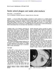
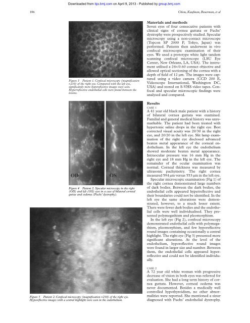
Figure 3 Patient 1. <strong>Confocal</strong> <strong>microscopy</strong> (magnification<br />
×210) of the right eye. Compared with the left eye,<br />
significantly more hyporeflective images were seen.<br />
Hyperreflective <strong>endothelial</strong> cells were found between the<br />
lesions.<br />
Figure 4 Patient 2. Specular <strong>microscopy</strong> <strong>in</strong> the right<br />
(OD) <strong>and</strong> left (OS) eyes <strong>in</strong> a case of bilateral <strong>cornea</strong>l<br />
guttae <strong>and</strong> oedema (Fuchs’ dystrophy).<br />
Figure 5 Patient 2. <strong>Confocal</strong> <strong>microscopy</strong> (magnification ×210) of the right eye.<br />
Hyporeflective images with a central highlight were seen <strong>in</strong> the endothelium.<br />
Materials <strong>and</strong> methods<br />
Seven eyes of four consecutive patients with<br />
cl<strong>in</strong>ical signs of <strong>cornea</strong> <strong>guttata</strong> or Fuchs’<br />
dystrophy were prospectively studied. Specular<br />
<strong>microscopy</strong> us<strong>in</strong>g a non-contact microscope<br />
(Topcon SP 2000 P, Tokyo, Japan) was<br />
performed. Patients then underwent <strong>in</strong> vivo<br />
confocal microscopic exam<strong>in</strong>ation of their<br />
eyes. We used a prototype white light t<strong>and</strong>em<br />
scann<strong>in</strong>g confocal microscope (LSU Eye<br />
Center, New Orleans, LA, USA). The <strong>in</strong>strument<br />
utilised a 24×/0.60 contact objective <strong>and</strong><br />
allowed optical section<strong>in</strong>g of the <strong>cornea</strong> with a<br />
depth of field of 12 µm. The images were captured<br />
us<strong>in</strong>g a video camera (CCD 200 E,<br />
Videoscope International, Wash<strong>in</strong>gton DC,<br />
USA) <strong>and</strong> stored on S-VHS video tapes. <strong>Confocal</strong><br />
<strong>and</strong> specular microscopic f<strong>in</strong>d<strong>in</strong>gs were<br />
analysed <strong>and</strong> compared.<br />
Results<br />
CASE 1<br />
A 41 year old black male patient with a history<br />
of bilateral <strong>cornea</strong> <strong>guttata</strong> was exam<strong>in</strong>ed.<br />
Familial <strong>and</strong> general medical history was unremarkable.<br />
The patient had been treated with<br />
hypertonic sal<strong>in</strong>e drops <strong>in</strong> the right eye. Best<br />
corrected visual acuity was 20/30 <strong>in</strong> the right<br />
eye, <strong>and</strong> 20/20 <strong>in</strong> the left eye. Slit lamp exam<strong>in</strong>ation<br />
of the right eye disclosed advanced<br />
beaten metal appearance of the <strong>cornea</strong>l endothelium.<br />
In the left eye the endothelium<br />
showed moderate beaten metal appearance.<br />
Intraocular pressure was 16 mm Hg <strong>in</strong> the<br />
right eye <strong>and</strong> 18 mm Hg <strong>in</strong> the left eye. The<br />
rema<strong>in</strong>der of the ocular exam<strong>in</strong>ation was<br />
normal. Corneal thickness was measured by<br />
ultrasonic pachymetry. The right <strong>cornea</strong><br />
measured 594 µm versus 553 µm <strong>in</strong> the left eye.<br />
Specular microscopic exam<strong>in</strong>ation (Fig 1) of<br />
the right <strong>cornea</strong> demonstrated large numbers<br />
of dark bodies. Between the dark bodies, the<br />
<strong>endothelial</strong> cells appeared hyperreflective <strong>and</strong><br />
their boundaries could not be identified. In the<br />
left eye the same alterations were demonstrated,<br />
however, to a much lesser extent.<br />
There were fewer dark bodies <strong>and</strong> the <strong>endothelial</strong><br />
cells were well <strong>in</strong>dividualised. They presented<br />
polymegathism <strong>and</strong> pleomorphism.<br />
In the left eye (Fig 2), confocal <strong>microscopy</strong><br />
demonstrated <strong>endothelial</strong> cells with polymegathism,<br />
pleomorphism, <strong>and</strong> few hyporeflective<br />
round images conta<strong>in</strong><strong>in</strong>g occasionally a central<br />
highlight. The right eye (Fig 3) presented more<br />
significant alterations. At the level of the<br />
endothelium, hyporeflective round images<br />
were found <strong>in</strong> larger size <strong>and</strong> number. Between<br />
them, the <strong>endothelial</strong> cells appeared hyperreflective<br />
<strong>and</strong> could not be identified <strong>in</strong>dividually.<br />
CASE 2<br />
A 72 year old white woman with progressive<br />
decrease of vision <strong>in</strong> both eyes was referred for<br />
evaluation. She had a long term history of <strong>cornea</strong><br />
<strong>guttata</strong>. However, <strong>cornea</strong>l oedema was<br />
never documented. Besides a medically well<br />
controlled hypothyroidism, no other abnormalities<br />
were reported. She mentioned a sister<br />
diagnosed with Fuchs’ <strong>endothelial</strong> dystrophy.