Incontinentia pigmenti - British Journal of Ophthalmology
Incontinentia pigmenti - British Journal of Ophthalmology
Incontinentia pigmenti - British Journal of Ophthalmology

Create successful ePaper yourself
Turn your PDF publications into a flip-book with our unique Google optimized e-Paper software.
Downloaded from<br />
bjo.bmj.com on June 16, 2013 - Published by group.bmj.com<br />
<strong>British</strong> <strong>Journal</strong> <strong>of</strong> <strong>Ophthalmology</strong>, 1987, 71, 629-634<br />
<strong>Incontinentia</strong> <strong>pigmenti</strong> (Bloch-Sulzberger syndrome):<br />
seven case reports from one family<br />
A SPALLONE<br />
From the St Gerardo Hospital, Department <strong>of</strong> <strong>Ophthalmology</strong>, Monza (Milan), Italy<br />
SUMMARY Seven members from a large family who showed signs <strong>of</strong> incontinentia <strong>pigmenti</strong> were<br />
examined. A clear X-linked dominant transmission was demonstrated, lethal in males. Study <strong>of</strong><br />
this family shows that vascular abnormalities <strong>of</strong> the retina and disorders <strong>of</strong> the retinal pigment<br />
epithelium are the most important ocular lesions in the Bloch-Sulzberger syndrome.<br />
<strong>Incontinentia</strong> <strong>pigmenti</strong> (Bloch-Sulzberger syndrome)<br />
is a genetic disease <strong>of</strong> the skin with generalised<br />
ectodermal and mesodermal dysplasia which may<br />
<strong>of</strong>ten involve the eyes (35% <strong>of</strong> the patients),' hair,<br />
teeth, and central nervous system. Skin lesions are<br />
Correspondence to Dr A Spallone.<br />
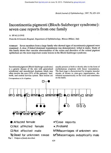
0: Affected female<br />
O:Not affected female<br />
O:Not affected male<br />
*:Dead for unknown cause<br />
Fig. 1 Pedigree <strong>of</strong>family described.<br />
usually present at birth or shortly after in the form <strong>of</strong><br />
erythematous eruptions with linear vesiculations.<br />
The final stage is characterised by irregular macular<br />
streaks <strong>of</strong> brown to slate-grey pigmentation, distributed<br />
asymmetrically on the torso and sometimes<br />
on the limbs.<br />
eCase reports<br />
*:Proband<br />
MMiscarriages <strong>of</strong> unknown sex<br />
MgMiscarriages autoptically male<br />
629
630<br />
Fig. 2 Case 1. Righteye.<br />
We have had the opportunity to study seven<br />
patients all <strong>of</strong> whom are members <strong>of</strong> one large family<br />
in which a total <strong>of</strong> 14 members were affected by<br />
incontinentia <strong>pigmenti</strong>. A clear X-linked dominant<br />
transmission was demonstrated, lethal in males (Fig.<br />
1). The following seven case reports illustrate the<br />
most important ocular lesions <strong>of</strong> the disease.<br />
Case reports<br />
Downloaded from<br />
bjo.bmj.com on June 16, 2013 - Published by group.bmj.com<br />
CASE 1<br />
This was the proband. A female child, 4 years old,<br />
birth weight 3 kg. The parents were not related. The<br />
dermatological symptoms <strong>of</strong> incontinentia <strong>pigmenti</strong><br />
appeared immediately after birth.<br />
Ocular examination showed nystagmus, oedematous<br />
cornea, and cataract (Fig. 2) in the right eye,<br />
microphthalmus with iridolenticular synechiae and<br />
complete cataract in the left eye. In this eye the iris<br />
was atrophic, with pigment irregularities (Fig. 3).<br />
Fig. 4 Case). Classic whorls and streaks <strong>of</strong>pigmenton<br />
torso and in right eye.<br />
Fig. 3 Case). Left eye.<br />
A Spallone<br />
Visual acuity was no perception <strong>of</strong> light in both eyes.<br />
Ultrasonography revealed the presence <strong>of</strong> bilateral<br />
retinal detachment with dense membranes in the<br />
vitreous. A general medical examination showed the<br />
classic whorls and streaks <strong>of</strong> pigment on the torso and<br />
on the right leg (Fig. 4). The grandmother had had<br />
18 pregnancies, 11 <strong>of</strong> which had been miscarriages.<br />
The seven fullterm pregnancies were all resulted in<br />
females. Five <strong>of</strong> these had ocular and dermatological<br />
problems; one died <strong>of</strong> an unknown cause, and the last<br />
one was healthy. The first <strong>of</strong> these five affected<br />
patients is the mother <strong>of</strong> our proband.<br />
CASE 2<br />
Mother <strong>of</strong> the proband, 37 years old, who has been<br />
affected by incontinentia <strong>pigmenti</strong> since birth.<br />
Ocular examination showed intermittent exotropia.<br />
The visual acuity was 1-0 in both eyes. Both<br />
lenses were clear. In the ocular fundus in the right eye<br />
no anomalies could be found; by contrast in the left<br />
eye the retinal vessels were anomalous. Towards the<br />
temporal equator both venules and arterioles became<br />
tortuous, slightly kinked, <strong>of</strong> an irregular calibre,<br />
increased in number, and with preretinal fibrosis.<br />
Apart from this area the peripheral temporal part <strong>of</strong><br />
the retina was completely avascular (Fig. 5). In the<br />
inferior nasal area there were small alterations <strong>of</strong> the<br />
retinal pigment epithelium. On general medical<br />
examination we noted some pigmentary alterations<br />
<strong>of</strong> the skin, the absence <strong>of</strong> the lateral upper incisors,<br />
and a conical appearance <strong>of</strong> the other teeth. The<br />
patient had had nine pregnancies; five <strong>of</strong> these had<br />
ended in miscarriages, two <strong>of</strong> which during necropsy<br />
were found to be males.<br />
CASE 3<br />
Sister <strong>of</strong> the proband, 14 years old. Immediately after<br />
birth the patient developed the dermatological signs<br />
<strong>of</strong> incontinentia <strong>pigmenti</strong>.
Downloaded from<br />
bjo.bmj.com on June 16, 2013 - Published by group.bmj.com<br />
<strong>Incontinentia</strong> <strong>pigmenti</strong> (Bloch-Sulzberger syndrome): seven case reportsfrom onefamily<br />
Fig. 5 Case2. Fluorescein angiogram <strong>of</strong>left eye. In the<br />
temporal equator the retinal vessels are tortuous, slightly<br />
kinked, and increased in number. The peripheral temporal<br />
part <strong>of</strong>the retina is completely avascular.<br />
Ophthalmological examination showed nystagmus<br />
and marked exotropia in the left eye. Visual acuity<br />
was 07 in the right eye and counting fingers in the left<br />
eye. The cornea and lens were both clear. The fundus<br />
in the right eye was normal. At the temporal equator<br />
Fig. 6 Case 3. Fluorescein angiogram <strong>of</strong>the left eye,<br />
showing abnormal vascularisation <strong>of</strong>temporalperipheral<br />
retina.<br />
Fig. 7 Case 6. Fundus <strong>of</strong>right eye: alteration <strong>of</strong>pigment<br />
epithelium and choriocapillaris with sharp edges.<br />
631<br />
<strong>of</strong> the left eye the vessels arborised and connected in<br />
the form <strong>of</strong> an arteriovenous anastomosis, with<br />
preretinal fibrosis (Fig. 6). From this equatorial zone<br />
<strong>of</strong> abnormal vascularisation to the ora serrata the<br />
peripheral temporal part <strong>of</strong> the retina was avascular.<br />
In the sector inferior to the optic disc a small defect <strong>of</strong><br />
the pigment epithelium was present.<br />
This patient suffered from epilepsy and had spastic<br />
paralysis. In addition one half <strong>of</strong> the body was noted<br />
to be shorter than the other.<br />
CASE 4<br />
Sister <strong>of</strong> the proband, 10 years old. <strong>Incontinentia</strong><br />
<strong>pigmenti</strong> from birth.<br />
At the age <strong>of</strong> 3 months leucokoria was diagnosed in<br />
the right eye. This eye was enucleated because <strong>of</strong><br />
suspected retinoblastoma. Visual acuity in the left<br />
eye as 0-8 with -3 sph. The ocular fundus in the left<br />
eye showed vascular abnormalities in the equatorial<br />
retina very similar to that <strong>of</strong> case 2. The peripheral<br />
temporal part <strong>of</strong> the retina was avascular. The typical<br />
pigmentary alterations <strong>of</strong> the skin were observed;<br />
dentition was incomplete, and some teeth were<br />
conical.<br />
CASE 5<br />
Sister <strong>of</strong> the proband, 8 years old, incontinentia<br />
<strong>pigmenti</strong> from birth. The anterior segment <strong>of</strong> the<br />
eyes was normal. Visual acuity was 0-6 in the right<br />
eye and 0.5 in the left eye. It was not possible to<br />
observe the ocular fundus in this patient.
632<br />
Downloaded from<br />
bjo.bmj.com on June 16, 2013 - Published by group.bmj.com<br />
A Spallone<br />
Fig. 8 Case 6. Fluorescein angiogram <strong>of</strong>right eye: in the Fig. 10 Case 6. Fluorescein angiogram <strong>of</strong>left eye. Below<br />
temporal periphery a circular alteration <strong>of</strong>the pigment the large atrophic area <strong>of</strong>the pigment epithelium are small<br />
epithelium and choriocapillaris is present. alterations <strong>of</strong>the retinal vascularisation.<br />
CASE 6<br />
Aunt <strong>of</strong> the proband, 34 years old. Vesicular lesions<br />
and hyperpigmentation <strong>of</strong> the skin occurred two days<br />
after birth.<br />
The right eye was exotropic. The visual acuity was<br />
0-2 in the right eye with -3 75 sph and 0-8 in the left<br />
eye with -8 sph. The corneas and lenses were both<br />
Fig. 9 Case 6. Fundus <strong>of</strong>left eye showing enormous<br />
atrophy <strong>of</strong>the pigment epithelium in the temporal quadrant.<br />
clear. Temporally to the macula a clearly defined<br />
change in the pigment epithelium and choriocapillaris<br />
with sharp edges was present (Fig. 7). In the temporal<br />
periphery another circular alteration <strong>of</strong> the pigment<br />
epithelium and choriocapillaris was visible (Fig. 8).<br />
This zone also showed mild anomalies <strong>of</strong> the retinal<br />
vascularisation. In the left eye it was possible to see<br />
a large region <strong>of</strong> atrophy <strong>of</strong> the pigment epithelium<br />
with sharp edges; it involved the whole temporal<br />
quadrant (Fig. 9). Just below this large area another<br />
small alteration <strong>of</strong> the retinal vascularisation was<br />
seen (Fig. 10). Apart from these changes, two other<br />
small atrophic patches were present. Toxoplasmosis<br />
was excluded by blood examinations. She had alopecia<br />
and abnormal teeth. The lateral incisors were<br />
missing (Fig. 11).<br />
CASE 7<br />
Daughter <strong>of</strong> case 6, 11 years old. <strong>Incontinentia</strong><br />
<strong>pigmenti</strong> from birth.<br />
Tilting <strong>of</strong> the head and strabismus in the right<br />
eye were noted at the age <strong>of</strong> 3 months. Cataract<br />
with retinal detachment were also diagnosed in this<br />
eye. Ophthalmological examination showed a blind<br />
microphthalmic right eye with cataract (Fig. 12).<br />
Ultrasonography revealed the presence <strong>of</strong> retinal<br />
detachment with dense membranes in the vitreous.<br />
Visual acuity was 1-0 with -1-50 sph in the left eye.<br />
In this eye the cornea and lens were both clear.<br />
The ocular fundus showed only a -small patch <strong>of</strong><br />
pigment epithelium atrophy without alteration <strong>of</strong> the
<strong>Incontinentia</strong> <strong>pigmenti</strong> (Bloch-Sulzbergersyndrome): seven case reports from onefamily<br />
Fig. 11 Case6. Abnormal configuration <strong>of</strong>theteeth.<br />
retinal circulation (Fig. 13). She had marked dental<br />
anomalies, alopecia, and the typical streaks <strong>of</strong> pigment<br />
on the torso.<br />
Discussion<br />
Downloaded from<br />
bjo.bmj.com on June 16, 2013 - Published by group.bmj.com<br />
The term incontinentia <strong>pigmenti</strong> was first used by<br />
Bloch2 in 1926, when during a histological examination<br />
he observed abnormalities <strong>of</strong> the pigment cells<br />
<strong>of</strong> the epithelium <strong>of</strong> the skin, which were thought to<br />
be 'incontinent' <strong>of</strong> melanin. In 1938 Sulzberger3<br />
found other ectodermal defects in association with<br />
this condition. In 1954 Franceshetti and Jadassohn4<br />
divided the disorder into two types: the classic<br />
incontinentia <strong>pigmenti</strong> or Bloch-Sulzberger variety,<br />
Fig. 12 Case 7. Microphthalmicrighteye with complicated<br />
cataract.<br />
633<br />
Fig. 13 Case 7. Fluorescein angiogram <strong>of</strong>left eye: small<br />
patch <strong>of</strong>pigment epithelium atrophy in the peripheral retina<br />
is present.<br />
which occurs almost exclusively in females, and the<br />
Naegli type (or reticular) with a dominant mode <strong>of</strong><br />
transmission, which involves both sexes without<br />
ocular malformations.<br />
Our family clearly belongs to the Bloch-Sulzberger<br />
variety, and it is certainly the largest family ever<br />
described. Ocular abnormalities are generally found<br />
in 35% <strong>of</strong> the cases, and they probably represent<br />
the most severe systemic anomalies associated with<br />
incontinentia <strong>pigmenti</strong>. Our patients confirm this last<br />
point, and they illustrate the whole range <strong>of</strong> ocular<br />
lesions. One patient (case 4) underwent enucleation<br />
at 6 months <strong>of</strong> age because <strong>of</strong> suspected retinoblastoma.<br />
Ourproband was completely blind. Myopia<br />
was present in three patients, strabismus with<br />
amblyopia in five, and cataract in three. It is interesting<br />
to note that cataracts were present only in the<br />
cases with retinal detachment, and they were probably<br />
caused by the retinal detachment. Microphthalmus<br />
was present in three cases, nystagmus in two. Ophthalmoscopically<br />
it was possible to see retinal abnormalities<br />
in four patients. In two other cases a dense<br />
cataract did not permit us to observe the fundus,<br />
but ultrasonography showed abnormalities <strong>of</strong> the<br />
vitreous, retinal detachment, and thickening <strong>of</strong> the<br />
choroid.<br />
In three <strong>of</strong> our cases, characteristic and well<br />
marked changes in the retinal pigment epithelium<br />
and choriocapillaris were visible during ophthalmoscopy<br />
as <strong>of</strong> large plaques, which are probably<br />
analogous to those observed on the skin. No history
634<br />
Downloaded from<br />
bjo.bmj.com on June 16, 2013 - Published by group.bmj.com<br />
<strong>of</strong> trauma, retinal detachment, or toxoplasmosis<br />
was obtained.5 In two patients these changes were<br />
bilateral, and in case 6 they were visible, covering all<br />
the temporal quadrant in the left eye. Previously only<br />
four papers have reported alterations <strong>of</strong> the retinal<br />
pigment epithelium in incontinentia <strong>pigmenti</strong>.69<br />
However, those alterations were limited, did not<br />
cover a wide area, and were less serious than ours.<br />
In addition to pigment epithelium the retinal<br />
vascularisation can be seriously affected. We observed<br />
retinal vessels were sharply interrupted in the<br />
temporal quadrant, with an arteriovenous shunt;<br />
they were tortuous and irregular in calibre, without<br />
signs <strong>of</strong> occlusion. From this zone to the ora serrata<br />
the retina was completely avascular. One can assume<br />
that the temporal retina had failed to develop,<br />
producing areas <strong>of</strong> capillary non-perfusion with preretinal<br />
fibrosis. Contraction <strong>of</strong> this preretinal fibrotic<br />
tissue results in retinal detachment and multiple<br />
convoluted infoldings <strong>of</strong> the retina resembling retinal<br />
dysplasia, retinopathy <strong>of</strong> prematurity, posterior<br />
hyperplastic primary vitreous, and other disorders.<br />
The retinal vascular abnormalities and the disorders<br />
<strong>of</strong> pigment epithelium are the most important<br />
ocular lesions in incontinentia <strong>pigmenti</strong>. Some<br />
authors8" have inferred a relation between retinal<br />
pigment epithelium anomalies and fibrovascular<br />
changes. However, our findings showed that this<br />
relationship is not so clear. In fact in case 6 there<br />
was a noticeable alteration <strong>of</strong> the retinal pigment<br />
epithelium and few vascular defects. By contrast in<br />
cases 2 and 3 we noted serious defects <strong>of</strong> the retinal<br />
A Spallone<br />
vessels and few alterations in the retinal pigment<br />
epithelium. The anomalies <strong>of</strong> the retinal vessels and<br />
retinal pigment epithelium may perhaps represent<br />
independent alterations in the development <strong>of</strong><br />
the retinal vascularisation and the retinal pigment<br />
epithelium, and not necessarily be related.<br />
References<br />
1 Carney RG. <strong>Incontinentia</strong> <strong>pigmenti</strong>: a world statistical analysis.<br />
Arch Dermatol 1976; 112: 535-42.<br />
2 Bloch B. Eigentumilche, bisher nicht beschriebene Pigmentaffektion<br />
(incontinentia <strong>pigmenti</strong>). Schweiz Med Wochenschr<br />
1926; 56: 404-5.<br />
3 Sulzberger MB. <strong>Incontinentia</strong> <strong>pigmenti</strong> (Bloch-Sulzberger);<br />
report <strong>of</strong> an additional case, with comment on possible relation<br />
to a new syndrome <strong>of</strong> familial and congenital anomalies. Arch<br />
Dermatol 1938; 38: 57-9.<br />
4 Franceschetti A, Jadassohn W. A propos de l'incontinentia<br />
<strong>pigmenti</strong>, delimitation de deux syndromes differents figurant<br />
sous le meme terme. Dermatologica 1954; 108: 1-28.<br />
5 Jensen VA. <strong>Incontinentia</strong> <strong>pigmenti</strong> (Bloch-Sulzberger) associated<br />
with proliferative eyeground changes and positive toxoplasmosis<br />
reaction. Acta Psychiatr Scand 1956; 31(suppl 108):<br />
197.<br />
6 Fischbein FI, Schub M, Lesko WS. <strong>Incontinentia</strong> <strong>pigmenti</strong>,<br />
pheochromocytoma and ocular abnormalities. Am J Ophthalmol<br />
1972; 73: 961-4.<br />
7 McCrary JA, Smith JL. Conjuntival and retinal incontinentia<br />
<strong>pigmenti</strong>. Arch Ophthalmol 1968; 79: 417-22.<br />
8 Rosenfeld SI, Smith ME. Ocular findings in incontinentia<br />
<strong>pigmenti</strong>. <strong>Ophthalmology</strong> 1985; 92: 543-6.<br />
9 Rabb EL. Ocular lesions in incontinentia <strong>pigmenti</strong>. J Pediatr<br />
Ophthalmol Strabismus 1983; 420: 42-8.<br />
10 Mensheha-Manhart 0. Rodriguez MM, Shields JA, Shannon<br />
GM, Mirabelli RP. Retinal pigment epithelium in incontinentia<br />
<strong>pigmenti</strong>. Am J Ophthalmol 1975; 79: 571.<br />
Accepted for publication 11 August 1986.
Downloaded from<br />
bjo.bmj.com on June 16, 2013 - Published by group.bmj.com<br />
References<br />
Email alerting<br />
service<br />
Notes<br />
<strong>Incontinentia</strong> <strong>pigmenti</strong><br />
(Bloch-Sulzberger syndrome):<br />
seven case reports from one<br />
family.<br />
A Spallone<br />
Br J Ophthalmol 1987 71: 629-634<br />
doi: 10.1136/bjo.71.8.629<br />
Updated information and services can be found at:<br />
http://bjo.bmj.com/content/71/8/629<br />
These include:<br />
Article cited in:<br />
http://bjo.bmj.com/content/71/8/629#related-urls<br />
To request permissions go to:<br />
http://group.bmj.com/group/rights-licensing/permissions<br />
To order reprints go to:<br />
http://journals.bmj.com/cgi/reprintform<br />
To subscribe to BMJ go to:<br />
http://group.bmj.com/subscribe/<br />
Receive free email alerts when new articles cite this<br />
article. Sign up in the box at the top right corner <strong>of</strong> the<br />
online article.