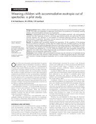
Incontinentia pigmenti - British Journal of Ophthalmology
Incontinentia pigmenti - British Journal of Ophthalmology
Incontinentia pigmenti - British Journal of Ophthalmology

You also want an ePaper? Increase the reach of your titles
YUMPU automatically turns print PDFs into web optimized ePapers that Google loves.
630<br />
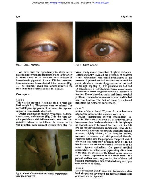
Fig. 2 Case 1. Righteye.<br />
We have had the opportunity to study seven<br />
patients all <strong>of</strong> whom are members <strong>of</strong> one large family<br />
in which a total <strong>of</strong> 14 members were affected by<br />
incontinentia <strong>pigmenti</strong>. A clear X-linked dominant<br />
transmission was demonstrated, lethal in males (Fig.<br />
1). The following seven case reports illustrate the<br />
most important ocular lesions <strong>of</strong> the disease.<br />
Case reports<br />
Downloaded from<br />
bjo.bmj.com on June 16, 2013 - Published by group.bmj.com<br />
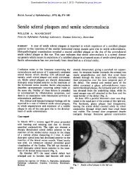
CASE 1<br />
This was the proband. A female child, 4 years old,<br />
birth weight 3 kg. The parents were not related. The<br />
dermatological symptoms <strong>of</strong> incontinentia <strong>pigmenti</strong><br />
appeared immediately after birth.<br />
Ocular examination showed nystagmus, oedematous<br />
cornea, and cataract (Fig. 2) in the right eye,<br />
microphthalmus with iridolenticular synechiae and<br />
complete cataract in the left eye. In this eye the iris<br />
was atrophic, with pigment irregularities (Fig. 3).<br />
Fig. 4 Case). Classic whorls and streaks <strong>of</strong>pigmenton<br />
torso and in right eye.<br />
Fig. 3 Case). Left eye.<br />
A Spallone<br />
Visual acuity was no perception <strong>of</strong> light in both eyes.<br />
Ultrasonography revealed the presence <strong>of</strong> bilateral<br />
retinal detachment with dense membranes in the<br />
vitreous. A general medical examination showed the<br />
classic whorls and streaks <strong>of</strong> pigment on the torso and<br />
on the right leg (Fig. 4). The grandmother had had<br />
18 pregnancies, 11 <strong>of</strong> which had been miscarriages.<br />
The seven fullterm pregnancies were all resulted in<br />
females. Five <strong>of</strong> these had ocular and dermatological<br />
problems; one died <strong>of</strong> an unknown cause, and the last<br />
one was healthy. The first <strong>of</strong> these five affected<br />
patients is the mother <strong>of</strong> our proband.<br />
CASE 2<br />
Mother <strong>of</strong> the proband, 37 years old, who has been<br />
affected by incontinentia <strong>pigmenti</strong> since birth.<br />
Ocular examination showed intermittent exotropia.<br />
The visual acuity was 1-0 in both eyes. Both<br />
lenses were clear. In the ocular fundus in the right eye<br />
no anomalies could be found; by contrast in the left<br />
eye the retinal vessels were anomalous. Towards the<br />
temporal equator both venules and arterioles became<br />
tortuous, slightly kinked, <strong>of</strong> an irregular calibre,<br />
increased in number, and with preretinal fibrosis.<br />
Apart from this area the peripheral temporal part <strong>of</strong><br />
the retina was completely avascular (Fig. 5). In the<br />
inferior nasal area there were small alterations <strong>of</strong> the<br />
retinal pigment epithelium. On general medical<br />
examination we noted some pigmentary alterations<br />
<strong>of</strong> the skin, the absence <strong>of</strong> the lateral upper incisors,<br />
and a conical appearance <strong>of</strong> the other teeth. The<br />
patient had had nine pregnancies; five <strong>of</strong> these had<br />
ended in miscarriages, two <strong>of</strong> which during necropsy<br />
were found to be males.<br />
CASE 3<br />
Sister <strong>of</strong> the proband, 14 years old. Immediately after<br />
birth the patient developed the dermatological signs<br />
<strong>of</strong> incontinentia <strong>pigmenti</strong>.