Incontinentia pigmenti - British Journal of Ophthalmology
Incontinentia pigmenti - British Journal of Ophthalmology
Incontinentia pigmenti - British Journal of Ophthalmology

Create successful ePaper yourself
Turn your PDF publications into a flip-book with our unique Google optimized e-Paper software.
632<br />
Downloaded from<br />
bjo.bmj.com on June 16, 2013 - Published by group.bmj.com<br />
A Spallone<br />
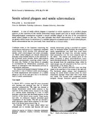
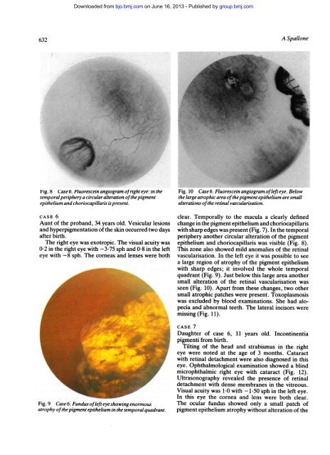
Fig. 8 Case 6. Fluorescein angiogram <strong>of</strong>right eye: in the Fig. 10 Case 6. Fluorescein angiogram <strong>of</strong>left eye. Below<br />
temporal periphery a circular alteration <strong>of</strong>the pigment the large atrophic area <strong>of</strong>the pigment epithelium are small<br />
epithelium and choriocapillaris is present. alterations <strong>of</strong>the retinal vascularisation.<br />
CASE 6<br />
Aunt <strong>of</strong> the proband, 34 years old. Vesicular lesions<br />
and hyperpigmentation <strong>of</strong> the skin occurred two days<br />
after birth.<br />
The right eye was exotropic. The visual acuity was<br />
0-2 in the right eye with -3 75 sph and 0-8 in the left<br />
eye with -8 sph. The corneas and lenses were both<br />
Fig. 9 Case 6. Fundus <strong>of</strong>left eye showing enormous<br />
atrophy <strong>of</strong>the pigment epithelium in the temporal quadrant.<br />
clear. Temporally to the macula a clearly defined<br />
change in the pigment epithelium and choriocapillaris<br />
with sharp edges was present (Fig. 7). In the temporal<br />
periphery another circular alteration <strong>of</strong> the pigment<br />
epithelium and choriocapillaris was visible (Fig. 8).<br />
This zone also showed mild anomalies <strong>of</strong> the retinal<br />
vascularisation. In the left eye it was possible to see<br />
a large region <strong>of</strong> atrophy <strong>of</strong> the pigment epithelium<br />
with sharp edges; it involved the whole temporal<br />
quadrant (Fig. 9). Just below this large area another<br />
small alteration <strong>of</strong> the retinal vascularisation was<br />
seen (Fig. 10). Apart from these changes, two other<br />
small atrophic patches were present. Toxoplasmosis<br />
was excluded by blood examinations. She had alopecia<br />
and abnormal teeth. The lateral incisors were<br />
missing (Fig. 11).<br />
CASE 7<br />
Daughter <strong>of</strong> case 6, 11 years old. <strong>Incontinentia</strong><br />
<strong>pigmenti</strong> from birth.<br />
Tilting <strong>of</strong> the head and strabismus in the right<br />
eye were noted at the age <strong>of</strong> 3 months. Cataract<br />
with retinal detachment were also diagnosed in this<br />
eye. Ophthalmological examination showed a blind<br />
microphthalmic right eye with cataract (Fig. 12).<br />
Ultrasonography revealed the presence <strong>of</strong> retinal<br />
detachment with dense membranes in the vitreous.<br />
Visual acuity was 1-0 with -1-50 sph in the left eye.<br />
In this eye the cornea and lens were both clear.<br />
The ocular fundus showed only a -small patch <strong>of</strong><br />
pigment epithelium atrophy without alteration <strong>of</strong> the