Senile scleral plaques and senile scleromalacia - British Journal of ...
Senile scleral plaques and senile scleromalacia - British Journal of ...
Senile scleral plaques and senile scleromalacia - British Journal of ...

Create successful ePaper yourself
Turn your PDF publications into a flip-book with our unique Google optimized e-Paper software.
<strong>British</strong> <strong>Journal</strong> <strong>of</strong> Ophthalmology, 1978, 62, 376-380<br />
<strong>Senile</strong> <strong>scleral</strong> <strong>plaques</strong> <strong>and</strong> <strong>senile</strong> <strong>scleromalacia</strong><br />
WILLEM A. MANSCHOT<br />
From the Ophthalmic Pathology Laboratory, Erasmus University, Rotterdam<br />
SUMMARY A case <strong>of</strong> <strong>senile</strong> <strong>scleral</strong> <strong>plaques</strong> is reported in which expulsion <strong>of</strong> a calcified plaque<br />
anterior to the insertion <strong>of</strong> the medial horizontal rectus muscle gave rise to <strong>senile</strong> <strong>scleromalacia</strong>.<br />
Histopathological examination revealed a second calcified plaque at the site <strong>of</strong> the contralateral<br />
<strong>senile</strong> <strong>scleral</strong> plaque in this eye. This case indicates that <strong>senile</strong> <strong>scleromalacia</strong> is a <strong>scleral</strong> disease<br />
sui generis which occurs by expulsion <strong>of</strong> a calcified plaque in advanced cases <strong>of</strong> <strong>senile</strong> <strong>scleral</strong> <strong>plaques</strong>.<br />
<strong>Senile</strong> <strong>scleromalacia</strong> has not previously been described as a clinical entity.<br />
Confusion exists in the literature concerning the<br />
nomenclature <strong>and</strong> nature <strong>of</strong> 2 apparently unrelated<br />
<strong>scleral</strong> lesions which develop with advanced age,<br />
namely, <strong>senile</strong> <strong>scleral</strong> <strong>plaques</strong> <strong>and</strong> <strong>senile</strong> <strong>scleromalacia</strong>.<br />
<strong>Senile</strong> <strong>scleral</strong> <strong>plaques</strong> are sharply demarcated<br />
slate-grey areas located anterior to the insertions <strong>of</strong><br />
the horizontal rectus muscles. <strong>Senile</strong> <strong>scleromalacia</strong><br />
describes spontaneously occurring <strong>scleral</strong> holes at<br />
the same site. Neither <strong>of</strong> these lesions is preceded<br />
or accompanied by inflammatory symptoms, <strong>and</strong><br />
there is no association with rheumatoid arthritis or<br />
<strong>scleromalacia</strong> perforans.<br />
The clinical <strong>and</strong> histopathological findings will be<br />
reported in a case <strong>of</strong> bilateral <strong>senile</strong> <strong>scleral</strong> <strong>plaques</strong>,<br />
in which sequestration <strong>and</strong> expulsion <strong>of</strong> a calcific<br />
plaque in one <strong>of</strong> the eyes gave rise to the development<br />
<strong>of</strong> <strong>senile</strong> <strong>scleromalacia</strong>.<br />
Case report<br />
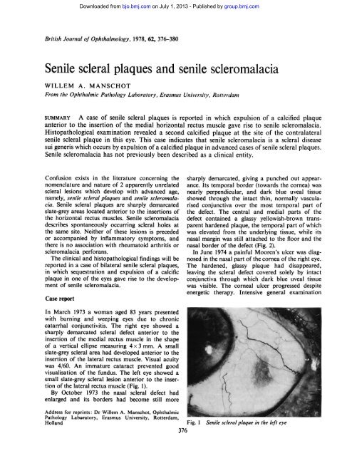
In March 1973 a woman aged 83 years presented<br />
with burning <strong>and</strong> weeping eyes due to chronic<br />
catarrhal conjunctivitis. The right eye showed a<br />
sharply demarcated <strong>scleral</strong> defect anterior to the<br />
insertion <strong>of</strong> the medial rectus muscle in the shape<br />
<strong>of</strong> a vertical ellipse measuring 4 x 3 mm. A small<br />
slate-grey <strong>scleral</strong> area had developed anterior to the<br />
insertion <strong>of</strong> the lateral rectus muscle. Visual acuity<br />
was 4/60. An immature cataract prevented good<br />
visualisation <strong>of</strong> the fundus. The left eye showed a<br />
small slate-grey <strong>scleral</strong> lesion anterior to the insertion<br />
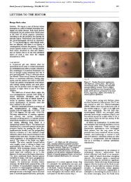
<strong>of</strong> the lateral rectus muscle (Fig. 1).<br />
By October 1973 the nasal <strong>scleral</strong> defect had<br />
enlarged <strong>and</strong> its borders had become still more<br />
Address for reprints: Dr Willem A. Manschot, Ophthalmic<br />
Pathology Laboratory, Erasmus University, Rotterdam,<br />
Holl<strong>and</strong><br />
Downloaded from<br />
bjo.bmj.com on July 1, 2013 - Published by group.bmj.com<br />
sharply demarcated, giving a punched out appearance.<br />
Its temporal border (towards the cornea) was<br />
nearly perpendicular, <strong>and</strong> dark blue uveal tissue<br />
showed through the intact thin, normally vascularised<br />
conjunctiva over the most temporal part <strong>of</strong><br />
the defect. The central <strong>and</strong> medial parts <strong>of</strong> the<br />
defect contained a glassy yellowish-brown transparent<br />
hardened plaque, the temporal part <strong>of</strong> which<br />
was elevated from the underlying tissue, while its<br />
nasal margin was still attached to the floor <strong>and</strong> the<br />
nasal border <strong>of</strong> the defect (Fig. 2).<br />
In June 1974 a painful Mooren's ulcer was diagnosed<br />
in the nasal part <strong>of</strong> the cornea <strong>of</strong> the right eye.<br />
The hardened, glassy plaque had disappeared,<br />
leaving the <strong>scleral</strong> defect covered solely by intact<br />
conjunctiva through which dark blue uveal tissue<br />
was visible. The corneal ulcer progressed despite<br />
energetic therapy. Intensive general examination<br />
Fig. 1 <strong>Senile</strong> sc/eral plaque in the left eye<br />
376
Seniile <strong>scleral</strong> <strong>plaques</strong> <strong>and</strong> <strong>senile</strong> <strong>scleromalacia</strong><br />
showed a large, vascularised thin area, the central<br />
margin <strong>of</strong> which appeared relatively thickened.<br />
Horizontal calottes were cut.<br />
Microscopical examination showed 2 symmetrically<br />
located <strong>scleral</strong> lesions on both sides over the<br />
pars plana <strong>of</strong> the ciliary body <strong>and</strong> the peripheral<br />
retina (Fig. 4).<br />
On the nasal side a <strong>scleral</strong> defect extended from<br />
2-5 mm in front <strong>of</strong> the ora serrata to 2-5 mm behind<br />
the ora (Fig. 5). The smooth floor <strong>of</strong> the defect was<br />
covered by epithelium <strong>and</strong> was formed by the<br />
deepest <strong>scleral</strong> layers which showed a normal<br />
fibrillar structure <strong>and</strong> birefringence without any<br />
sign <strong>of</strong> hyalinisation or necrosis. The thickness <strong>of</strong><br />
the defect measured about one-third to one-sixth <strong>of</strong><br />
_>.-X
378<br />
Downloaded from<br />
bjo.bmj.com on July 1, 2013 - Published by group.bmj.com<br />
Fig. 6 Higher magnification <strong>of</strong><br />
lesion (b) in Fig. 4. The <strong>senile</strong><br />
<strong>scleral</strong> plaque contains a<br />
calcified centre, <strong>and</strong> there is loss<br />
<strong>of</strong> cellularity in the surrounding<br />
tissue. (H <strong>and</strong> E x 23)<br />
plasma cells. The peripheral part <strong>of</strong> the <strong>scleral</strong> floor<br />
<strong>of</strong> the defect was covered by scar tissue, while the<br />
epithelium had formed a proliferative fold between<br />
the scar tissue <strong>and</strong> the remaining sclera.<br />
The site <strong>of</strong> the <strong>scleral</strong> lesion in the temporal part<br />
<strong>of</strong> the sclera corresponded to that <strong>of</strong> the <strong>scleral</strong><br />
defect on the nasal side. In the sections its horizontal<br />
diameter was about 4 mm with its major part<br />
located in front <strong>of</strong> the ora serrata. The affected<br />
sclera showed no thinning, but its centre contained<br />
a calcific plaque (Fig. 6), which, despite repeated<br />
decalcifying procedures, stained strongly by the<br />
von Kossa method <strong>and</strong> with alizarin red. The<br />
<strong>scleral</strong> tissue all around the calcific plaque showed<br />
a total loss <strong>of</strong> cellularity, normal birefringence, <strong>and</strong><br />
no hyalinisation.<br />
The nasal half <strong>of</strong> the cornea contained a large<br />
ulcer. Part <strong>of</strong> its peripheral slope was rather steep<br />
but elsewhere it shelved more gradually, while its<br />
central margin showed the characteristic overhanging<br />
lip <strong>of</strong> a Mooren's ulcer. The thickness <strong>of</strong> the<br />
overhanging lip was about half that <strong>of</strong> the cornea,<br />
while the lamellar cleft between the overhanging lip<br />
<strong>and</strong> the floor <strong>of</strong> the ulcer was partially filled by<br />
proliferating squamous epithelium in which mitotic<br />
figures were present. At one site the stromal necrosis<br />
had extended to Descemet's membrane, but generally<br />
the membrane was covered by a few stromal lamellae,<br />
a layer <strong>of</strong> heavily vascularised scar tissue <strong>of</strong><br />
variable thickness, <strong>and</strong> a rather thick layer <strong>of</strong><br />
epithelium.<br />
Willem A. Manischot<br />
Fig. 5 Higher magnification <strong>of</strong><br />
lesion (a) in Fig. 4. The floor is<br />
formed by the deepest <strong>scleral</strong><br />
layers <strong>and</strong> is covered by<br />
epithelium. No hyalinisation or<br />
necrosis is present. (H <strong>and</strong> E<br />
x 21)..<br />
The iris stroma <strong>and</strong> the ciliary body contained<br />
mononuclear inflammatory cells. The lens cortex<br />
showed vacuolisation. The posterior segment was<br />
not remarkable except for a diffuse intra<strong>scleral</strong><br />
scattering <strong>of</strong> calcific granules <strong>of</strong> the type not infrequently<br />
encountered in elderly people.<br />
Discussion<br />
<strong>Senile</strong> <strong>scleral</strong> <strong>plaques</strong> have been described by a<br />
great variety <strong>of</strong> terms namely, 'localised areas <strong>of</strong><br />
calcareous degeneration in the sclera' (Katz, 1929),<br />
'<strong>senile</strong> degeneration <strong>of</strong> the sclera' (Pillat, 1933),<br />
'<strong>senile</strong> thinning <strong>of</strong> the sclera' (Kiss, 1934; Graves,<br />
1937, 1939, 1941), 'circumscribed <strong>scleromalacia</strong> at<br />
high age' (Kyrieleis, 1939), '<strong>scleral</strong> <strong>plaques</strong>' (Culler,<br />
1939), 'hyaline <strong>scleral</strong> <strong>plaques</strong>' (Bosh<strong>of</strong>f, 1942),<br />
'<strong>senile</strong> hyaline <strong>scleral</strong> <strong>plaques</strong>' (Roper, 1945), <strong>and</strong><br />
'focal <strong>senile</strong> translucency <strong>of</strong> the sclera' (Cogan <strong>and</strong><br />
Kuwabara, 1959). Since advanced age appears to be<br />
the most important predisposing factor <strong>and</strong> hyalinisation<br />
<strong>of</strong> the <strong>scleral</strong> tissue does not occur, the<br />
designation <strong>senile</strong> <strong>scleral</strong> <strong>plaques</strong> seems to be the<br />
most appropriate.<br />
<strong>Senile</strong> <strong>scleral</strong> <strong>plaques</strong> are not rare but may escape<br />
notice through absence <strong>of</strong> subjective symptoms.<br />
The <strong>plaques</strong> appear as symmetrical, sharply demarcated,<br />
glassy slate-grey coloured areas just anterior<br />
to the insertions <strong>of</strong> the horizontal rectus muscles.<br />
Only in one instance has a different site been recorded,<br />
Gasteiger (1937) describing a third narrow
Downloaded from<br />
bjo.bmj.com on July 1, 2013 - Published by group.bmj.com<br />
<strong>Senile</strong> <strong>scleral</strong> <strong>plaques</strong> <strong>and</strong> <strong>senile</strong> <strong>scleromalacia</strong><br />
plaque in front <strong>of</strong> the insertion <strong>of</strong> the inferior rectus<br />
muscle. Their shape is that <strong>of</strong> a vertical angular<br />
ellipse, with an average horizontal width <strong>of</strong> 2 mm<br />
<strong>and</strong> an average vertical height <strong>of</strong> 5 to 6 mm. The<br />
overlying conjunctiva is thin but appears normal<br />
for the patient's age. The <strong>plaques</strong> are translucent,<br />
as previously mentioned by Rol<strong>and</strong>i (1915), Pillat<br />
(1933), Gasteiger (1937), <strong>and</strong> Kyrieleis (1939). This<br />
translucency was emphasised by Cogan <strong>and</strong> Kuwabara<br />
(1959), who coined the name 'focal <strong>senile</strong><br />
translucency <strong>of</strong> the sclera'. The fact that <strong>senile</strong><br />
<strong>scleral</strong> <strong>plaques</strong> are restricted to the region immediately<br />
in front <strong>of</strong> the insertion <strong>of</strong> the horizontal<br />
rectus muscles is thought to be due to the constant<br />
stress <strong>and</strong> strain on the <strong>scleral</strong> fibres exerted by<br />
muscular action. One <strong>of</strong> the causes <strong>of</strong> the transparency<br />
<strong>of</strong> the <strong>plaques</strong> may be local dehydration<br />
(Fischer, 1926).<br />
Histopathology. Histopathological findings in<br />
single cases have been reported by Urrets Zavalia<br />
et al. (1937), Culler (1939), <strong>and</strong> Kyrieleis (1939). A<br />
detailed study <strong>of</strong> 30 specimens by Cogan <strong>and</strong><br />
Kuwabara (1959) showed that, contrary to previous<br />
reports, the thickness <strong>of</strong> the sclera at the site <strong>of</strong> the<br />
<strong>senile</strong> <strong>plaques</strong> is either normal or slightly increased.<br />
Furthermore the <strong>plaques</strong> were seen to be characterised<br />
by early loss <strong>of</strong> cellularity, as Kyrieleis had<br />
already noted, although the collagenous fibres had<br />
a normal fibrillar structure <strong>and</strong> birefringence with<br />
no sign <strong>of</strong> hyalinisation.<br />
Another characteristic feature is calcification in<br />
the centres <strong>of</strong> the translucent areas. Calcified<br />
<strong>plaques</strong> were found in less than half the cases studied,<br />
their prevalence in lesions with extensive areas <strong>of</strong><br />
translucency suggesting that calcification is probably<br />
a sequel to the loss <strong>of</strong> cellularity. In all recorded<br />
cases the calcified <strong>plaques</strong> overlay the pars plana <strong>of</strong><br />
the ciliary body <strong>and</strong> were usually composed <strong>of</strong><br />
calcium phosphate, although in an exceptional case<br />
Cogan <strong>and</strong> Kuwabara (1959) demonstrated calcium<br />
sulphate. Findings suggestive <strong>of</strong> calcification had<br />
previously been reported by a number <strong>of</strong> observers<br />
(Rol<strong>and</strong>i, 1915; Urrets Zavalia et al., 1937; Culler,<br />
1939; Kyrieleis, 1939).<br />
As commented by Cogan <strong>and</strong> Kuwabara (1959),<br />
calcification could be a secondary, <strong>and</strong> probably<br />
late, sequel to the loss <strong>of</strong> cellularity. My own case<br />
demonstrates that sometimes this stage is followed<br />
by a further complication, namely, sequestration<br />
<strong>and</strong> expulsion <strong>of</strong> the calcified plaque, leaving a<br />
<strong>scleral</strong> hole which mimics the clinical picture <strong>of</strong><br />
<strong>scleromalacia</strong> perforans. The expulsion <strong>of</strong> the calcified<br />
plaque in the present case was documented by<br />
photography. It is interesting that Kyrieleis (1939)<br />
was <strong>of</strong> the opinion that expulsion <strong>of</strong> the almost<br />
completely sequestrated intra<strong>scleral</strong> calcific plaque<br />
379<br />
in his patient would almost certainly have caused a<br />
<strong>scleromalacia</strong> 'perforans'. The fact that Cogan <strong>and</strong><br />
Kuwabara could not find <strong>scleral</strong> indentations or<br />
abnormal <strong>scleral</strong> thinning in any <strong>of</strong> the cases in<br />
their series indicates that expulsion <strong>of</strong> the calcified<br />
<strong>plaques</strong> is rare. In none <strong>of</strong> their cases had the<br />
<strong>senile</strong> <strong>scleral</strong> plaque progressed to the ultimate stage<br />
<strong>of</strong> <strong>senile</strong> <strong>scleromalacia</strong>.<br />
<strong>Senile</strong> <strong>scleromalacia</strong> is essentially different from<br />
<strong>scleromalacia</strong> perforans, being characterised by (1)<br />
advanced age <strong>of</strong> the patient, (2) location just<br />
anterior to the insertions <strong>of</strong> the horizontal rectus<br />
muscles alone, (3) a vertical, irregular, oval or<br />
kidney-shaped punched out <strong>scleral</strong> defect, covered<br />
by thin conjunctival tissue <strong>and</strong> possibly having a<br />
glassy, yellowish-grey hardened plaque in the floor,<br />
(4) the presence <strong>of</strong> <strong>senile</strong> <strong>scleral</strong> <strong>plaques</strong> in the<br />
ipsilateral <strong>and</strong> contralateral eye, (5) development<br />
by expulsion <strong>of</strong> a sequestrated calcified plaque, (6)<br />
no evidence <strong>of</strong> (rheumatoid) <strong>scleral</strong> nodules or<br />
rheumatic disease, <strong>and</strong> (7) histopathological finding<br />
<strong>of</strong> a flat intact thin <strong>scleral</strong> floor covered by a continuous<br />
healthy layer <strong>of</strong> epithelium, <strong>and</strong> having<br />
smooth sharp margins <strong>and</strong> minimal inflammatory<br />
reaction with absence <strong>of</strong> necrosis. The floor <strong>and</strong> the<br />
surrounding <strong>scleral</strong> tissue show normal fibrillar<br />
architecture <strong>and</strong> birefringence with loss <strong>of</strong> cellularity<br />
<strong>and</strong> absence <strong>of</strong> hyalinisation. In most cases a<br />
<strong>senile</strong> <strong>scleral</strong> plaque is present anterior to the<br />
insertion <strong>of</strong> the opposite horizontal rectus muscle.<br />
Scleromalacia perforans (van der Hoeve, 1934), on<br />
the contrary, is characterised by (1) less advanced<br />
age <strong>of</strong> the patient, (2) location anywhere in the<br />
anterior sclera, (3) the emergence <strong>of</strong> large holes in<br />
the sclera at the base <strong>of</strong> which the uvea appears to<br />
be bare, (4) absence <strong>of</strong> <strong>senile</strong> <strong>scleral</strong> <strong>plaques</strong>, (5)<br />
gradual development from a necrobiotic rheumatoid<br />
nodule, <strong>and</strong> (6) characteristic histopathology (Verhoeff<br />
<strong>and</strong> King, 1938; Ashton <strong>and</strong> Hobbs, 1952;<br />
Anderson <strong>and</strong> Margolis, 1952): the entire thickness<br />
<strong>of</strong> the affected sclera is necrotic so that the defect<br />
is devoid <strong>of</strong> a floor <strong>and</strong> the margins are ill defined.<br />
The necrotic granulomatous inflammation <strong>of</strong>ten<br />
extends into the underlying uvea <strong>and</strong> the surrounding<br />
sclera. Rheumatoid nodules may be found<br />
elsewhere in the sclera.<br />
Despite these differences the early clinical manifestations<br />
<strong>of</strong> <strong>senile</strong> <strong>scleromalacia</strong> <strong>and</strong> <strong>scleromalacia</strong><br />
perforans may be confusingly similar, both being<br />
initially painless with minimal inflammatory reaction.<br />
It is probable that many cases <strong>of</strong> <strong>senile</strong> <strong>scleromalacia</strong><br />
have been misdiagnosed as <strong>scleromalacia</strong><br />
perforans, if only because <strong>senile</strong> <strong>scleromalacia</strong> has<br />
never been described as a separate entity, despite<br />
some authors having stressed the essential differences<br />
between <strong>senile</strong> <strong>scleral</strong> <strong>plaques</strong> <strong>and</strong> sclero-
380<br />
malacia perforans (Roper, 1945; Anderson <strong>and</strong><br />
Margolis, 1952).<br />
The present case, however, lends strong support<br />
to the prediction made by Kyrieleis almost 40 years<br />
ago that <strong>senile</strong> <strong>scleromalacia</strong> is a disease sui generis<br />
which occurs by expulsion <strong>of</strong> a calcified plaque in<br />
advanced cases <strong>of</strong> <strong>senile</strong> scieral <strong>plaques</strong>.<br />
So far the simultaneous occurrence <strong>of</strong> <strong>senile</strong><br />
scieral <strong>plaques</strong> <strong>and</strong> Mooren's ulcer has not been<br />
reported <strong>and</strong> is unexplained. On the basis <strong>of</strong> a<br />
single case it is probably safest to refrain from<br />
speculation.<br />
The author thanks Dr V. W. Antic, who provided<br />
the clinical data, Mr P. van der Heul, technician,<br />
<strong>and</strong> Miss P. Delfos, photographer.<br />
References<br />
Downloaded from<br />
bjo.bmj.com on July 1, 2013 - Published by group.bmj.com<br />
Anderson, B., <strong>and</strong> Margolis, G. (1952). Scleromalacia.<br />
Americant <strong>Journal</strong> <strong>of</strong> Ophthalmology, 35, 917-931.<br />
Ashton, N., <strong>and</strong> Hobbs, H. E. (1952). Effect <strong>of</strong> cortisone on<br />
rheumatoid nodules <strong>of</strong> the sclera (<strong>scleromalacia</strong> perforans).<br />
<strong>British</strong> <strong>Journal</strong> <strong>of</strong> Ophthalmnology, 36, 373-384.<br />
Bosh<strong>of</strong>f, P. H. (1942). Hyaline <strong>scleral</strong> <strong>plaques</strong>. Archives <strong>of</strong><br />
Ophthalmology, 28, 503-506.<br />
Cogan, D. G., <strong>and</strong> Kuwabara, T. (1959). Focal <strong>senile</strong> translucency<br />
<strong>of</strong> the sclera. Archives <strong>of</strong> Ophthalnology, 62,<br />
604-610.<br />
Willeni A. Manschol<br />
Culler, A. M. (1939). The pathology <strong>of</strong> <strong>scleral</strong> <strong>plaques</strong>.<br />
<strong>British</strong> Joiurtnal <strong>of</strong> Ophthalnmology, 23, 44-50.<br />
Fischer, F. P. (1926). Experimentelle Untersuchungen an der<br />
Lederhaut. Archiv fuir Aiugenheilkunde, 97, 467-492.<br />
Gasteiger, H. (1937). Ueber <strong>senile</strong> Entartung der Lederhaut<br />
an den Ansatzstellen der geraden Augenmuskeln. Klinische<br />
Moniatsblatter fiir Auigenlheilkiunde, 98, 767-772.<br />
Graves, B. (1937). Bilateral mesial superficial deficiency <strong>of</strong><br />
the sclera. <strong>British</strong> Journial <strong>of</strong> Ophthalmology, 21, 534-538.<br />
Graves, B. (1939). Bilateral mesial superficial 'deficiency' <strong>of</strong><br />
the sclera (<strong>scleral</strong> <strong>plaques</strong>). <strong>British</strong> Joutrnial <strong>of</strong> Ophthailmnology,<br />
23, 191-204.<br />
Graves, B. (1941). Bilateral (mesial) deficiency <strong>of</strong> the sclera:<br />
<strong>scleral</strong> <strong>plaques</strong>. <strong>British</strong> Jouirnal <strong>of</strong> Ophthalmology, 25, 35-38.<br />
Hoeve, J., van der (1934). Scleromalacia perforans. Archives<br />
<strong>of</strong> Ophthalmology, 11, 111-118.<br />
Katz, D. (1929). A localised area <strong>of</strong> calcareous degeneration<br />
in the sclera. Archives <strong>of</strong> Ophthalmnology, 2, 30-33.<br />
Kiss, J. (1934). Fall von <strong>senile</strong>r Skleraverdulnnung. Klinische<br />
Monatsblatter fiir Auigentheilkuttde, 92, 121-122.<br />
Kyrieleis, W. (1939). Ueber umschriebenen Lederhautschwund<br />
(Skleromalazie) in hoherem Lebensalter. Klinische<br />
Montatsblaitter fir A ugemiheilkunde, 103, 441-452.<br />
Pillat, A. (1933). Ueber eine eigenartige <strong>senile</strong> Entartung der<br />
Lederhaut an den Ansatzstellen der geraden Augen-<br />
Muskeln. Zeitschrift fiir Augenheilkuinde, 82, 113-123.<br />
Rol<strong>and</strong>i, S. (1915). cited by Roper, K. L. (1945).<br />
Roper, K. L. (1945). <strong>Senile</strong> hyaline <strong>scleral</strong> <strong>plaques</strong>. Archives<br />
<strong>of</strong> Ophthalmology, 34, 283-291.<br />
Urrets Zavalia, A., Maldonado Allende, I., <strong>and</strong> Obregon<br />
Oliva, R. (1937). Cited by Roper, K. L. (1945).<br />
Verhoeff, F. H., <strong>and</strong> King, M. J. (1938). Scleromalacia<br />
perforans. Archives <strong>of</strong> Ophthalmology, 20, 1013-1035.
Downloaded from<br />
bjo.bmj.com on July 1, 2013 - Published by group.bmj.com<br />
Email alerting<br />
service<br />
Notes<br />
<strong>Senile</strong> <strong>scleral</strong> <strong>plaques</strong> <strong>and</strong> <strong>senile</strong><br />
<strong>scleromalacia</strong>.<br />
W. A. Manschot<br />
Br J Ophthalmol 1978 62: 376-380<br />
doi: 10.1136/bjo.62.6.376<br />
Updated information <strong>and</strong> services can be found at:<br />
http://bjo.bmj.com/content/62/6/376<br />
These include:<br />
To request permissions go to:<br />
http://group.bmj.com/group/rights-licensing/permissions<br />
To order reprints go to:<br />
http://journals.bmj.com/cgi/reprintform<br />
To subscribe to BMJ go to:<br />
http://group.bmj.com/subscribe/<br />
Receive free email alerts when new articles cite this<br />
article. Sign up in the box at the top right corner <strong>of</strong> the<br />
online article.