Senile scleral plaques and senile scleromalacia - British Journal of ...
Senile scleral plaques and senile scleromalacia - British Journal of ...
Senile scleral plaques and senile scleromalacia - British Journal of ...

You also want an ePaper? Increase the reach of your titles
YUMPU automatically turns print PDFs into web optimized ePapers that Google loves.
Downloaded from<br />
bjo.bmj.com on July 1, 2013 - Published by group.bmj.com<br />
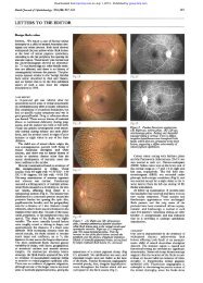
<strong>Senile</strong> <strong>scleral</strong> <strong>plaques</strong> <strong>and</strong> <strong>senile</strong> <strong>scleromalacia</strong><br />
plaque in front <strong>of</strong> the insertion <strong>of</strong> the inferior rectus<br />
muscle. Their shape is that <strong>of</strong> a vertical angular<br />
ellipse, with an average horizontal width <strong>of</strong> 2 mm<br />
<strong>and</strong> an average vertical height <strong>of</strong> 5 to 6 mm. The<br />
overlying conjunctiva is thin but appears normal<br />
for the patient's age. The <strong>plaques</strong> are translucent,<br />
as previously mentioned by Rol<strong>and</strong>i (1915), Pillat<br />
(1933), Gasteiger (1937), <strong>and</strong> Kyrieleis (1939). This<br />
translucency was emphasised by Cogan <strong>and</strong> Kuwabara<br />
(1959), who coined the name 'focal <strong>senile</strong><br />
translucency <strong>of</strong> the sclera'. The fact that <strong>senile</strong><br />
<strong>scleral</strong> <strong>plaques</strong> are restricted to the region immediately<br />
in front <strong>of</strong> the insertion <strong>of</strong> the horizontal<br />
rectus muscles is thought to be due to the constant<br />
stress <strong>and</strong> strain on the <strong>scleral</strong> fibres exerted by<br />
muscular action. One <strong>of</strong> the causes <strong>of</strong> the transparency<br />
<strong>of</strong> the <strong>plaques</strong> may be local dehydration<br />
(Fischer, 1926).<br />
Histopathology. Histopathological findings in<br />
single cases have been reported by Urrets Zavalia<br />
et al. (1937), Culler (1939), <strong>and</strong> Kyrieleis (1939). A<br />
detailed study <strong>of</strong> 30 specimens by Cogan <strong>and</strong><br />
Kuwabara (1959) showed that, contrary to previous<br />
reports, the thickness <strong>of</strong> the sclera at the site <strong>of</strong> the<br />
<strong>senile</strong> <strong>plaques</strong> is either normal or slightly increased.<br />
Furthermore the <strong>plaques</strong> were seen to be characterised<br />
by early loss <strong>of</strong> cellularity, as Kyrieleis had<br />
already noted, although the collagenous fibres had<br />
a normal fibrillar structure <strong>and</strong> birefringence with<br />
no sign <strong>of</strong> hyalinisation.<br />
Another characteristic feature is calcification in<br />
the centres <strong>of</strong> the translucent areas. Calcified<br />
<strong>plaques</strong> were found in less than half the cases studied,<br />
their prevalence in lesions with extensive areas <strong>of</strong><br />
translucency suggesting that calcification is probably<br />
a sequel to the loss <strong>of</strong> cellularity. In all recorded<br />
cases the calcified <strong>plaques</strong> overlay the pars plana <strong>of</strong><br />
the ciliary body <strong>and</strong> were usually composed <strong>of</strong><br />
calcium phosphate, although in an exceptional case<br />
Cogan <strong>and</strong> Kuwabara (1959) demonstrated calcium<br />
sulphate. Findings suggestive <strong>of</strong> calcification had<br />
previously been reported by a number <strong>of</strong> observers<br />
(Rol<strong>and</strong>i, 1915; Urrets Zavalia et al., 1937; Culler,<br />
1939; Kyrieleis, 1939).<br />
As commented by Cogan <strong>and</strong> Kuwabara (1959),<br />
calcification could be a secondary, <strong>and</strong> probably<br />
late, sequel to the loss <strong>of</strong> cellularity. My own case<br />
demonstrates that sometimes this stage is followed<br />
by a further complication, namely, sequestration<br />
<strong>and</strong> expulsion <strong>of</strong> the calcified plaque, leaving a<br />
<strong>scleral</strong> hole which mimics the clinical picture <strong>of</strong><br />
<strong>scleromalacia</strong> perforans. The expulsion <strong>of</strong> the calcified<br />
plaque in the present case was documented by<br />
photography. It is interesting that Kyrieleis (1939)<br />
was <strong>of</strong> the opinion that expulsion <strong>of</strong> the almost<br />
completely sequestrated intra<strong>scleral</strong> calcific plaque<br />
379<br />
in his patient would almost certainly have caused a<br />
<strong>scleromalacia</strong> 'perforans'. The fact that Cogan <strong>and</strong><br />
Kuwabara could not find <strong>scleral</strong> indentations or<br />
abnormal <strong>scleral</strong> thinning in any <strong>of</strong> the cases in<br />
their series indicates that expulsion <strong>of</strong> the calcified<br />
<strong>plaques</strong> is rare. In none <strong>of</strong> their cases had the<br />
<strong>senile</strong> <strong>scleral</strong> plaque progressed to the ultimate stage<br />
<strong>of</strong> <strong>senile</strong> <strong>scleromalacia</strong>.<br />
<strong>Senile</strong> <strong>scleromalacia</strong> is essentially different from<br />
<strong>scleromalacia</strong> perforans, being characterised by (1)<br />
advanced age <strong>of</strong> the patient, (2) location just<br />
anterior to the insertions <strong>of</strong> the horizontal rectus<br />
muscles alone, (3) a vertical, irregular, oval or<br />
kidney-shaped punched out <strong>scleral</strong> defect, covered<br />
by thin conjunctival tissue <strong>and</strong> possibly having a<br />
glassy, yellowish-grey hardened plaque in the floor,<br />
(4) the presence <strong>of</strong> <strong>senile</strong> <strong>scleral</strong> <strong>plaques</strong> in the<br />
ipsilateral <strong>and</strong> contralateral eye, (5) development<br />
by expulsion <strong>of</strong> a sequestrated calcified plaque, (6)<br />
no evidence <strong>of</strong> (rheumatoid) <strong>scleral</strong> nodules or<br />
rheumatic disease, <strong>and</strong> (7) histopathological finding<br />
<strong>of</strong> a flat intact thin <strong>scleral</strong> floor covered by a continuous<br />
healthy layer <strong>of</strong> epithelium, <strong>and</strong> having<br />
smooth sharp margins <strong>and</strong> minimal inflammatory<br />
reaction with absence <strong>of</strong> necrosis. The floor <strong>and</strong> the<br />
surrounding <strong>scleral</strong> tissue show normal fibrillar<br />
architecture <strong>and</strong> birefringence with loss <strong>of</strong> cellularity<br />
<strong>and</strong> absence <strong>of</strong> hyalinisation. In most cases a<br />
<strong>senile</strong> <strong>scleral</strong> plaque is present anterior to the<br />
insertion <strong>of</strong> the opposite horizontal rectus muscle.<br />
Scleromalacia perforans (van der Hoeve, 1934), on<br />
the contrary, is characterised by (1) less advanced<br />
age <strong>of</strong> the patient, (2) location anywhere in the<br />
anterior sclera, (3) the emergence <strong>of</strong> large holes in<br />
the sclera at the base <strong>of</strong> which the uvea appears to<br />
be bare, (4) absence <strong>of</strong> <strong>senile</strong> <strong>scleral</strong> <strong>plaques</strong>, (5)<br />
gradual development from a necrobiotic rheumatoid<br />
nodule, <strong>and</strong> (6) characteristic histopathology (Verhoeff<br />
<strong>and</strong> King, 1938; Ashton <strong>and</strong> Hobbs, 1952;<br />
Anderson <strong>and</strong> Margolis, 1952): the entire thickness<br />
<strong>of</strong> the affected sclera is necrotic so that the defect<br />
is devoid <strong>of</strong> a floor <strong>and</strong> the margins are ill defined.<br />
The necrotic granulomatous inflammation <strong>of</strong>ten<br />
extends into the underlying uvea <strong>and</strong> the surrounding<br />
sclera. Rheumatoid nodules may be found<br />
elsewhere in the sclera.<br />
Despite these differences the early clinical manifestations<br />
<strong>of</strong> <strong>senile</strong> <strong>scleromalacia</strong> <strong>and</strong> <strong>scleromalacia</strong><br />
perforans may be confusingly similar, both being<br />
initially painless with minimal inflammatory reaction.<br />
It is probable that many cases <strong>of</strong> <strong>senile</strong> <strong>scleromalacia</strong><br />
have been misdiagnosed as <strong>scleromalacia</strong><br />
perforans, if only because <strong>senile</strong> <strong>scleromalacia</strong> has<br />
never been described as a separate entity, despite<br />
some authors having stressed the essential differences<br />
between <strong>senile</strong> <strong>scleral</strong> <strong>plaques</strong> <strong>and</strong> sclero-