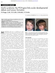
Senile scleral plaques and senile scleromalacia - British Journal of ...
Senile scleral plaques and senile scleromalacia - British Journal of ...
Senile scleral plaques and senile scleromalacia - British Journal of ...

Create successful ePaper yourself
Turn your PDF publications into a flip-book with our unique Google optimized e-Paper software.
378<br />
Downloaded from<br />
bjo.bmj.com on July 1, 2013 - Published by group.bmj.com<br />
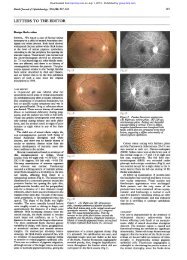
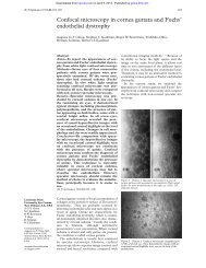
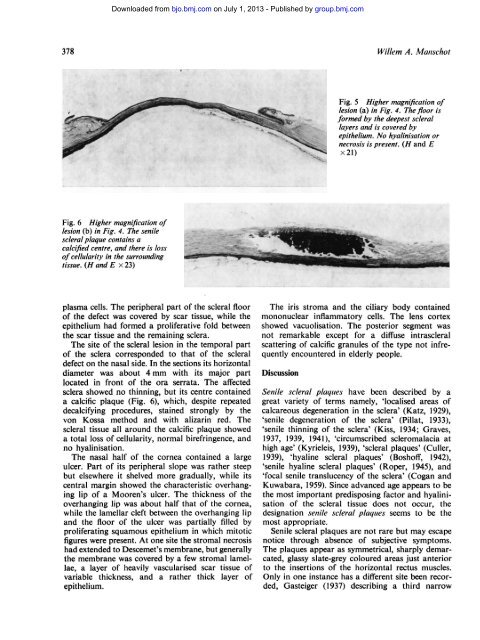
Fig. 6 Higher magnification <strong>of</strong><br />
lesion (b) in Fig. 4. The <strong>senile</strong><br />
<strong>scleral</strong> plaque contains a<br />
calcified centre, <strong>and</strong> there is loss<br />
<strong>of</strong> cellularity in the surrounding<br />
tissue. (H <strong>and</strong> E x 23)<br />
plasma cells. The peripheral part <strong>of</strong> the <strong>scleral</strong> floor<br />
<strong>of</strong> the defect was covered by scar tissue, while the<br />
epithelium had formed a proliferative fold between<br />
the scar tissue <strong>and</strong> the remaining sclera.<br />
The site <strong>of</strong> the <strong>scleral</strong> lesion in the temporal part<br />
<strong>of</strong> the sclera corresponded to that <strong>of</strong> the <strong>scleral</strong><br />
defect on the nasal side. In the sections its horizontal<br />
diameter was about 4 mm with its major part<br />
located in front <strong>of</strong> the ora serrata. The affected<br />
sclera showed no thinning, but its centre contained<br />
a calcific plaque (Fig. 6), which, despite repeated<br />
decalcifying procedures, stained strongly by the<br />
von Kossa method <strong>and</strong> with alizarin red. The<br />
<strong>scleral</strong> tissue all around the calcific plaque showed<br />
a total loss <strong>of</strong> cellularity, normal birefringence, <strong>and</strong><br />
no hyalinisation.<br />
The nasal half <strong>of</strong> the cornea contained a large<br />
ulcer. Part <strong>of</strong> its peripheral slope was rather steep<br />
but elsewhere it shelved more gradually, while its<br />
central margin showed the characteristic overhanging<br />
lip <strong>of</strong> a Mooren's ulcer. The thickness <strong>of</strong> the<br />
overhanging lip was about half that <strong>of</strong> the cornea,<br />
while the lamellar cleft between the overhanging lip<br />
<strong>and</strong> the floor <strong>of</strong> the ulcer was partially filled by<br />
proliferating squamous epithelium in which mitotic<br />
figures were present. At one site the stromal necrosis<br />
had extended to Descemet's membrane, but generally<br />
the membrane was covered by a few stromal lamellae,<br />
a layer <strong>of</strong> heavily vascularised scar tissue <strong>of</strong><br />
variable thickness, <strong>and</strong> a rather thick layer <strong>of</strong><br />
epithelium.<br />
Willem A. Manischot<br />
Fig. 5 Higher magnification <strong>of</strong><br />
lesion (a) in Fig. 4. The floor is<br />
formed by the deepest <strong>scleral</strong><br />
layers <strong>and</strong> is covered by<br />
epithelium. No hyalinisation or<br />
necrosis is present. (H <strong>and</strong> E<br />
x 21)..<br />
The iris stroma <strong>and</strong> the ciliary body contained<br />
mononuclear inflammatory cells. The lens cortex<br />
showed vacuolisation. The posterior segment was<br />
not remarkable except for a diffuse intra<strong>scleral</strong><br />
scattering <strong>of</strong> calcific granules <strong>of</strong> the type not infrequently<br />
encountered in elderly people.<br />
Discussion<br />
<strong>Senile</strong> <strong>scleral</strong> <strong>plaques</strong> have been described by a<br />
great variety <strong>of</strong> terms namely, 'localised areas <strong>of</strong><br />
calcareous degeneration in the sclera' (Katz, 1929),<br />
'<strong>senile</strong> degeneration <strong>of</strong> the sclera' (Pillat, 1933),<br />
'<strong>senile</strong> thinning <strong>of</strong> the sclera' (Kiss, 1934; Graves,<br />
1937, 1939, 1941), 'circumscribed <strong>scleromalacia</strong> at<br />
high age' (Kyrieleis, 1939), '<strong>scleral</strong> <strong>plaques</strong>' (Culler,<br />
1939), 'hyaline <strong>scleral</strong> <strong>plaques</strong>' (Bosh<strong>of</strong>f, 1942),<br />
'<strong>senile</strong> hyaline <strong>scleral</strong> <strong>plaques</strong>' (Roper, 1945), <strong>and</strong><br />
'focal <strong>senile</strong> translucency <strong>of</strong> the sclera' (Cogan <strong>and</strong><br />
Kuwabara, 1959). Since advanced age appears to be<br />
the most important predisposing factor <strong>and</strong> hyalinisation<br />
<strong>of</strong> the <strong>scleral</strong> tissue does not occur, the<br />
designation <strong>senile</strong> <strong>scleral</strong> <strong>plaques</strong> seems to be the<br />
most appropriate.<br />
<strong>Senile</strong> <strong>scleral</strong> <strong>plaques</strong> are not rare but may escape<br />
notice through absence <strong>of</strong> subjective symptoms.<br />
The <strong>plaques</strong> appear as symmetrical, sharply demarcated,<br />
glassy slate-grey coloured areas just anterior<br />
to the insertions <strong>of</strong> the horizontal rectus muscles.<br />
Only in one instance has a different site been recorded,<br />
Gasteiger (1937) describing a third narrow