Senile scleral plaques and senile scleromalacia - British Journal of ...
Senile scleral plaques and senile scleromalacia - British Journal of ...
Senile scleral plaques and senile scleromalacia - British Journal of ...

You also want an ePaper? Increase the reach of your titles
YUMPU automatically turns print PDFs into web optimized ePapers that Google loves.
Seniile <strong>scleral</strong> <strong>plaques</strong> <strong>and</strong> <strong>senile</strong> <strong>scleromalacia</strong><br />
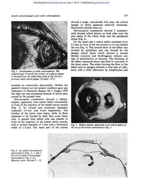
showed a large, vascularised thin area, the central<br />
margin <strong>of</strong> which appeared relatively thickened.<br />
Horizontal calottes were cut.<br />
Microscopical examination showed 2 symmetrically<br />
located <strong>scleral</strong> lesions on both sides over the<br />
pars plana <strong>of</strong> the ciliary body <strong>and</strong> the peripheral<br />
retina (Fig. 4).<br />
On the nasal side a <strong>scleral</strong> defect extended from<br />
2-5 mm in front <strong>of</strong> the ora serrata to 2-5 mm behind<br />
the ora (Fig. 5). The smooth floor <strong>of</strong> the defect was<br />
covered by epithelium <strong>and</strong> was formed by the<br />
deepest <strong>scleral</strong> layers which showed a normal<br />
fibrillar structure <strong>and</strong> birefringence without any<br />
sign <strong>of</strong> hyalinisation or necrosis. The thickness <strong>of</strong><br />
the defect measured about one-third to one-sixth <strong>of</strong><br />
_>.-X