Congenital divided naevus of the eyelids - British Journal of ...
Congenital divided naevus of the eyelids - British Journal of ...
Congenital divided naevus of the eyelids - British Journal of ...

Create successful ePaper yourself
Turn your PDF publications into a flip-book with our unique Google optimized e-Paper software.
<strong>British</strong> <strong>Journal</strong> <strong>of</strong> Ophthalmology, 1988, 72, 198-201<br />
<strong>Congenital</strong> <strong>divided</strong> <strong>naevus</strong> <strong>of</strong> <strong>the</strong> <strong>eyelids</strong><br />
P J McDONNELL' AND B J MAYOU2<br />
From <strong>the</strong> 'Department <strong>of</strong> Ophthalmology and <strong>the</strong> 2Department <strong>of</strong> Plastic, Surgery, St Thomas's Hospital,<br />
London<br />
SUMMARY A newborn infant presenting with a <strong>divided</strong> congenital melanocytic <strong>naevus</strong> <strong>of</strong> <strong>the</strong> eyelid<br />
is described. Because <strong>of</strong> <strong>the</strong> severe disfigurement, risk <strong>of</strong> later malignant change in <strong>the</strong> lesion, and<br />
<strong>the</strong> possibility <strong>of</strong> deprivation amblyopia, early surgical treatment is recommended for all medium<br />
and large congenital melanocytic naevi <strong>of</strong> <strong>the</strong> eyelid. Surgery in <strong>the</strong> first few months <strong>of</strong> life gives <strong>the</strong><br />
best cosmetic results.<br />
Divided <strong>naevus</strong> <strong>of</strong> <strong>the</strong> <strong>eyelids</strong> is a rare form <strong>of</strong><br />
congenital melanocytic <strong>naevus</strong> that involves <strong>the</strong><br />
upper and lower <strong>eyelids</strong> <strong>of</strong> one eye. The <strong>naevus</strong> is<br />
present in contiguous areas <strong>of</strong> <strong>the</strong> upper and lower lid<br />
margins, so that when <strong>the</strong> <strong>eyelids</strong> are closed <strong>the</strong> eye<br />
appears to be covered by one large <strong>naevus</strong>.<br />
<strong>Congenital</strong> <strong>divided</strong> <strong>naevus</strong> <strong>of</strong> <strong>the</strong> <strong>eyelids</strong> was first<br />
described in 1919 by Fuchs.' Since <strong>the</strong>n about 30<br />
cases have been reported. All were children or adults<br />
at <strong>the</strong> time <strong>of</strong> presentation. We believe this is <strong>the</strong> first<br />
case report <strong>of</strong> a baby with <strong>divided</strong> <strong>naevus</strong> presenting<br />
at birth to <strong>the</strong> ophthalmologist and plastic surgeon,<br />
and receiving early surgical treatment.<br />
Correspondence to Mr P J McDonnell, FRCS, South Wing Eye<br />
Department, St Thomas's Hospital, Lambeth Palace Road, London<br />
SE1 7EH.<br />
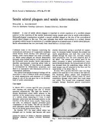
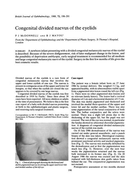
Fig. 1 Clinical photograph showing <strong>the</strong> thickened and<br />
darkly pigmented <strong>naevus</strong> involving <strong>the</strong> upper and lower lids<br />
and medial canthus <strong>of</strong><strong>the</strong> left eye.<br />
Case report<br />
The patient was a female infant born on 27 June<br />
1986 by normal delivery. She weighed 3-2 kg, and<br />
appeared healthy, with no abnormalities visible apart<br />
from a pigmented skin lesion round <strong>the</strong> left eye (Fig.<br />
1). There were no o<strong>the</strong>r pigmented skin lesions and<br />
no relevant family history. The lesion had a vertical<br />
diameter <strong>of</strong> 2 cm and a horizontal diameter <strong>of</strong> 2-5 cm.<br />
The skin was darkly pigmented and thickened and<br />
involved <strong>the</strong> medial three-quarters <strong>of</strong> <strong>the</strong> upper and<br />
lower lid and <strong>the</strong> medial canthus. There was also<br />
some pigmentation <strong>of</strong> <strong>the</strong> tarsal conjunctiva <strong>of</strong> both<br />
lids. The bulbar conjunctiva and <strong>the</strong> sclera were<br />
normal. There was a slight left ptosis due to <strong>the</strong><br />
thickening <strong>of</strong> <strong>the</strong> upper lid, but <strong>the</strong> pupil was not<br />
covered. The rest <strong>of</strong> <strong>the</strong> eye was normal; in particular<br />
<strong>the</strong> fundus showed no abnormal choroidal pigmentation.<br />
A diagnosis <strong>of</strong> congenital <strong>divided</strong> melanocytic<br />
<strong>naevus</strong> <strong>of</strong> <strong>the</strong> <strong>eyelids</strong> was made.<br />
On 10 July 1986 dermabrasion <strong>of</strong> <strong>the</strong> <strong>naevus</strong> was<br />
carried out under general anaes<strong>the</strong>tic, and a punch<br />
biopsy <strong>of</strong> <strong>the</strong> skin was taken. Histological examination<br />
showed a congenital melanocytic <strong>naevus</strong> with<br />
prominent junctional activity and marked pigmentation<br />
(Fig. 2). The <strong>naevus</strong> was markedly debulked by<br />
<strong>the</strong> dermabrasion and at first <strong>the</strong> pigmentation was<br />
much less (Fig. 3). However, over <strong>the</strong> next four<br />
weeks <strong>the</strong> pigmentation returned and <strong>the</strong> <strong>naevus</strong><br />
bulk increased to its former size (Fig. 4). On 20<br />
August 1986 <strong>the</strong> <strong>naevus</strong> was excised and <strong>the</strong> skin<br />
replaced with full-thickness skin grafts taken from<br />
behind <strong>the</strong> ear. The conjunctival pigmentation was<br />
not excised.<br />
Postoperative assessment at two months showed<br />
198
__: .'.U.' .......... :.'<br />
<strong>Congenital</strong> <strong>divided</strong> <strong>naevus</strong> <strong>of</strong> <strong>the</strong> <strong>eyelids</strong><br />
199<br />
rII~~~~~~~~~~~~~~~~~o<br />
Fig. 2 Photomicrograph <strong>of</strong>punch<br />
biopsy specimen showing nests <strong>of</strong><br />
<strong>naevus</strong> cells (arrows) in <strong>the</strong> dermis.<br />
j19,~~~~~~~~~~~~~~~~~~~~~~~~~~r fl44t<br />
p ,n Haematoxylin and eosin.<br />
4. ZK -<br />
$l tr-<br />
#,~~~~~~~~~ ,'4 S.~~~~~~~~~~~~~~~:<br />
.4.'# -w~~~~~~~~~~~~~~~~~~~~~~~~~' AM1<br />
good lid closure and a satisfactory graft, with no childhood and early adult life, and <strong>the</strong> lesion may be<br />
recurrence <strong>of</strong> pigmentation (Fig. 5). The visual axis very disfiguring. Divided naevi <strong>of</strong> <strong>the</strong> <strong>eyelids</strong> are<br />
was clear and no deviation was demonstrated on usually medium sized and involve contiguous<br />
cover test.<br />
portions <strong>of</strong> <strong>the</strong> upper and lower lid. They are very<br />
rare, and only about 30 cases have been reported.<br />
Discussion<br />
In <strong>the</strong> first series to be reported Fuchs' described<br />
six patients, <strong>the</strong> youngest <strong>of</strong> whom was 4 years, with<br />
<strong>Congenital</strong> melanocytic naevi occur in about 1% <strong>of</strong> <strong>divided</strong> naevi. No patients received active treatment.<br />
<strong>the</strong> population. They may be <strong>divided</strong> into three In 1937 Collenza3 reported on two patients with<br />
groups2 according to size: (1) small (less than 1-5 cm <strong>divided</strong> naevi: a 19-year-old girl and a 26-year-old<br />
in diameter), (2) medium (1-5 cm to 20 cm in man, both <strong>of</strong> whom were treated by excision <strong>of</strong> <strong>the</strong><br />
diameter), and (3) large (greater than 20 cm in <strong>naevus</strong> and repair with full-thickness postauricular<br />
diameter). The medium and large naevi have similar skin grafts. Callahan,' and Harrison and Okun5<br />
clinical features, which include a thickened irregular reported single cases. The most recent series was<br />
surface, increased pigmentation giving a dark brown<br />
colour, and varying amounts <strong>of</strong> abnormal hair<br />
growth. The small naevi are usually flat and only<br />
lightly pigmented. <strong>Congenital</strong> melanocytic naevi<br />
<strong>of</strong>ten become larger, thicker, and more hairy in<br />
'i7el/i<br />
.. A::<br />
v|.<br />
.: -::<br />
Fig. 3 Appearance <strong>of</strong><strong>the</strong> left eye one week after<br />
Fig. 4 Five weeks after dermabrasion <strong>the</strong>re is a marked<br />
dermabrasion with minimal pigmentation visible.<br />
return <strong>of</strong><strong>the</strong> pigmentation.
200<br />
P2 J McDonnell and B J Mayou<br />
N<br />
Fig. 5 Appearance <strong>of</strong><strong>the</strong> left eyefollowing excision <strong>of</strong><strong>the</strong><br />
<strong>naevus</strong> andfull thickness skin grafts to <strong>the</strong> lids.<br />
reported by Ehlers6 in 1969. He described 10 cases,<br />
<strong>the</strong> youngest patient being a 15-year-old boy: two<br />
patients had simple excision, two needed fullthickness<br />
skin grafts to repair <strong>the</strong> defect, two were<br />
treated with cryo<strong>the</strong>rapy, and <strong>the</strong> rest received no<br />
treatment. There are no cases in <strong>the</strong> literature <strong>of</strong> a<br />
patient presenting at birth to <strong>the</strong> ophthalmologist.<br />
The <strong>divided</strong> <strong>naevus</strong> is thought to arise during fetal<br />
development at a time when <strong>the</strong> <strong>eyelids</strong> are fused.<br />
The <strong>eyelids</strong> first appear as ectodermal protrusions at<br />
6 weeks <strong>of</strong> fetal development (12 mm stage). They<br />
grow towards each o<strong>the</strong>r and gradually fuse, with <strong>the</strong><br />
process being complete at 9 weeks (40 mm stage).<br />
Lipid <strong>the</strong>n starts to appear in <strong>the</strong> junctional zone at<br />
about 15 weeks, but <strong>the</strong> <strong>eyelids</strong> stay fused until about<br />
24 weeks (<strong>the</strong> 150 mm stage), when <strong>the</strong>y gradually<br />
separate.'<br />
Studies have shown that melanocytes migrate from<br />
<strong>the</strong> neural crest and reach human fetal epidermis<br />
about <strong>the</strong> 12- to 14-week stage,8 and it seems likely<br />
that <strong>the</strong> <strong>divided</strong> melanocytic <strong>naevus</strong> arises just before<br />
<strong>the</strong> 15th week <strong>of</strong> fetal development.<br />
<strong>Congenital</strong> melanocytic naevi can cause a number<br />
<strong>of</strong> problems for <strong>the</strong> patient. The appearance <strong>of</strong> <strong>the</strong><br />
<strong>naevus</strong> is very disfiguring and, if in a prominent<br />
position, <strong>of</strong>ten causes problems <strong>of</strong> cosmesis, particularly<br />
in childhood and adolescence. There is a definite<br />
risk <strong>of</strong> malignant change in <strong>the</strong> <strong>naevus</strong> giving rise to<br />
malignant melanoma, which is best documented for<br />
large naevi but is thought to exist for medium sized<br />
naevi as well.9 The reported incidence <strong>of</strong> malignant<br />
change is very variable, ranging from 2% to 30%<br />
depending on <strong>the</strong> length <strong>of</strong> follow-up, with an<br />
average <strong>of</strong> 14% for a whole lifetime.<br />
A melanocytic <strong>naevus</strong> involving <strong>the</strong> <strong>eyelids</strong> may<br />
affect visual development if <strong>the</strong> increased bulk <strong>of</strong> <strong>the</strong><br />
upper lid causes a mechanical ptosis and occlusion <strong>of</strong><br />
<strong>the</strong> visual axis.'<br />
Treatment <strong>of</strong> <strong>the</strong> congenital melanocytic <strong>naevus</strong> is<br />
controversial, and in <strong>the</strong> form <strong>of</strong> <strong>the</strong> <strong>divided</strong> <strong>naevus</strong><br />
special problems arise. In <strong>the</strong> past, before <strong>the</strong> malignant<br />
potential <strong>of</strong> <strong>the</strong>se lesions was recognised, <strong>the</strong><br />
management consisted <strong>of</strong> observation and sometimes<br />
late surgical removal and reconstruction for cosmetic<br />
reasons. However, with <strong>the</strong> realisation <strong>of</strong> <strong>the</strong> risk <strong>of</strong><br />
malignant change and <strong>the</strong> observation that most<br />
cases <strong>of</strong> prepubertal melanoma arising in congenital<br />
melanocytic <strong>naevus</strong> occur in <strong>the</strong> first 3 to 5 years <strong>of</strong><br />
life, <strong>the</strong> emphasis has changed to a more aggressive<br />
surgical management <strong>of</strong> <strong>the</strong>se patients. "<br />
The cosmetic appearance <strong>of</strong> surgical scars is<br />
usually poor in childhood and improves throughout<br />
adult life. However, <strong>the</strong> first few months <strong>of</strong> life are<br />
characterised by excellent healing <strong>of</strong> surgical scars,<br />
and this explains <strong>the</strong> surgical preference for cleft lip<br />
repair before <strong>the</strong> third month <strong>of</strong> life. We <strong>the</strong>refore<br />
favour very early reconstructive surgery to achieve<br />
<strong>the</strong> best cosmetic result. Dermabrasion has been<br />
reported to be a successful technique for removing<br />
congenital melanocytic <strong>naevus</strong> if <strong>the</strong> lesion is treated<br />
early in life and <strong>the</strong> <strong>naevus</strong> cells are still limited to <strong>the</strong><br />
superficial dermis.'2'3 This technique uses a metal<br />
burr attachment to a dental drill which rotates at high<br />
speed and is used to abrade <strong>the</strong> epidermis and<br />
superficial dermis. Unfortunately in our patient <strong>the</strong><br />
punch biopsy done at <strong>the</strong> time <strong>of</strong> <strong>the</strong> dermabrasion<br />
revealed that <strong>the</strong> <strong>naevus</strong> cells were already deep in in<br />
<strong>the</strong> dermis, and it was not surprising that <strong>the</strong> lesion<br />
recurred fairly quickly. If <strong>the</strong> <strong>naevus</strong> involves <strong>the</strong><br />
deep dermis and subcutaneous tissue, <strong>the</strong>n <strong>the</strong><br />
treatment consists <strong>of</strong> full-thickness excision followed<br />
by repair with full-thickness skin graft or split skin<br />
graft depending on <strong>the</strong> size <strong>of</strong> <strong>the</strong> defect.<br />
Large naevi involving <strong>the</strong> <strong>eyelids</strong> may have to be<br />
treated in stages. ' Divided naevi which are excised<br />
are most conveniently repaired with a full-thickness<br />
postauricular skin graft. Five cases <strong>of</strong> <strong>divided</strong> naevi<br />
treated by this procedure have been reported.36 The<br />
present report is to <strong>the</strong> best <strong>of</strong> our knowledge <strong>the</strong> first<br />
case <strong>of</strong> <strong>divided</strong> congenital melanocytic <strong>naevus</strong> <strong>of</strong> <strong>the</strong><br />
<strong>eyelids</strong> presenting at birth and treated surgically in<br />
early life.<br />
References<br />
I Fuchs A. Ueber geteilte Naevi der Augenlider. Klin Monatsbl<br />
Augenheilkd 1919; 63: 678-83.<br />
2 Rigel DS, Friedman RJ. The management <strong>of</strong> patients with<br />
dysplastic and congenital nevi. Dermatol Clin 1985; 3: 251-5.<br />
3 Collenza D. Nevi divisi pigmentari delle palpebre. Boll Oculist<br />
1937; 16: 435-60.<br />
4 Callahan A. The removal <strong>of</strong> adjacent nevi <strong>of</strong> <strong>the</strong> <strong>eyelids</strong>. Am J<br />
Ophthalmol 1946; 29: 563-5.<br />
5 Harrison R, Okun M. Divided nevus. Arch Dermatol 1960; 82:<br />
235-6.<br />
6 Ehlers N. Divided nevus. Acta Ophthalmol (Kbh) 1969; 47:<br />
1004-11.
<strong>Congenital</strong> <strong>divided</strong> <strong>naevus</strong> <strong>of</strong> <strong>the</strong> <strong>eyelids</strong><br />
7 Andersen H, Ehlers N, Matthiessen ME. Histochemistry and<br />
development <strong>of</strong> <strong>the</strong> human <strong>eyelids</strong>. Acta Ophthalmol (Kbh)<br />
1965; 43: 642-68.<br />
8 Breathnach AS, Wyllie LM. Electron microscopy <strong>of</strong> melanocytes<br />
and Langerhans cells in human fetal epidermis at fourteen<br />
weeks. J Invest Dermatol 1965; 44: 51-60.<br />
9 Kaplan EN. The risk <strong>of</strong> malignancy in large congenital nevi. Plast<br />
Reconstr Surg 1974; 53: 421-8.<br />
10 Antinone RL, Helveston EM, Bennett JE, Keener P. Giant<br />
hairy nevus: preventable cause <strong>of</strong> amblyopia. J Pediatr<br />
Ophthalmol 1976; 13: 192-5.<br />
11 Arons MS, Hurwitz S. <strong>Congenital</strong> nevocellular nevus: a review<br />
201<br />
<strong>of</strong> <strong>the</strong> treatment controversy and a report <strong>of</strong> 46 cases. Plast<br />
Reconstr Surg 1983; 72: 355-65.<br />
12 Johnson HA. Permanent removal <strong>of</strong> pigmentation from giant<br />
hairy naevi by dermabrasion in early life. BrJ Plast Surg 1977; 30:<br />
321-3.<br />
13 Miller CJ, Becker DW. Removing pigmentation by dermabrading<br />
naevi in infancy. Br J Plast Surg 1979; 32: 124-6.<br />
14 de Dulanto F, Camacho-Martinez F, Sanchez-Muros J, de<br />
Cosme L. A giant hairy pigmented nevus on <strong>the</strong> face: excision<br />
and reconstruction in stages. J Dermatol Surg Oncol 1979; 5:<br />
215-8.<br />
Accepted for publication 7January 1987.