Confocal microscopy in cornea guttata and Fuchs' endothelial ...
Confocal microscopy in cornea guttata and Fuchs' endothelial ...
Confocal microscopy in cornea guttata and Fuchs' endothelial ...

You also want an ePaper? Increase the reach of your titles
YUMPU automatically turns print PDFs into web optimized ePapers that Google loves.
Downloaded from<br />
bjo.bmj.com on April 9, 2013 - Published by group.bmj.com<br />
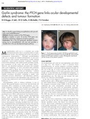
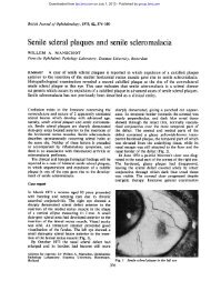
<strong>Confocal</strong> <strong>microscopy</strong> <strong>in</strong> <strong>cornea</strong> <strong>guttata</strong> <strong>and</strong> Fuchs’ <strong>endothelial</strong> dystrophy 189<br />
is rarely dependent on the patient’s ability to<br />
rema<strong>in</strong> still.<br />
Cornea <strong>guttata</strong> <strong>and</strong> Fuchs’ dystrophy are<br />
rarely a diagnostic dilemma, <strong>and</strong> most of the<br />
time are readily diagnosed simply by slit lamp<br />
exam<strong>in</strong>ation. Based on our experience, we<br />
th<strong>in</strong>k that confirmation of the diagnosis is best<br />
accomplished by specular <strong>microscopy</strong>, because<br />
it is easier to use. However, confocal <strong>microscopy</strong><br />
is a worthwhile alternative, especially <strong>in</strong><br />
cases of <strong>cornea</strong>l oedema.<br />
The authors do not have any commercial or proprietary <strong>in</strong>terest<br />
<strong>in</strong> the products or companies cited <strong>in</strong> this article. The authors<br />
do not have any f<strong>in</strong>ancial <strong>in</strong>terest or receive payment as a consultant,<br />
reviewer, or evaluator.<br />
This study was supported <strong>in</strong> part by US Public Health Service<br />
grants EY00346 (SCK), EY02580 (HEK), <strong>and</strong> EY02377<br />
(HEK) from the National Eye Institute, National Institutes of<br />
Health, Bethesda, MD; Department of the Army, Cooperative<br />
Agreement DAMD17-93-V-3013. (This does not necessarily<br />
reflect the position or the policy of the government, <strong>and</strong> no oYcial<br />
endorsement should be <strong>in</strong>ferred) (RWB, HEK); an<br />
unrestricted departmental grant from Research to Prevent<br />
Bl<strong>in</strong>dness, Inc, New York City; Société Académique Vaudoise<br />
(AGYC), Switzerl<strong>and</strong>; the Swiss National Research Foundation<br />
(AGYC), Switzerl<strong>and</strong>; <strong>and</strong> Verrey Foundation (AGYC),<br />
Switzerl<strong>and</strong>.<br />
1 War<strong>in</strong>g GO 3rd, Rodrigues MM, Laibson PR. Corneal dystrophies.<br />
II. Endothelial dystrophies. Surv Ophthalmol<br />
1978;23:147–68.<br />
2 Krachmer JH, Purcell JJ Jr, Young CW, et al. A study of<br />
sixty-four families with <strong>cornea</strong>l <strong>endothelial</strong> dystrophy. Arch<br />
Ophthalmol 1978;96:2035–9.<br />
3 Beuerman RW. <strong>Confocal</strong> <strong>microscopy</strong>: Into the cl<strong>in</strong>ic. Cornea<br />
1995;14:1–2.<br />
4 Cavanagh HD, Petroll WM, Alizadeh H, et al. Cl<strong>in</strong>ical <strong>and</strong><br />
diagnostic use of <strong>in</strong> vivo confocal <strong>microscopy</strong> <strong>in</strong> patients<br />
with <strong>cornea</strong>l disease. Ophthalmology 1993;100:1444–54.<br />
5 Cavanagh HD, McCulley JP. In vivo confocal <strong>microscopy</strong><br />
<strong>and</strong> acanthamoeba keratitis. Am J Ophthalmol 1996;121:<br />
207–8.<br />
6 Chew SJ, Beuerman RW, Assoul<strong>in</strong>e M, et al. Early diagnosis<br />
of <strong>in</strong>fectious keratitis with <strong>in</strong> vivo real time confocal <strong>microscopy</strong>.<br />
CLAO J 1992;18:197–201.<br />
7 Chiou AGY, Cadez R, B÷hnke M. Diagnosis of DieVenbachia<br />
<strong>in</strong>duced <strong>cornea</strong>l <strong>in</strong>jury by confocal <strong>microscopy</strong>. Br J<br />
Ophthalmol 1997;81:168–9.<br />
8 Florakis GJ, Moazami G, Schubert H, et al. Scann<strong>in</strong>g slit<br />
confocal <strong>microscopy</strong> of fungal keratitis. Arch Ophthalmol<br />
1997;115:1461–3.<br />
9 Kaufman SC, Chew SJ, Capps SC, et al. <strong>Confocal</strong><br />
<strong>microscopy</strong> of <strong>cornea</strong>l penetration by tarantula hairs. Scann<strong>in</strong>g<br />
1994;16:312–5.<br />
10 Kaufman SC, Beuerman RW, Goldberg D. A new form of<br />
primary, localized, <strong>cornea</strong>l amyloidosis: A case report with<br />
confocal <strong>microscopy</strong>. Metab Pediatr Syst Ophthalmol 1995;<br />
18:1–4.<br />
11 Pfister DR, Cameron JD, Krachmer JH, et al. <strong>Confocal</strong><br />
<strong>microscopy</strong> f<strong>in</strong>d<strong>in</strong>gs of acanthamoeba keratitis. Am J Ophthalmol<br />
1996;121:119–28.<br />
12 Shah GK, Pfister D, Probst LE, et al. Diagnosis of<br />
microsporidial keratitis by confocal <strong>microscopy</strong> <strong>and</strong> the<br />
chromatrope sta<strong>in</strong>. Am J Ophthalmol 1996;121:89–91.<br />
13 Sutph<strong>in</strong> JE, Kantor AL, Mathers WD, et al. Evaluation of<br />
<strong>in</strong>fectious crystall<strong>in</strong>e keratitis with confocal <strong>microscopy</strong> <strong>in</strong> a<br />
case series. Cornea 1997;16:21–6.<br />
14 Kaufman SC, Beuerman RW, Kaufman HE. Diagnosis of<br />
advanced Fuchs’ <strong>endothelial</strong> dystrophy with the confocal<br />
microscope. Am J Ophthalmol 1993;116:652–3.<br />
15 La<strong>in</strong>g RA, Leibowitz HM, Oak SS, et al. Endothelial mosaic<br />
<strong>in</strong> Fuchs’ dystrophy. A qualitative evaluation with the<br />
specular microscope. Arch Ophthalmol 1981;99:80–3.<br />
16 Bigar F, Schimmelpfennig B, Hurzeler R. Cornea <strong>guttata</strong> <strong>in</strong><br />
donor material. Arch Ophthalmol 1978;96:653–5.<br />
17 War<strong>in</strong>g GO, Font RL, Rodrigues MM, et al. Alterations of<br />
Descemet’s membrane <strong>in</strong> <strong>in</strong>terstitial keratitis. Am J<br />
Ophthalmol 1976;81:773–85.<br />
18 Pouliquen Y, Dhermy P, Renard G, et al. Comb<strong>in</strong>ed macular<br />
dystrophy <strong>and</strong> <strong>cornea</strong> <strong>guttata</strong>: An electron microscopic<br />
study. Albrecht Von Graefes Arch Kl<strong>in</strong> Exp Ophthalmol 1980;<br />
212:149–58.<br />
19 Cibis GW, Krachmer JA, Phelps CD, et al. The cl<strong>in</strong>ical<br />
spectrum of posterior polymorphous dystrophy. Arch Ophthalmol<br />
1977;95:1529–37.<br />
20 Brooks AM, Grant GB, Gillies WE. The identification of<br />
<strong>cornea</strong>l guttae. Cornea 1991;10:249–60.<br />
21 Krachmer JH, Schnitzer JI, Fratk<strong>in</strong> J. Cornea<br />
pseudo<strong>guttata</strong>: a cl<strong>in</strong>ical <strong>and</strong> histopathologic description of<br />
<strong>endothelial</strong> cell edema. Arch Ophthalmol 1981;99:1377–81.