

Journal of Cell and Molecular Biology - ResearchGate
Journal of Cell and Molecular Biology - ResearchGate
Journal of Cell and Molecular Biology - ResearchGate

You also want an ePaper? Increase the reach of your titles
YUMPU automatically turns print PDFs into web optimized ePapers that Google loves.
II SFM were grown in 100 – 500 ml Erlenmeyer<br />
flasks on an orbital shaker at 29ºC. The suspension<br />
culture was infected with the virus carrying the B17<br />
gene when the cells were in mid-exponential<br />
growth <strong>and</strong> the density <strong>of</strong> cells is between 1 – 3 x<br />
10 6 cells/ml. 2 plaque-forming units (pfu) were<br />
added to each cell in suspension, a parameter that is<br />
usually called multiplicity <strong>of</strong> infection, MOI.<br />
Harvesting is done 24 to 48 hours later. The media<br />
is then centrifuged at 250xg for 5 minutes, <strong>and</strong> the<br />
supernatant is collected <strong>and</strong> stored in 2 μg/ml<br />
PMSF or aprotinin, 2 mM EDTA, <strong>and</strong> 0.05% NaN3<br />
at 4 ºC.<br />
The transfection with the B17 gene was done<br />
with a pDLST8 plasmid containing the B17 cDNA<br />
sequence into a recombinant donor plasmid. The<br />
donor plasmid was then hosted for one day in<br />
competent DH10Bac E. coli cells, <strong>and</strong><br />
subsequently transposed for antibiotic selection for<br />
2 days in E. coli (Lac7) containing a recombinant<br />
bacmid. In day 4, the recombinant bacmid DNA<br />
was introduced into the Sf-9 cells for recombinant<br />
virus particles to be produced the next two days. A<br />
viral titer was done by plaque assay to determine<br />
the number <strong>of</strong> pfus in the stock <strong>and</strong> to concentrate,<br />
if necessary. The viral stock was then used to infect<br />
other suspension cultures.<br />
Aliquots <strong>of</strong> the stored media stock were<br />
incubated for two hours in glass tubes containing<br />
protein-G Sepharose in the ratio 4:1 to remove<br />
media IgGs prior to the last incubation for two<br />
hours in an immuno-adsorbant column. This<br />
immobilization column contained a similar bed<br />
volume <strong>of</strong> protein-G sepharose coupled with antiapo<br />
B IgG. The anti-B IgG was crosslinked to the<br />
protein-G Sepharose with DMP in TEA or a basic<br />
Na-phosphate buffer, <strong>and</strong> the blocking was<br />
achieved with ethanolamine. The immobilized B17<br />
was then eluted with an acidic glycine buffer (pH<br />
2.5), <strong>and</strong> the eluate was brought a neutral pH by<br />
adding a volume <strong>of</strong> tris (pH 8) amounting to 10%<br />
<strong>of</strong> the total elution volume. The eluate was then<br />
tested for purity, dialyzed against a K-phosphate<br />
buffer (pH 7.4) <strong>and</strong> stored at 4ºC.<br />
The concentration <strong>of</strong> B17 in solution was<br />
initially determined by the Lowry method using<br />
bovine serum albumin as the st<strong>and</strong>ard (Lowry et<br />
al., 1951). Alternatively, a multiplicity factor was<br />
determined so that the concentration can be<br />
approximated from the direct measurement <strong>of</strong> the<br />
protein UV absorbance at 280 nm, A280. This was<br />
achieved by preparing two sets <strong>of</strong> samples with the<br />
same amount <strong>of</strong> protein in each sample. One set<br />
was diluted with differing volumes <strong>of</strong> chemical<br />
Expression, purification <strong>and</strong> quantification <strong>of</strong> B17 39<br />
denaturant (e.g. urea), while the other was diluted<br />
with the same volumes <strong>of</strong> the sample buffer. The<br />
absorbance A280 was then measured for both sets<br />
<strong>and</strong> the one with denaturant was compared with the<br />
reported A280 <strong>of</strong> tryptophan <strong>and</strong> tyrosine. A<br />
multiplicity factor <strong>of</strong> 2 was then determined such<br />
that the concentration <strong>of</strong> B17 in that solution is<br />
equal to A280 x 2 (mg/ml).<br />
Results<br />
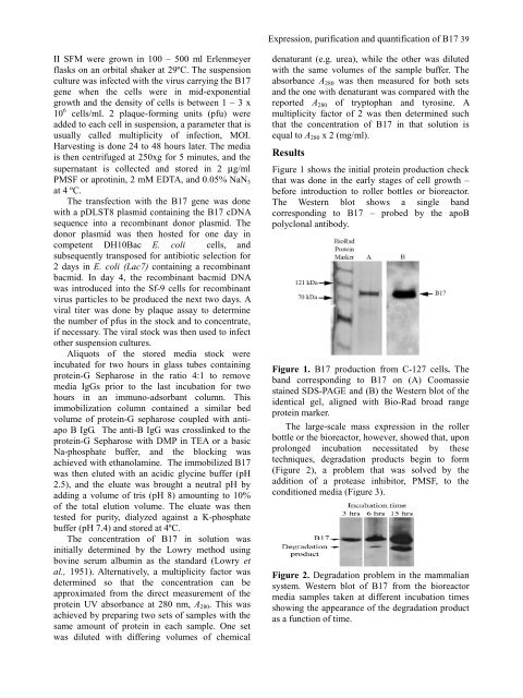
Figure 1 shows the initial protein production check<br />
that was done in the early stages <strong>of</strong> cell growth –<br />
before introduction to roller bottles or bioreactor.<br />
The Western blot shows a single b<strong>and</strong><br />
corresponding to B17 – probed by the apoB<br />
polyclonal antibody.<br />
Figure 1. B17 production from C-127 cells. The<br />
b<strong>and</strong> corresponding to B17 on (A) Coomassie<br />
stained SDS-PAGE <strong>and</strong> (B) the Western blot <strong>of</strong> the<br />
identical gel, aligned with Bio-Rad broad range<br />
protein marker.<br />
The large-scale mass expression in the roller<br />
bottle or the bioreactor, however, showed that, upon<br />
prolonged incubation necessitated by these<br />
techniques, degradation products begin to form<br />
(Figure 2), a problem that was solved by the<br />
addition <strong>of</strong> a protease inhibitor, PMSF, to the<br />
conditioned media (Figure 3).<br />
Figure 2. Degradation problem in the mammalian<br />
system. Western blot <strong>of</strong> B17 from the bioreactor<br />
media samples taken at different incubation times<br />
showing the appearance <strong>of</strong> the degradation product<br />
as a function <strong>of</strong> time.


