Glaucoma Masqueraders – Our Clinical Experience – Has ... - KSOS
Glaucoma Masqueraders – Our Clinical Experience – Has ... - KSOS
Glaucoma Masqueraders – Our Clinical Experience – Has ... - KSOS

You also want an ePaper? Increase the reach of your titles
YUMPU automatically turns print PDFs into web optimized ePapers that Google loves.
March 2008 Meenakshi Dhar et al. - <strong>Glaucoma</strong> Masquerades 31<br />
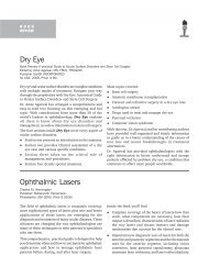
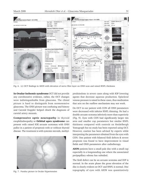
Fig. 6. (c) OCT findings in AION with elevation of nerve fibre layer on ONH scan and raised RNFL thickness<br />
In Ocular Ischemic syndrome OCT did not provide<br />
any corroborative evidence, rather, the OCT changes<br />
were indistinguishable from glaucoma. The clinial<br />
picture is hard to distinguish from normotensive<br />
glaucoma. The ONH picture was confusing and history<br />
and Carotid Doppler helped clinch the diagnosis of<br />
carotid artery stenosis.<br />
Compressive optic neuropathy in thyroid<br />
exophthalmopathy or Orbital apex syndrome can<br />
present with raised IOP, arcuate scotomas with ONH<br />
pallor in a patient of proptosis with or without thyroid<br />
disease. The treatment is with systemic steroids, methyl-<br />
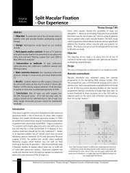
Fig. 7. Fundus picture in Ocular Hypertension<br />
prednisolone in severe cases along with IOP lowering<br />
agents that decrease aqueous production. Episcleral<br />
venous pressure is raised in these cases, thus medication<br />
that acts on the outflow mechanism may not work.<br />
On OCT in our patient with CON all ONH parameters<br />
were decreased with inferior RNFL thinning. He had a<br />
double arcuate scotoma inferiorly more than superiorly<br />
(Fig. 9). Eyes with CON had significantly larger rim<br />
area and smaller cup parameters but similar RNFL<br />
thickness compared with controls on Heidelbergh<br />
Tomograph but no study has been reported using OCT.<br />
However, caution has been advised by experts while<br />
interpreting the parameters obtained from the eyes with<br />
CON. One patient with bilateral field defects & severe<br />
proptosis was found to have improvement in visual<br />
fields and ONH parameters after radiotherapy.<br />
AION patients have a small pale disc with a small cup<br />
especially in a longstanding one where the assosciated<br />
peripapillary edema has subsided.<br />
The field defect can be an arcuate scotoma and IOP is<br />
normal. In the acute phase the gross elevation of the<br />
disc is clearly evident on OCT and RNFL is normal. Disc<br />
topography of eyes with AION was quantitatively