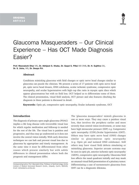
Glaucoma Masqueraders – Our Clinical Experience – Has ... - KSOS
Glaucoma Masqueraders – Our Clinical Experience – Has ... - KSOS
Glaucoma Masqueraders – Our Clinical Experience – Has ... - KSOS

You also want an ePaper? Increase the reach of your titles
YUMPU automatically turns print PDFs into web optimized ePapers that Google loves.
22 Kerala Journal of Ophthalmology Vol. XX, No. 1<br />
ORIGINAL<br />
ARTICLE<br />
<strong>Glaucoma</strong> <strong>Masqueraders</strong> <strong>–</strong> <strong>Our</strong> <strong>Clinical</strong><br />
<strong>Experience</strong> <strong>–</strong> <strong>Has</strong> OCT Made Diagnosis<br />
Easier?<br />
Prof. Meenakshi Dhar MS, Dr. Abhijeet S. Khake, Dr. Gopal S. Pillai MD DNB, Dr. H. Sujithra DO,<br />
Dr. S. Jisha, MS, Dr. Deepa P.A.<br />
Abstract<br />
Introduction<br />
Conditions mimicking glaucoma with field changes or optic nerve head changes similar to<br />
glaucoma can puzzle the clinician. We present a series of 17 patients with optic nerve head<br />
pit, optic nerve head drusen, ONH coloboma, ocular ischemic syndrome, compressive optic<br />
neuropathy, and ocular hypertensives with high cup disc ratio in myopic optic discs which<br />
appear glaucomatous but with no field loss. OCT helped us to differentiate some of these.<br />
The clinical presentation, visual field analysis, OCT picture and also features clinching the<br />
diagnosis in these patients is discussed in detail.<br />
Keywords: Optic pit, compressive optic neuropathy, Ocular ischaemic syndrome, OCT<br />
The diagnosis of primary open angle glaucoma (POAG)<br />
implies a life long disease with irreversible visual loss<br />
for which regular medication and followup is needed<br />
for the rest of the life. The visual loss is painless and<br />
progressive, and thus may go undetected as it does not<br />
involve the central vision initially. With early detection<br />
techniques we can halt and prevent visual loss due to<br />
glaucoma by appropriate and timely management. At<br />
the same time it must be differentiated from other<br />
causes which present similarly but with subtle<br />
differences in clinical presentation where both the<br />
prognosis and management differ.<br />
Corresponding Author, Prof. Meenakshi Dhar, Professor of Ophthalmology, Amrita<br />
Institute of Medical Sciences& Research Centre,, Edapally, Cochin, 682026,<br />
Email mdhar@aims.amrita.edu<br />
The ‘glaucoma masqueraders’ mimick glaucoma in<br />
one or more ways. They may cause a painless visual<br />
loss, that involves the periphery earlier and more<br />
severely than central vision involvement, or some may<br />
have high intraocular pressure (IOP) e.g. Compressive<br />
optic neuropathy (CON),Ocular hypertension (OHT).<br />
Others may have optic nerve head (ONH) changes<br />
similar to glaucomatous optic neuropathy<br />
(e.g. Optic pit and isolated ONH coloboma). Still<br />
others may have visual field defects simulating or<br />
mimicking glaucoma. Superior arcuate scotoma may<br />
be seen in optic pit, anterior ischemic optic neuropathy<br />
(AION), compressive optic neuropathy. <strong>Glaucoma</strong> field<br />
loss affects the nasal quadrant initially and may mask<br />
an unusual visual field presentation of a pituitary tumor.<br />
Differentiating a case of normotensive glaucoma from<br />
OHT can be a diagnostic dilemma.
March 2008 Meenakshi Dhar et al. - <strong>Glaucoma</strong> Masquerades 23<br />
Materials and Methods<br />
This is a nonrandomised, noncomparative, institution<br />
based, clinical observational case series study of 17<br />
cases selected from the out-patient department from<br />
December 2004 to 2007 June.<br />
Those patients who had at least 1-2 features of POAG,<br />
either a high IOP, or an enlarged cup, or an arcuate<br />
scotoma or had a suspicious small disc with pallor were<br />
included.<br />
All patients underwent a complete glaucoma evaluation<br />
including applanation tonometry, pachymetry,<br />
gonioscopy, automated perimetry with 30-2 on<br />
Humphrey, and a dilated stereoscopic optic nerve head<br />
evaluation.<br />
OCT was done for all patients using Fast RNFL, Fast<br />
optic nerve head scan and Macular protocol on the<br />
Stratus OCT Version 4 machine.<br />
Observations<br />
The fundus findings, the HFA visual field, and OCT<br />
findings were noted and correlated for all the patients<br />
included in the study.<br />
Fig. 1. (b) HFA 30-2 of a case of Optic Pit<br />
Table 1: Conditions mimicking glaucoma<br />
No. of cases<br />
Optic pit 6<br />
Optic Coloboma 1<br />
Ocular Ischemic Syndrome (OIS) 2<br />
Compressive Optic Neuropathy (CON) 3<br />
AION 1<br />
Orbital apex syndrome 1<br />
Myopia and OHT 1<br />
Optic nerve head drusen 1<br />
Optic neuritis/ Angle closure glaucoma 1<br />
Of the 17 patients (Table 1), 6 had optic nerve head<br />
pit-3 unilateral and 3 bilateral. All the 6 patients<br />
with optic pit had an arcuate scotoma (Fig. 1b)<br />
Fig. 1. (a) Fundus photo of a case of Optic Pit
24 Kerala Journal of Ophthalmology Vol. XX, No. 1<br />
Table 2: BCVA of patients of Optic Pit<br />
Diagnosis Vision<br />
Right Eye Left Eye<br />
Patient 1 Optic pit 6/6 6/6<br />
Patient 2 Optic pit 5/60 ph 6/24 6/6<br />
Patient 3 Optic pit 6/6 6/18p<br />
Patient 4 Optic pit 5/ 60-6/36 6/6<br />
Patient 5 Optic pit 6/6 6/6<br />
Patient 6 Optic pit 6/6 6/6<br />
Patient 7 Optic pit 6/6 6/6<br />
(c)<br />
with normal IOP which did not progress in the 12<br />
months of followup. Central vision was decreased in<br />
3 patients and three had bilateral pits. Pits were seen<br />
on the temporal aspect of the disc. The optic nerve head<br />
showed characteristic features of an optic pit (Fig. 1a).<br />
On OCT the pits could be seen on the temporal aspect<br />
of the cup on a horizontal line scan. There was RNFL<br />
loss around the disc (Fig. 1c, d, e) and normal optic<br />
nerve head parameters were found in all of our patients.<br />
One case has cystoid changes and schisis cavity<br />
formation in the peripapillary retina. The serous<br />
macular detachment on OCT communicated with the<br />
optic pit.<br />
Two, had Ocular Ischemic syndrome (OIS) and three<br />
had Compressive optic neuropathy. One each had optic<br />
nerve head drusen, Optic nerve coloboma, AION, orbital<br />
apex syndrome, misdiagnosed optic neuritis.<br />
The patient with Optic nerve head coloboma had<br />
an isolated coloboma with no anterior coloboma<br />
(Fig. 2a).The patient had a superior arcuate scotoma<br />
(Fig. 2c) with a suspicious looking disc and a normal<br />
intraocular pressure. There was a white excavation in<br />
the disc which was decentered inferiorly, with a thin<br />
inferior rim and a normal superior rim (Fig. 2a and b).<br />
OCT in this case showed inferior retinal fibre layer<br />
(RNFL) thinning (Fig. 2d) and signs of a connection<br />
Fig. 1. (c,d,e) OCT of a case of Optic Pit showing (c)<strong>–</strong>RNFL, (d)<strong>–</strong>ONH, (e)<strong>–</strong>line scan[clockwise]<br />
(d)<br />
(e)
March 2008 Meenakshi Dhar et al. - <strong>Glaucoma</strong> Masquerades 25<br />
Fig. 2. (a) Fundus photograph showing an isolated optic disc<br />
coloboma with a white excavation on disc, decentered<br />
inferiorly with a thin inferior rim and a normal<br />
superior rim<br />
Fig. 2. (b) Red free fundus photograph of optic disc coloboma<br />
Fig. 2 (c) HFA left eye showing a superior arcuate<br />
scotoma<br />
between the perineural space and the inner retinal<br />
layers on the temporal optic disc border (Fig. 2e), as<br />
well as schisis-like changes extending from the disc to<br />
the macula, with cystoid degeneration and two lamellar<br />
holes. There was increased retinal nerve fibre layer<br />
thickness and reduced macular thickness.<br />
The patient with Optic nerve head drusen had<br />
bilateral visible superotemporal drusen that lead to<br />
bilateral inferior arcuate scotoma. The drusen unless<br />
seen stereoscopically could be mistaken for notching<br />
of the cup. On stereoscopic examination it presented as<br />
a raised lesion. On OCT both eyes showed a superotemporal<br />
thinning on RNFL scan, and all parameters on ONH<br />
analysis were raised. (Fig. 3 a, b and c).<br />
The two patients with Ocular Ischaemic syndrome<br />
had a high CD ratio, with a deep cup and extensive<br />
field loss with normal IOP’s. On OCT patient had RNFL<br />
loss, and decreased ONH parameters like VIRA, HIRW<br />
and rim area 2<br />
History and Carotid Doppler helped clinch the diagnosis<br />
of Carotid artery stenosis (Fig. 4 a and b).<br />
Three patients with Compressive optic neuropathy<br />
(Fig. 5) in thyroid exophthalmopathy and one with<br />
Fig. 2 (d) OCT showing Inferior Retinal Nerve Fibre Layer (RNFL)<br />
thinning in an isolated Optic nerve head coloboma
26 Kerala Journal of Ophthalmology Vol. XX, No. 1<br />
Fig. 2. (e) ONH findings of the isolated ONH coloboma<br />
Fig. 3. (a) Fundus picture of drusen of ONH<br />
Fig. 3. (b) HFA showing Inferior arcuate scotoma in drusen of ONH<br />
Orbital apex syndrome presented with raised IOP,<br />
arcuate scotomas with ONH pallor associated with<br />
proptosis. The patients were managed on systemic<br />
steroids <strong>–</strong> (Methylprednisolone in severe cases) along<br />
with IOP lowering agents that decrease aqueous<br />
production.<br />
On OCT in our patients all ONH parameters were<br />
decreased with inferior RNFL thinning with corresponding<br />
field changes (Fig. 5b & c). One had a double arcuate<br />
scotoma inferiorly more than superiorly (Fig. 5d).<br />
The AION patient [Fig. 6a, b & c] had a small pale disc<br />
with a small cup, indicating a resolved neuropathy with<br />
resorption of the peripapillary oedema.<br />
The field defect was an arcuate scotoma and IOP was<br />
normal. In the acute phase the gross elevation of the<br />
disc was clearly evident on OCT and RNFL was normal.<br />
Also one had high intraocular pressure, and high cup<br />
disc ratio in a myopic optic discs which appeared<br />
glaucomatous. This patient had no field loss even on<br />
followup and normal OCT parameters. This patient had<br />
ocular hypertension with a borderline thick cornea,<br />
open angles, no field defects and normal OCT. The<br />
patient is on follow-up regularly and medication has<br />
not been started for last 1 year (Fig. 7).<br />
Incomplete and partial Binasal hemianopia was<br />
seen in one patient. It appeared like glaucomatous optic<br />
neuropathy as the CD ratio was high. The IOP was<br />
normal, and had been mistakenly treated as bilateral<br />
POAG. After pituitary surgery on the adenoma, the field
March 2008 Meenakshi Dhar et al. - <strong>Glaucoma</strong> Masquerades 27<br />
Fig. 3. (c) OCT findings in drusen of ONH<br />
defects dramatically disappeared. In this patient neither<br />
RNFL loss nor ONH abnormalities were seen on OCT.<br />
Discussion<br />
Optic Pit The visual field defects in an optic pit is a<br />
superior arcuate scotoma similar to glaucoma, but it is<br />
nonprogressive, and management does not include an<br />
IOP lowering agent. The danger to visual loss is due to<br />
neurosensory detachment or retinoschisis. It is only the<br />
discerning trained eye of a clinician, that picks up the<br />
optic pit on stereoscopic fundus examination rather<br />
than diagnose it as optic cupping. OCT has made the<br />
diagnosis easier, as on OCT the pit is clearly visible on<br />
the temporal aspect of the cup on a horizontal line scan,<br />
and so is the associated neurosensory detachment.<br />
RNFL and optic nerve head parameters are normal.<br />
Optic pit is one condition where OCT helps in<br />
differentiating it from glaucoma. According to literature<br />
15 % have bilateral disease, while three out of six of<br />
our patients had bilateral disease. The cystoid changes<br />
and schisis cavity formation in the peripapillary retina<br />
can be seen. The serous macular detachment on OCT<br />
can be seen communicating with the optic pit.
28 Kerala Journal of Ophthalmology Vol. XX, No. 1<br />
Fig. 4. (a) Fundus findings in a case of Ocular Ischemic<br />
Syndrome (OIS)<br />
Fig. 4. (b) OCT findings in OIS<br />
Optic Nerve head coloboma when isolated, i.e.<br />
with no anterior or retinal coloboma, presents with a<br />
superior arcuate scotoma in a suspicious looking disc<br />
in a patient with normal intraocular pressure. It is<br />
caused by the failure of complete closure of the<br />
proximal end of the embryonic fissure. It is<br />
characterized by a white excavation in the disc which<br />
is decentered inferiorly. The inferior rim is usually thin<br />
or absent whereas the superior rim is relatively normal.<br />
OCT showed signs of a connection between the perineural<br />
space and the inner retinal layers on the temporal optic<br />
disc border, as well as schisis-like changes extending<br />
from the disc to the macula, with cystoid degeneration<br />
and two lamellar holes in their nasal portion. There is
March 2008 Meenakshi Dhar et al. - <strong>Glaucoma</strong> Masquerades 29<br />
Fig. 5. (a) Fundus findings in compressive optic neuropathy<br />
(CON)<br />
Fig. 5. (b) OCT findings in thyroid ophthalmopathy causing CON<br />
Fig. 5. (c) Corresponding field defects in the same patient<br />
increased retinal nerve fibre layer thickness and reduced<br />
macular thickness.<br />
Optic Nerve Head Drusen (OND)<br />
Optic disc drusen occurs in about 1 % of the population<br />
and are found more frequently in Caucasians. 75 %<br />
are bilateral. Inherited or sporadic, it is a form of calcific<br />
degeneration in some of the axons of the optic nerve.
30 Kerala Journal of Ophthalmology Vol. XX, No. 1<br />
Fig. 5. (d) Biarcuate scotoma in a patient with compressive<br />
optic neuropathy<br />
Fig. 6. (a) Left eye Fundus showing findings of AION<br />
Fig. 6. (b) Unilateral inferior field defect in AION<br />
Visual acuity is often not affected, but the visual fields<br />
of these patients can be abnormal and deteriorate over<br />
time. The drusen may be buried or evident. The disc picture<br />
shows an elevated disc that may be confused with<br />
papilledema (Fig. 3a). The lesion may be progressive.<br />
Buried drusens are less symptomatic. There is no existing<br />
treatment for optic nerve head drusen. Proper diagnosis<br />
and patient education is the best-available modality of<br />
care. Patients need to be aware of potential complications<br />
which, while rare, can affect vision. Visual-field testing<br />
can aid in monitoring for subtle changes in vision.<br />
OCT has shown a thinning of peripapillary retinal nerve<br />
fiber layer. The lowest values were found in the superior<br />
and inferior quadrants. Calcification, picked up both<br />
on USG B scan and CT scan, is the characteristic feature<br />
for diagnosis for ONH drusen.<br />
Visual field defects are uncommon in eyes with buried<br />
OND. Eyes with buried OND may have focal RNFL<br />
defects but have normal average RNFL thickness.<br />
Coexistence of ONH drusen and POAG in eyes can be<br />
most effectively picked up by OCT earliest where the<br />
disc may appear elevated on examination but RNFL<br />
thinning may be picked up even prior to appearance of<br />
visual field defects.
March 2008 Meenakshi Dhar et al. - <strong>Glaucoma</strong> Masquerades 31<br />
Fig. 6. (c) OCT findings in AION with elevation of nerve fibre layer on ONH scan and raised RNFL thickness<br />
In Ocular Ischemic syndrome OCT did not provide<br />
any corroborative evidence, rather, the OCT changes<br />
were indistinguishable from glaucoma. The clinial<br />
picture is hard to distinguish from normotensive<br />
glaucoma. The ONH picture was confusing and history<br />
and Carotid Doppler helped clinch the diagnosis of<br />
carotid artery stenosis.<br />
Compressive optic neuropathy in thyroid<br />
exophthalmopathy or Orbital apex syndrome can<br />
present with raised IOP, arcuate scotomas with ONH<br />
pallor in a patient of proptosis with or without thyroid<br />
disease. The treatment is with systemic steroids, methyl-<br />
Fig. 7. Fundus picture in Ocular Hypertension<br />
prednisolone in severe cases along with IOP lowering<br />
agents that decrease aqueous production. Episcleral<br />
venous pressure is raised in these cases, thus medication<br />
that acts on the outflow mechanism may not work.<br />
On OCT in our patient with CON all ONH parameters<br />
were decreased with inferior RNFL thinning. He had a<br />
double arcuate scotoma inferiorly more than superiorly<br />
(Fig. 9). Eyes with CON had significantly larger rim<br />
area and smaller cup parameters but similar RNFL<br />
thickness compared with controls on Heidelbergh<br />
Tomograph but no study has been reported using OCT.<br />
However, caution has been advised by experts while<br />
interpreting the parameters obtained from the eyes with<br />
CON. One patient with bilateral field defects & severe<br />
proptosis was found to have improvement in visual<br />
fields and ONH parameters after radiotherapy.<br />
AION patients have a small pale disc with a small cup<br />
especially in a longstanding one where the assosciated<br />
peripapillary edema has subsided.<br />
The field defect can be an arcuate scotoma and IOP is<br />
normal. In the acute phase the gross elevation of the<br />
disc is clearly evident on OCT and RNFL is normal. Disc<br />
topography of eyes with AION was quantitatively
32 Kerala Journal of Ophthalmology Vol. XX, No. 1<br />
characterized by small and shallow cupping and a relatively<br />
large rim area compared to eyes with OAG matched for<br />
age and VFD. In eyes with AION, significant correlation<br />
with VFD was found only for the RNFL thickness<br />
evaluated with SLP but not for the HRT II parameters.<br />
Conclusion<br />
OCT can help in confirming the diagnosis in certain<br />
cases, and prevent overdiagnosis of POAG in clinical<br />
settings similar to glaucoma. Thus, patients of optic<br />
nerve head pit, coloboma, drusen, CON, AION, Ocular<br />
Ischemic Syndrome and ocular hypertensives can be<br />
managed better. OCT has the advantage vis a vis GDX<br />
that apart from the glaucoma evaluation, the retina<br />
can be simultaneously evaluated. CON and OIS have<br />
indistinguishable findings from glaucoma on OCT. The<br />
OCT helps in distinguishing optic pit, ONH coloboma,<br />
ONH drusen and AION.<br />
References<br />
1. Graefes Arch Clin Exp Ophthalmol. 2005 Jan;243(1):<br />
76-81. Epub 2004 Aug 4. STRATUS optical coherence<br />
tomography in unilateral colobomatous excavation of<br />
the optic disc and secondary retinoschisis. Meirelles RL,<br />
Aggio FB, Costa RA, Farah ME.<br />
2. Can J Ophthalmol. 2007 Apr;42(2):307-9 Optical<br />
coherence tomographic characteristics in morning glory<br />
disc anomaly. Srinivasan G, Venkatesh P, Garg S.<br />
3. Karacorlu SA, Karacorlu M, Ozdemir H, Burumcek E,<br />
Esgin H Optical coherence tomography in optic pit<br />
maculopathy. 1: Int Ophthalmol. 2007 Oct;27(5):293-<br />
7. Epub 2007 May 9<br />
4. Nagai-Kusuhara A, Nakamura M, Kanamori A,<br />
Nakanishi Y, Kusuhara S, Negi A. Evaluation of optic<br />
nerve head configuration in various types of optic<br />
neuropathy with Heidelberg Retina Tomograph. Eye.<br />
2007 May 25; [Epub ahead of print]<br />
5. Wilkins JM, Pomeranz HD Visual manifestations of<br />
visible and buried optic disc drusen J Neuroophthalmol.<br />
2004 Jun;24(2):125-9. Bernardczyk-Meller J,<br />
Wasilewicz R, Pecold-Stepniewska H, Wasiewicz-Rager<br />
J. [OCT and PVEP examination in eyes with visible<br />
idiopathic optic disc drusen] Klin Monatsbl<br />
Augenheilkd. 2006 Dec;223(12):993-6<br />
6. Saito H, Tomidokoro A, Sugimoto E, Aihara M, Tomita<br />
G, Fujie K, Wakakura M, Araie M Optic disc topography<br />
and peripapillary retinal nerve fiber layer thickness in<br />
nonarteritic ischemic optic neuropathy and open-angle<br />
glaucoma. Ophthalmology. 2006 Aug;113(8):1340-4.<br />
Epub 2006 Jun 22<br />
7. Amselem L, Montero J, Diaz-Llopis M, Pulido JS,<br />
Bakri SJ, Palomares P, Garcia-Delpech S. Intravitreal<br />
bevacizumab (Avastin) injection in ocular ischemic<br />
syndrome. Am J Ophthalmol. 2007 Jul;144(1):<br />
122-4<br />
8. Leal BC, Moura FC, Monteiro ML. Retinal nerve fiber<br />
layer loss documented by Stratus OCT in patients with<br />
pituitary adenoma: case report. Arq Bras Oftalmol. 2006<br />
Mar-Apr;69(2):251-4. Epub 2006 May<br />
9. Malhotra R, Gregory-Evans K.Management of ocular<br />
ischaemic syndrome. Br J Ophthalmol. 2000<br />
Dec;84(12):1428-31.