Glaucoma Masqueraders – Our Clinical Experience – Has ... - KSOS
Glaucoma Masqueraders – Our Clinical Experience – Has ... - KSOS
Glaucoma Masqueraders – Our Clinical Experience – Has ... - KSOS

You also want an ePaper? Increase the reach of your titles
YUMPU automatically turns print PDFs into web optimized ePapers that Google loves.
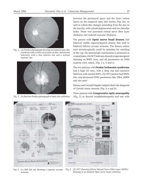
March 2008 Meenakshi Dhar et al. - <strong>Glaucoma</strong> Masquerades 25<br />
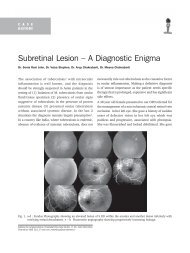
Fig. 2. (a) Fundus photograph showing an isolated optic disc<br />
coloboma with a white excavation on disc, decentered<br />
inferiorly with a thin inferior rim and a normal<br />
superior rim<br />
Fig. 2. (b) Red free fundus photograph of optic disc coloboma<br />
Fig. 2 (c) HFA left eye showing a superior arcuate<br />
scotoma<br />
between the perineural space and the inner retinal<br />
layers on the temporal optic disc border (Fig. 2e), as<br />
well as schisis-like changes extending from the disc to<br />
the macula, with cystoid degeneration and two lamellar<br />
holes. There was increased retinal nerve fibre layer<br />
thickness and reduced macular thickness.<br />
The patient with Optic nerve head drusen had<br />
bilateral visible superotemporal drusen that lead to<br />
bilateral inferior arcuate scotoma. The drusen unless<br />
seen stereoscopically could be mistaken for notching<br />
of the cup. On stereoscopic examination it presented as<br />
a raised lesion. On OCT both eyes showed a superotemporal<br />
thinning on RNFL scan, and all parameters on ONH<br />
analysis were raised. (Fig. 3 a, b and c).<br />
The two patients with Ocular Ischaemic syndrome<br />
had a high CD ratio, with a deep cup and extensive<br />
field loss with normal IOP’s. On OCT patient had RNFL<br />
loss, and decreased ONH parameters like VIRA, HIRW<br />
and rim area 2<br />
History and Carotid Doppler helped clinch the diagnosis<br />
of Carotid artery stenosis (Fig. 4 a and b).<br />
Three patients with Compressive optic neuropathy<br />
(Fig. 5) in thyroid exophthalmopathy and one with<br />
Fig. 2 (d) OCT showing Inferior Retinal Nerve Fibre Layer (RNFL)<br />
thinning in an isolated Optic nerve head coloboma