Glaucoma Masqueraders – Our Clinical Experience – Has ... - KSOS
Glaucoma Masqueraders – Our Clinical Experience – Has ... - KSOS
Glaucoma Masqueraders – Our Clinical Experience – Has ... - KSOS

You also want an ePaper? Increase the reach of your titles
YUMPU automatically turns print PDFs into web optimized ePapers that Google loves.
March 2008 Meenakshi Dhar et al. - <strong>Glaucoma</strong> Masquerades 27<br />
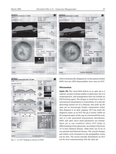
Fig. 3. (c) OCT findings in drusen of ONH<br />
defects dramatically disappeared. In this patient neither<br />
RNFL loss nor ONH abnormalities were seen on OCT.<br />
Discussion<br />
Optic Pit The visual field defects in an optic pit is a<br />
superior arcuate scotoma similar to glaucoma, but it is<br />
nonprogressive, and management does not include an<br />
IOP lowering agent. The danger to visual loss is due to<br />
neurosensory detachment or retinoschisis. It is only the<br />
discerning trained eye of a clinician, that picks up the<br />
optic pit on stereoscopic fundus examination rather<br />
than diagnose it as optic cupping. OCT has made the<br />
diagnosis easier, as on OCT the pit is clearly visible on<br />
the temporal aspect of the cup on a horizontal line scan,<br />
and so is the associated neurosensory detachment.<br />
RNFL and optic nerve head parameters are normal.<br />
Optic pit is one condition where OCT helps in<br />
differentiating it from glaucoma. According to literature<br />
15 % have bilateral disease, while three out of six of<br />
our patients had bilateral disease. The cystoid changes<br />
and schisis cavity formation in the peripapillary retina<br />
can be seen. The serous macular detachment on OCT<br />
can be seen communicating with the optic pit.