Instructions for Philips PW1830.pdf
Instructions for Philips PW1830.pdf
Instructions for Philips PW1830.pdf

Create successful ePaper yourself
Turn your PDF publications into a flip-book with our unique Google optimized e-Paper software.
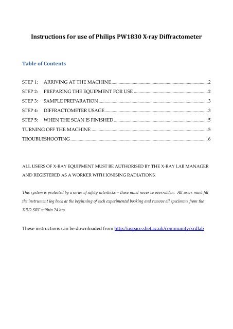
<strong>Instructions</strong> <strong>for</strong> use of <strong>Philips</strong> PW1830 X-ray Diffractometer<br />
Table of Contents<br />
STEP 1: ARRIVING AT THE MACHINE .................................................................................. 2<br />
STEP 2: PREPARING THE EQUIPMENT FOR USE ............................................................... 2<br />
STEP 3: SAMPLE PREPARATION ............................................................................................. 3<br />
STEP 4: DIFFRACTOMETER USAGE ........................................................................................ 3<br />
STEP 5: WHEN THE SCAN IS FINISHED ................................................................................ 5<br />
TURNING OFF THE MACHINE ................................................................................................... 5<br />
TROUBLESHOOTING ..................................................................................................................... 6<br />
ALL USERS OF X-RAY EQUIPMENT MUST BE AUTHORISED BY THE X-RAY LAB MANAGER<br />
AND REGISTERED AS A WORKER WITH IONISING RADIATIONS.<br />
This system is protected by a series of safety interlocks – these must never be overridden. All users must fill<br />
the instrument log book at the beginning of each experimental booking and remove all specimens from the<br />
XRD SRF within 24 hrs.<br />
These instructions can be downloaded from http://uspace.shef.ac.uk/community/xrdlab
STEP 1: ARRIVING AT THE MACHINE<br />
Make sure the machine is on:<br />
1. Are the power setting displays showing 40kV and 30mA? If so – the machine is on,<br />
and most likely ready to use - proceed to Step 3!<br />
2. If the kV and mA displays are blank, the machine is switched off. Check <strong>for</strong> any<br />
notices that the machine is not working. If there are none, proceed to Step 2 to turn<br />
the instrument on.<br />
3. If the displays are reading 0kV and 0mA, check the LCD display next to the<br />
‘Shutters’ button. If this is reading ‘Error 42’ there is a problem with the cooling<br />
water supply – most likely the water filter needs changing!<br />
STEP 2: PREPARING THE EQUIPMENT FOR USE<br />
1. Make sure water supply on (anticlockwise).<br />
- Check flow between 3.5 L/min and 6 L/min on flow rate meter mounted on the<br />
rear of the diffractometer.<br />
2. Make sure that the power is on at the isolator switch.<br />
3. Press ‘POWER ON’.<br />
- When switched ON, the generator will per<strong>for</strong>m a SELF TEST; once completed the<br />
message ‚TARGET 15kV 10mA‛ should appear on the LCD display.<br />
4. Switch on HT by pushing ‘HT ON‘ button.<br />
- The generator should go automatically to 15 kV and 10 mA.<br />
5. If the shutter is open, press CLOSE SHUTTER, ‘4’ and then ENTER.<br />
6. Turn the voltage (kV) and current (mA) up to the working values:<br />
- Turn the ‘kV’ dial to 40.<br />
- Turn the ‘kV’ dial to 30.<br />
REMEMBER: THE mA SHOULD ALWAYS BE LOWER THAN THE kV!
STEP 3: SAMPLE PREPARATION<br />
Note that ALL sample prep should be done in I6b, not in I6. All samples MUST be<br />
labelled with your name! Unlabelled samples will be treated as hazardous waste and<br />
disposed of immediately. When prepared, proceed to Step 4.<br />
For powder samples:<br />
1. Make sure your powder is well-ground.<br />
2. Select the biggest Aluminium specimen tray that you can fully fill. The correct<br />
holders have the aperture <strong>for</strong> powder towards the TOP of the holder.<br />
3. Remove the back cover, and place on to a glass microscope slide. Completely fill<br />
the aperture with powder. Use a microscope slide to ensure that the specimen<br />
surface is level with the top of the holder. Replace the back cover, and carefully<br />
turn over. Remove the glass slide very carefully to expose your specimen surface.<br />
4. Label the specimen holder with your name using a marker pen.<br />
For bulk samples:<br />
1. Make sure your sample has at least one surface which is level and smooth.<br />
2. Remove or add Apiezon putty to the brass specimen holder as necessary, and<br />
mould the putty so that your sample can sit on top of a mound of putty.<br />
3. Place the sample on to the putty and press down lightly to help it stick.<br />
4. Turn the holder upside down; gently press down on both sides on to a flat surface.<br />
The top of the sample MUST BE LEVEL with the top of the holder.<br />
Key point #1: incorrect specimen height WILL result in specimen height displacement<br />
errors in your data, e.g. if the sample is too high, your Bragg reflections will be shifted to<br />
higher angles (and too low, peaks shifted to lower angles). This can make phase analysis<br />
very difficult!<br />
Key point #2: the Apiezon putty is partly crystalline, so will give peaks in your diffraction<br />
pattern should it get in to the beam. Make sure your specimen is mounted on the putty,<br />
and not embedded too deeply into it!
STEP 4: DIFFRACTOMETER USAGE<br />
1. Make sure the computer is switched on.<br />
2. Load sample in to the diffractometer. Old samples must be removed, tipped in to<br />
aluminium foil, labelled with the date and name of whoever ran the sample, then<br />
placed in to the used specimen box <strong>for</strong> collection.<br />
3. Place chamber door onto goniometer.<br />
4. Ensure that all shielding is correctly positioned and close doors.<br />
5. Make sure the shutter is closed, then launch the ‘HBX’ software, if not running.<br />
- Note: If you try to launch the HBX software, and it appears to already be running,<br />
simply press Ctrl, Alt and Delete, and close the HBX software be<strong>for</strong>e trying again.<br />
6. Set the scanning parameters:<br />
(i) Starting angle, in degrees 2θ (> 3º).<br />
(ii) Finish angle, in degrees 2θ (< 160º).<br />
(iii) Step size, in degrees 2θ (> 0.01º; 0.02 is a good standard, though 0.05 is often<br />
best <strong>for</strong> quick, good quality data <strong>for</strong> phase analyses. Use 0.01 <strong>for</strong> cell refinement).<br />
(iv) Scan speed in degrees per minute (between 0.1 and 2 degrees/minute).<br />
(v) Enter comment, if desired, <strong>for</strong> your own reference.<br />
(vi) Enter sample name (8 or less characters, no ‘special’ characters).<br />
(vii) Save your data in your folder on the hard drive by selecting ‘Options’/’Data<br />
Path and Type’ and then selecting your data folder.<br />
7. Press ‘Begin Scan’ on the PC.<br />
8. When prompted, open the shutter by holding the SHUTTERS button, and then<br />
pressing the ‘OPEN’ button <strong>for</strong> shutter 2.<br />
Note: if the shutter fails to open, the most common cause is the enclosure door being poorly<br />
fitted.<br />
9. Use mini-monitor to check <strong>for</strong> any radiation leaks.<br />
10. MAKE SURE THAT YOU FILL IN THE LOG BOOK.<br />
The scan may be stopped at any time with ‘STOP+SAVE’ or ‘ABORT SCAN’.
STEP 5: WHEN THE SCAN IS FINISHED<br />
1. Close the shutter: On the front of the diffractometer, press CLOSE SHUTTER,<br />
enter ‘4’ and then press ENTER.<br />
2. The data will be saved automatically to the hard drive. If you cannot find your<br />
data, it is most likely operator error! Use of special characters (&, +, etc) will cause<br />
data not to be saved – or perhaps the ‘Data Path and Type’ wasn’t selected<br />
(correctly). Try using the ‘Search’ function in Windows to look <strong>for</strong> your file.<br />
3. Remove sample.<br />
NOTE: Used samples MUST be removed from the XRD Lab within 24 hours of being run!<br />
TURNING OFF THE MACHINE<br />
The diffractometers are normally in use around the clock, seven days a week. If <strong>for</strong> some<br />
reason it is necessary to turn the machines off:<br />
1. Lower mA then kV if not starting new scan immediately. To do this:<br />
- Press ‘mA’ and enter 10, and press ‘ENTER’.<br />
- Press ‘kV’ and enter 15, and press ‘ENTER’.<br />
REMEMBER: THE mA SHOULD ALWAYS BE LOWER THAN THE kV!<br />
2. Press ‘FUNCTION’, enter ‘2’ then press ‘ENTER’.<br />
3. Press ‘STANDBY’.<br />
The equipment is now on standby mode. To isolate the instrument completely:<br />
1. Turn the isolator switch on the wall to ‘OFF’.<br />
2. Turn the water off (clockwise).
TROUBLESHOOTING<br />
The shutter will not open! Try putting the chamber door on again – the round button on<br />
The ‘No waterflow’ light is<br />
lit, and the machine<br />
appears to be off.<br />
The shutter is closing<br />
automatically of its own<br />
accord!<br />
the side of the X-ray tube housing must be fully pressed in<br />
be<strong>for</strong>e the instrument will recognise that it is safe to unleash<br />
the X-rays.<br />
If this button is fully in, check all the housing doors are fully<br />
closed and all interlocks in contact.<br />
There is also an interlock underneath the ‘top hat’<br />
arrangement (just on the chamber side be<strong>for</strong>e the secondary<br />
beam monochromator) – try pinching that closed.<br />
Finally, if the problem still persists, it is likely that one or<br />
both of the ‘Shutter Open’ bulbs has blown. Contact the Lab<br />
Manager.<br />
Last revised by N. Reeves-McLaren, 5 h January 2012.<br />
Low water pressure. Report to the Lab Manager.<br />
Probably someone has pressed the Shutter 2 ‘Clock’ button.<br />
Press the ‘∞‘ button, and try again.