ABSTRACTS - British Journal of Ophthalmology
ABSTRACTS - British Journal of Ophthalmology
ABSTRACTS - British Journal of Ophthalmology
You also want an ePaper? Increase the reach of your titles
YUMPU automatically turns print PDFs into web optimized ePapers that Google loves.
Downloaded from bjo.bmj.com on August 22, 2013 - Published by<br />
group.bmj.com<br />
CONTUSION OF GLOBE AFTER SHOT INJURY<br />
523<br />
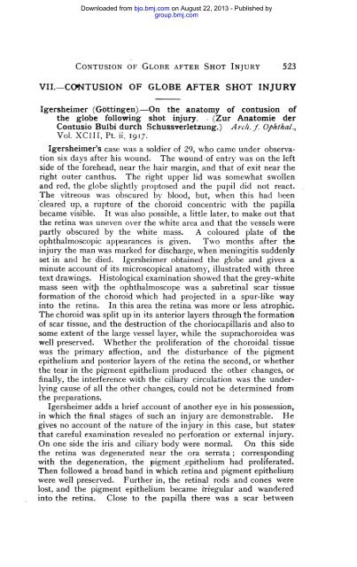
VII.-CONTUSION OF GLOBE AFTER SHOT INJURY<br />
Igersheimer (Gottingen).-On the anatomy <strong>of</strong> contusion <strong>of</strong><br />
the globe following shot injury. . (Zur Anatomie der<br />
Contusio Bulbi durch Schussverletzung.) Arch. f. Ophkal.,<br />
Vol. XCIII, Pt. ii, 1917.<br />
Igersheimer's case was a soldier <strong>of</strong> 29, who came under observation<br />
six days after his wound. The wound-<strong>of</strong> entry was on the left<br />
side <strong>of</strong> the forehead, near the hair margin, and that <strong>of</strong> exit near the<br />
right outer canthus. The right upper lid was somewhat swollen<br />
and red, the globe slightly proptosed and the pupil did not react.<br />
The vitreous was obscured by blood, but, when this had been<br />
cleared up, a rupture <strong>of</strong> the choroid concentric with the papilla<br />
became visible. It was also possible, a little later, to make out that<br />
the retina was uneven over the white area and that the vessels were<br />
partly obscured by the white mass. A coloured plate <strong>of</strong> the<br />
ophthalmoscopic appearances is given. Two months after the<br />
injury the man was marked for discharge, when meningitis suddenly<br />
set in and he died. Igersheimer obtained the globe and gives a<br />
minute account <strong>of</strong> its microscopical anatomy, illustrated with three<br />
text drawings.<br />
Histological examination showed that the grey-white<br />
mass seen with the ophthalmoscope was a §ubretinal scar tissue<br />
formation <strong>of</strong> the choroid which had projected in a spur-like way<br />
into the retina. In this area the retina was more or less atrophic.<br />
The choroid was split up in its anterior layers through the formation<br />
<strong>of</strong> scar tissue, and the destruction <strong>of</strong> the choriocapillaris and also to<br />
some extent <strong>of</strong> the large vessel layer, while the suprachoroidea was<br />
well preserved. Whether the proliferation <strong>of</strong> the choroidal. tissue<br />
was the primary affection, and the disturbance <strong>of</strong> the pigment<br />
epithelium and posterior layers <strong>of</strong> the retina the second, or whether<br />
the tear in the pigment epithelium produced the other changes, or<br />
finally, the interference with the ciliary circulation was the underlying<br />
cause <strong>of</strong> all the other changes, could not be determined from<br />
the preparations.<br />
Igersheimer adds a brief account <strong>of</strong> another eye in his possession,<br />
in which the final stages <strong>of</strong> such an injury are demonstrable. He<br />
gives no account <strong>of</strong> the nature <strong>of</strong> the injury in this case, but states,<br />
that careful examination revealed no perforation or external injury.<br />
On one side the iris and ciliary body were normal. On this side<br />
the retina was degenerated near the ora serrata; corresponding<br />
with the degeneration, the pigment epithelium had proliferated.<br />
Then followed a broad band in which retina and pigment epithelium.<br />
were well preserved. Further in, the retinal rods and cones were<br />
lost, and the pigment epithelium became iriegular and wandered<br />
into the retina. Close to the papilla there was a scar between


