DNA triplex structures in neurodegenerative disorder, Friedreich's ...
DNA triplex structures in neurodegenerative disorder, Friedreich's ...
DNA triplex structures in neurodegenerative disorder, Friedreich's ...
You also want an ePaper? Increase the reach of your titles
YUMPU automatically turns print PDFs into web optimized ePapers that Google loves.
528 Moganty R Rajeswari<br />
symptoms and die ma<strong>in</strong>ly due to heart failure. It has been<br />
proved that these neurological manifestations result from primary<br />
degeneration of dorsal root ganglion associated with<br />
axonal degeneration <strong>in</strong> posterior columns, sp<strong>in</strong>ocerebellar<br />
tracts and corticosp<strong>in</strong>al tracts <strong>in</strong> sp<strong>in</strong>al cord. Neuro-imag<strong>in</strong>g<br />
reveals th<strong>in</strong>n<strong>in</strong>g of the cervical sp<strong>in</strong>al cord. In more advanced<br />
stages of the disease, cerebellar atrophy and cerebellar vermis<br />
atrophy are also seen. However, there is no accurate measurement<br />
of cl<strong>in</strong>ical progression of disease. International<br />
Cooperative Ataxia Rat<strong>in</strong>g Scale (Trouillas et al., 1997) has<br />
been widely used to assess cl<strong>in</strong>ical manifestations of FRDA,<br />
but none of the scales is very specific. Some of the ICARS<br />
parameters and radiological MR imag<strong>in</strong>g data used <strong>in</strong> data of<br />
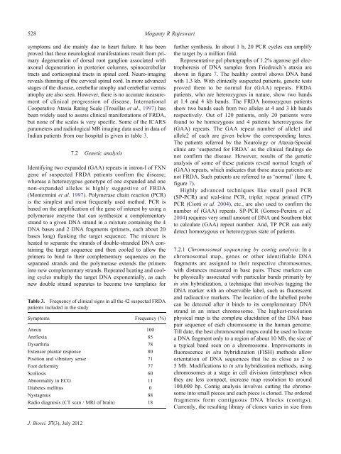
Indian patients from our hospital is given <strong>in</strong> table 3.<br />
7.2 Genetic analysis<br />
Identify<strong>in</strong>g two expanded (GAA) repeats <strong>in</strong> <strong>in</strong>tron-I of FXN<br />
gene of suspected FRDA patients confirm the disease;<br />
whereas a heterozygous genotype of one expanded and one<br />
non-expanded alleles is highly suggestive of FRDA<br />
(Monterm<strong>in</strong>i et al. 1997). Polymerase cha<strong>in</strong> reaction (PCR)<br />
is the simplest and most frequently used method. PCR is<br />
based on the amplification of the gene of <strong>in</strong>terest by us<strong>in</strong>g a<br />
polymerase enzyme that can synthesize a complementary<br />
strand to a given <strong>DNA</strong> strand <strong>in</strong> a mixture conta<strong>in</strong><strong>in</strong>g the 4<br />
<strong>DNA</strong> bases and 2 <strong>DNA</strong> fragments (primers, each about 20<br />
bases long) flank<strong>in</strong>g the target sequence. The mixture is<br />
heated to separate the strands of double-stranded <strong>DNA</strong> conta<strong>in</strong><strong>in</strong>g<br />
the target sequence and then cooled to allow the<br />
primers to b<strong>in</strong>d to their complementary sequences on the<br />
separated strands and the polymerase extends the primers<br />
<strong>in</strong>to new complementary strands. Repeated heat<strong>in</strong>g and cool<strong>in</strong>g<br />
cycles multiply the target <strong>DNA</strong> exponentially, as each<br />
new double strand separates to become two templates for<br />
Table 3. Frequency of cl<strong>in</strong>ical signs <strong>in</strong> all the 42 suspected FRDA<br />
patients <strong>in</strong>cluded <strong>in</strong> the study<br />
Symptoms Frequency (%)<br />
Ataxia 100<br />
Areflexia 85<br />
Dysarthria 78<br />
Extensor plantar response 80<br />
Position and vibratory sense 71<br />
Foot deformity 77<br />
Scoliosis 60<br />
Abnormality <strong>in</strong> ECG 11<br />
Diabetes mellitus 0<br />
Nystagmus 88<br />
Radio diagnosis (CT scan / MRI of bra<strong>in</strong>) 18<br />
further synthesis. In about 1 h, 20 PCR cycles can amplify<br />
the target by a million fold.<br />
Representative gel photographs of 1.2% agarose gel electrophoresis<br />
of <strong>DNA</strong> samples from Friedreich’s ataxia are<br />
shown <strong>in</strong> figure 7. The healthy control shows <strong>DNA</strong> band<br />
with 1.3 kb. With cl<strong>in</strong>ically suspected patients, genetic tests<br />
proved them to be normal for (GAA) repeats. FRDA<br />
patients, who are heterozygous <strong>in</strong> nature, show two bands<br />
at 1.4 and 4 kb bands. The FRDA homozygous patients<br />
show two bands each from two alleles at 4 and 3 kb bands<br />
respectively. Out of 120 patients, only 20 patients were<br />
found to be homozygous and 4 patients heterozygous for<br />
(GAA) repeats. The GAA repeat number of allele1 and<br />
allele2 of each are given below the correspond<strong>in</strong>g lanes.<br />
The patients referred by the Neurology or Ataxia-Special<br />
cl<strong>in</strong>ic are ‘suspected for FRDA’ as the cl<strong>in</strong>ical f<strong>in</strong>d<strong>in</strong>gs do<br />
not confirm the disease. However, results of the genetic<br />
analysis of some of these patients reveal normal length of<br />
(GAA) repeats, which <strong>in</strong>dicates that those ataxia patients are<br />
not FRDA. Such patients are referred to as ‘normal’ (lane 4,<br />
figure 7).<br />
Highly advanced techniques like small pool PCR<br />
(SP-PCR) and real-time PCR, triplet repeat primed (TP)<br />
PCR (Ciotti et al. 2004), etc., are also used to confirm the<br />
number of (GAA) repeats. SP-PCR (Gomes-Pereira et al.<br />
2004) requires very small amount of <strong>DNA</strong> and Southern blot<br />
to calculate (GAA) repeat number. And, TP PCR can only<br />
detect homozygous or heterozygous state of patients.<br />
7.2.1 Chromosomal sequenc<strong>in</strong>g by contig analysis: In a<br />
chromosomal map, genes or other identifiable <strong>DNA</strong><br />
fragments are assigned to their respective chromosomes,<br />
with distances measured <strong>in</strong> base pairs. These markers can<br />
be physically associated with particular bands primarily by<br />
<strong>in</strong> situ hybridization, a technique that <strong>in</strong>volves tagg<strong>in</strong>g the<br />
<strong>DNA</strong> marker with an observable label, such as fluorescent<br />
and radioactive markers. The location of the labelled probe<br />
can be detected after it b<strong>in</strong>ds to its complementary <strong>DNA</strong><br />
strand <strong>in</strong> an <strong>in</strong>tact chromosome. The highest-resolution<br />
physical map is the complete elucidation of the <strong>DNA</strong> base<br />
pair sequence of each chromosome <strong>in</strong> the human genome.<br />
Till date, the best chromosomal maps could be used to locate<br />
a <strong>DNA</strong> fragment only to a region of about 10 Mb, the size of<br />
a typical band seen on a chromosome. Improvements <strong>in</strong><br />
fluorescence <strong>in</strong> situ hybridization (FISH) methods allow<br />
orientation of <strong>DNA</strong> sequences that lie as close as 2 to<br />
5 Mb. Modifications to <strong>in</strong> situ hybridization methods, us<strong>in</strong>g<br />
chromosomes at a stage <strong>in</strong> cell division (<strong>in</strong>terphase) when<br />
they are less compact, <strong>in</strong>crease map resolution to around<br />
100,000 bp. Contig analysis <strong>in</strong>volves cutt<strong>in</strong>g the chromosome<br />
<strong>in</strong>to small pieces and each piece is cloned. The ordered<br />
fragments form contiguous <strong>DNA</strong> blocks (contigs).<br />
Currently, the result<strong>in</strong>g library of clones varies <strong>in</strong> size from<br />
J. Biosci. 37(3), July 2012


