DNA triplex structures in neurodegenerative disorder, Friedreich's ...
DNA triplex structures in neurodegenerative disorder, Friedreich's ...
DNA triplex structures in neurodegenerative disorder, Friedreich's ...

Create successful ePaper yourself
Turn your PDF publications into a flip-book with our unique Google optimized e-Paper software.
<strong>DNA</strong> <strong>triplex</strong>es <strong>in</strong> <strong>neurodegenerative</strong> <strong>disorder</strong> 523<br />
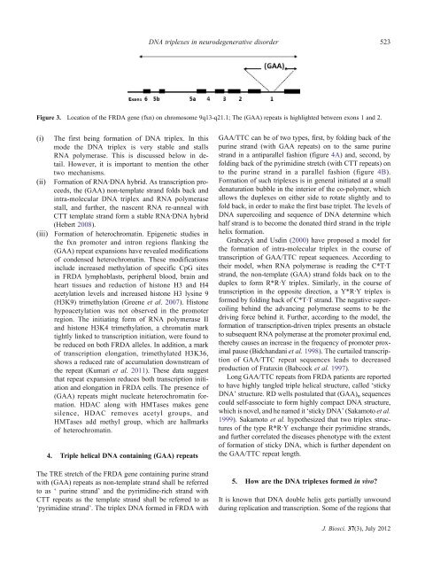
Figure 3. Location of the FRDA gene (fxn) on chromosome 9q13-q21.1; The (GAA) repeats is highlighted between exons 1 and 2.<br />
(i)<br />
(ii)<br />
(iii)<br />
The first be<strong>in</strong>g formation of <strong>DNA</strong> <strong>triplex</strong>. In this<br />
mode the <strong>DNA</strong> <strong>triplex</strong> is very stable and stalls<br />
RNA polymerase. This is discussed below <strong>in</strong> detail.<br />
However, it is important to mention the other<br />
two mechanisms.<br />
Formation of RNA∙<strong>DNA</strong> hybrid. As transcription proceeds,<br />
the (GAA) non-template strand folds back and<br />
<strong>in</strong>tra-molecular <strong>DNA</strong> <strong>triplex</strong> and RNA polymerase<br />
stall, and further, the nascent RNA re-anneal with<br />
CTT template strand form a stable RNA∙<strong>DNA</strong> hybrid<br />
(Hebert 2008).<br />
Formation of heterochromat<strong>in</strong>. Epigenetic studies <strong>in</strong><br />
the fxn promoter and <strong>in</strong>tron regions flank<strong>in</strong>g the<br />
(GAA) repeat expansions have revealed modifications<br />
of condensed heterochromat<strong>in</strong>. These modifications<br />
<strong>in</strong>clude <strong>in</strong>creased methylation of specific CpG sites<br />
<strong>in</strong> FRDA lymphoblasts, peripheral blood, bra<strong>in</strong> and<br />
heart tissues and reduction of histone H3 and H4<br />
acetylation levels and <strong>in</strong>creased histone H3 lys<strong>in</strong>e 9<br />
(H3K9) trimethylation (Greene et al. 2007). Histone<br />
hypoacetylation was not observed <strong>in</strong> the promoter<br />
region. The <strong>in</strong>itiat<strong>in</strong>g form of RNA polymerase II<br />
and histone H3K4 trimethylation, a chromat<strong>in</strong> mark<br />
tightly l<strong>in</strong>ked to transcription <strong>in</strong>itiation, were found to<br />
be reduced on both FRDA alleles. In addition, a mark<br />
of transcription elongation, trimethylated H3K36,<br />
shows a reduced rate of accumulation downstream of<br />
the repeat (Kumari et al. 2011). These data suggest<br />
that repeat expansion reduces both transcription <strong>in</strong>itiation<br />
and elongation <strong>in</strong> FRDA cells. The presence of<br />
(GAA) repeats might nucleate heterochromat<strong>in</strong> formation.<br />
HDAC along with HMTases makes gene<br />
silence, HDAC removes acetyl groups, and<br />
HMTases add methyl group, which are hallmarks<br />
of heterochromat<strong>in</strong>.<br />
4. Triple helical <strong>DNA</strong> conta<strong>in</strong><strong>in</strong>g (GAA) repeats<br />
The TRE stretch of the FRDA gene conta<strong>in</strong><strong>in</strong>g pur<strong>in</strong>e strand<br />
with (GAA) repeats as non-template strand shall be referred<br />
to as ‘ pur<strong>in</strong>e strand’ and the pyrimid<strong>in</strong>e-rich strand with<br />
CTT repeats as the template strand shall be referred to as<br />
‘pyrimid<strong>in</strong>e strand’. The <strong>triplex</strong> <strong>DNA</strong> formed <strong>in</strong> FRDA with<br />
GAA/TTC can be of two types, first, by fold<strong>in</strong>g back of the<br />
pur<strong>in</strong>e strand (with GAA repeats) on to the same pur<strong>in</strong>e<br />
strand <strong>in</strong> a antiparallel fashion (figure 4A) and, second, by<br />
fold<strong>in</strong>g back of the pyrimid<strong>in</strong>e stretch (with CTT repeats) on<br />
to the pur<strong>in</strong>e strand <strong>in</strong> a parallel fashion (figure 4B).<br />
Formation of such <strong>triplex</strong>es is <strong>in</strong> general <strong>in</strong>itiated at a small<br />
denaturation bubble <strong>in</strong> the <strong>in</strong>terior of the co-polymer, which<br />
allows the duplexes on either side to rotate slightly and to<br />
fold back, <strong>in</strong> order to make the first base triplet. The levels of<br />
<strong>DNA</strong> supercoil<strong>in</strong>g and sequence of <strong>DNA</strong> determ<strong>in</strong>e which<br />
half strand is to become the donated third strand <strong>in</strong> the triple<br />
helix formation.<br />
Grabczyk and Usd<strong>in</strong> (2000) have proposed a model for<br />
the formation of <strong>in</strong>tra-molecular <strong>triplex</strong> <strong>in</strong> the course of<br />
transcription of GAA/TTC repeat sequences. Accord<strong>in</strong>g to<br />
their model, when RNA polymerase is read<strong>in</strong>g the C*T∙T<br />
strand, the non-template (GAA) strand folds back on to the<br />
duplex to form R*R∙Y <strong>triplex</strong>. Similarly, <strong>in</strong> the course of<br />
transcription <strong>in</strong> the opposite direction, a Y*R∙Y <strong>triplex</strong> is<br />
formed by fold<strong>in</strong>g back of C*T∙T strand. The negative supercoil<strong>in</strong>g<br />
beh<strong>in</strong>d the advanc<strong>in</strong>g polymerase seems to be the<br />
driv<strong>in</strong>g force beh<strong>in</strong>d it. Further, accord<strong>in</strong>g to the model, the<br />
formation of transcription-driven <strong>triplex</strong> presents an obstacle<br />
to subsequent RNA polymerase at the promoter proximal end,<br />
thereby causes an <strong>in</strong>crease <strong>in</strong> the frequency of promoter proximal<br />
pause (Bidchandani et al. 1998). The curtailed transcription<br />
of GAA/TTC repeat sequences leads to decreased<br />
production of Fratax<strong>in</strong> (Babcock et al. 1997).<br />
Long GAA/TTC repeats from FRDA patients are reported<br />
to have highly tangled triple helical structure, called ‘sticky<br />
<strong>DNA</strong>’ structure. RD wells postulated that (GAA) n sequences<br />
could self-associate to form highly compact <strong>DNA</strong> structure,<br />
which is novel, and he named it ‘sticky <strong>DNA</strong>’ (Sakamoto et al.<br />
1999). Sakamoto et al. hypothesized that two <strong>triplex</strong> <strong>structures</strong><br />
of the type R*R∙Y exchange their pyrimid<strong>in</strong>e strands,<br />
and further correlated the diseases phenotype with the extent<br />
of formation of sticky <strong>DNA</strong>, which is further dependent on<br />
the GAA/TTC repeat length.<br />
5. How are the <strong>DNA</strong> <strong>triplex</strong>es formed <strong>in</strong> vivo?<br />
It is known that <strong>DNA</strong> double helix gets partially unwound<br />
dur<strong>in</strong>g replication and transcription. Some of the regions that<br />
J. Biosci. 37(3), July 2012