Photon-Induced Near Field Electron Microscopy - California Institute ...
Photon-Induced Near Field Electron Microscopy - California Institute ...
Photon-Induced Near Field Electron Microscopy - California Institute ...

Create successful ePaper yourself
Turn your PDF publications into a flip-book with our unique Google optimized e-Paper software.
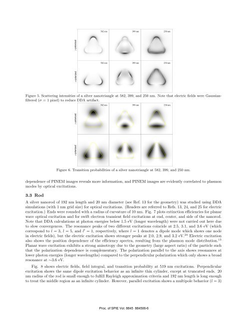
582 nm 399 nm 250 nm<br />
582 nm 399 nm 250 nm<br />
x-polarized<br />
y-polarized<br />
Figure 5. Scattering intensities of a silver nanotriangle at 582, 399, and 250 nm. Note that electric fields were Gaussianfiltered<br />
(σ = 1 pixel) to reduce DDA artifact.<br />
582 nm 399 nm 250 nm<br />
x-polarized y-polarized<br />
Figure 6. Transition probabilities of a silver nanotriangle at 582, 399, and 250 nm.<br />
dependence of PINEM images reveals more information, and PINEM images are evidently correlated to plasmon<br />
modes by optical excitations.<br />
3.3 Rod<br />
A silver nanorod of 192 nm length and 20 nm diameter (see Ref. 13 for the geometry) was studied using DDA<br />
simulations (with 1 nm grid size) for optical excitations. (Readers are referred to Refs. 13, 24, and 25 for electric<br />
excitation.) Ends were rounded with a radius of curvature of 10 nm. Fig. 7 plots extinction efficiencies for planar<br />
wave optical excitation and for swift electron transient field excitations at end, center, and side of the nanorod.<br />
Note that DDA calculations at photon energies below 1.5 eV (longer wavelength) were not carried out here due<br />
to slow convergences. The resonance peaks of two different excitations coincide at 2.5, 3.1, and 3.6 eV (which<br />
correspond to l = 3, l = 5, and l ′ = 1, respectively, where l = 1 denotes a dipole mode which shows one node<br />
in electric fields), but the electric excitation shows stronger peaks at 2.0, 2.9, and 3.2 eV. 24 Electric excitation<br />
also shows the position dependence of the efficiency spectra, resulting from the plasmon mode distribution. 13<br />
Planar wave excitation exhibits a strong anisotropy due to the geometry (large aspect ratio) of the particle such<br />
that the polarization dependence is complementary. The polarization parallel to the axis shows resonances at<br />
lower photon energies (longer wavelengths) compared to the perpendicular polarization which only shows a broad<br />
resonance at ∼3.6 eV.<br />
Fig. 8 shows electric fields, field integral, and transition probability at 519 nm excitations. Perpendicular<br />
excitation shows the same dipole excitation behavior as an infinite thin cylinder, except at truncated ends. 20<br />
nm radius of the rod is small enough to fulfill Rayleigh approximation criteria and 192 nm length is long enough<br />
to treat the middle region as an infinite cylinder. However, parallel excitation shows a multipole behavior (l = 3)<br />
Proc. of SPIE Vol. 8845 884506-6