
Create successful ePaper yourself
Turn your PDF publications into a flip-book with our unique Google optimized e-Paper software.
Software Flexibility<br />
Easier <strong>Image</strong> Sharing,<br />
Better <strong>Image</strong> Processing<br />
NewTom NNT analysis software<br />
canal, NNT is designed to<br />
professionals the capacity to<br />
is the perfect complement to<br />
deliver high quality images<br />
view images exported with<br />
<strong>Cone</strong> <strong>Beam</strong> <strong>3D</strong> imaging. NNT<br />
that can be placed into<br />
DICOM 3.0 without having to<br />
supports the identification of<br />
user-defined templates,<br />
purchase NNT.<br />
root inclination, position of<br />
deliverable digitally, on paper,<br />
<strong>Image</strong> data can be burned onto<br />
impacted and supernumerary<br />
or on film. NNT gathers<br />
CD or DVD for imaging centers<br />
teeth, resorption, hyperplastic<br />
any combination of images<br />
and referring doctors, allowing<br />
growth, and tooth structure<br />
onto one screen for custom<br />
dental and medical<br />
anomalies. With the ability<br />
to mark the mandibular<br />
reports. NNT is delivered with<br />
a standard viewer, giving other<br />
professionals the opportunity to<br />
easily share images.<br />
Figure 1<br />
Figure<br />
Superior Third-Party Compatibility<br />
NewTom images are<br />
and utilized in a myriad of<br />
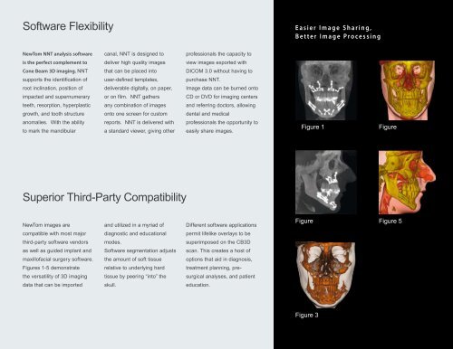
Different software applications<br />
Figure<br />
Figure 5<br />
compatible with most major<br />
diagnostic and educational<br />
permit lifelike overlays to be<br />
third-party software vendors<br />
modes.<br />
superimposed on the CB<strong>3D</strong><br />
as well as guided implant and<br />
Software segmentation adjusts<br />
scan. This creates a host of<br />
maxillofacial surgery software.<br />
the amount of soft tissue<br />
options that aid in diagnosis,<br />
Figures 1-5 demonstrate<br />
relative to underlying hard<br />
treatment planning, pre-<br />
the versatility of <strong>3D</strong> imaging<br />
tissue by peering “into” the<br />
surgical analyses, and patient<br />
data that can be imported<br />
skull.<br />
education.<br />
Figure 3