Download the PDF
Download the PDF
Download the PDF
Create successful ePaper yourself
Turn your PDF publications into a flip-book with our unique Google optimized e-Paper software.
CET<br />
CONTINUING<br />
EDUCATION &<br />
TRAINING<br />
THIS ISSUE CET: FREE<br />
✔<br />
✔<br />
Approved for Optometrists Approved for DOs<br />
To gain more standard CET points for this year’s PAYL series, purchase your credits at<br />
www.otbookshop.co.uk and <strong>the</strong>n enter online at: www.otcet.co.uk to take your exams<br />
✘<br />
Sponsored by:<br />
26<br />
Module 12 Part 5<br />
Non-Retinal Complications of<br />
Diabetes Mellitus<br />
COURSE CODE: C-10557/O<br />
08/05/09 CET<br />
Shaheen P Shah MRCOphth MSc<br />
Diabetes mellitus (DM) is a chronic disease that occurs when a) <strong>the</strong><br />
pancreas does not produce enough insulin (insulin dependent diabetes or<br />
Type 1 diabetes), which is typically of early onset or b) when <strong>the</strong> body<br />
cannot effectively use <strong>the</strong> insulin it produces (maturity onset or noninsulin<br />
dependent diabetes). Type 1 diabetes can be regarded as an<br />
autoimmune disorder in which <strong>the</strong>re is destruction of <strong>the</strong> pancreatic islet<br />
cells in a genetically susceptible host. 1<br />
The world is currently experiencing a global epidemic of this incurable<br />
disease and <strong>the</strong> human and economic costs are enormous. Predictions<br />
estimate a doubling of <strong>the</strong> number affected from 180 million reaching an<br />
estimated 366 million by 2030. 2 The global increase in diabetes is attributed to increased life expectancy,<br />
urbanisation and changes in lifestyle and diet. Although previously recognised as a disease of <strong>the</strong> developed<br />
world, diabetes is becoming increasingly prevalent in <strong>the</strong> Developing World. In India alone, <strong>the</strong> number<br />
affected is expected to reach 80 million by 2030. Unfortunately, <strong>the</strong> majority of <strong>the</strong> burden from this disease<br />
is also now predicted to lie in low income countries and in those of working age. 3<br />
Hyperglycaemia, or raised blood sugar,<br />
is a common effect of uncontrolled DM<br />
and over time, it leads to serious<br />
damage to many of <strong>the</strong> body's systems.<br />
The eyes, often indicators of systemic<br />
disease, are particularly vulnerable to<br />
<strong>the</strong> pathophysiological changes that<br />
occur as a result of DM and clinical<br />
trials have demonstrated that tight<br />
control of blood glucose, HbA1c and<br />
blood pressure can reduce <strong>the</strong> risk of<br />
blindness. 4<br />
Diabetic retinopathy, which is <strong>the</strong><br />
leading cause of blindness in people<br />
under <strong>the</strong> age of 65 years in high income<br />
countries, is <strong>the</strong> most well-known<br />
ocular complication of DM. 5 In addition<br />
to diabetic retinopathy, <strong>the</strong>re are a<br />
number of o<strong>the</strong>r complications<br />
associated with this metabolic disorder<br />
that can be devastating to vision and<br />
ocular health. 6 This review aims to<br />
describe <strong>the</strong>se o<strong>the</strong>r, less well-known,<br />
complications.<br />
Cataract<br />
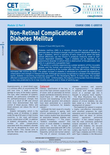
Cataract, opacification of <strong>the</strong> lens, is<br />
one of <strong>the</strong> most common causes of loss<br />
of vision, with an estimated 16 million<br />
people worldwide bilaterally blind as a<br />
result (Figure 1). 7 DM influences <strong>the</strong><br />
function and morphology of <strong>the</strong> lens 8<br />
and advanced glycation end products<br />
have been postulated as a possible<br />
pathogenic mechanism for diabetic<br />
cataract. 9<br />
Although <strong>the</strong> main cause of a visually<br />
significant cataract is ageing, both<br />
clinical epidemiological studies and<br />
basic science studies have documented<br />
an association between diabetes and<br />
4, 5, 10-20<br />
cataract formation. The classic<br />
“diabetic snowflake cataract” that<br />
occurs in acute diabetes is nowadays<br />
rarely seen. In fact, <strong>the</strong> subtype of<br />
cataract most associated with DM is <strong>the</strong><br />
posterior sub-capsular form and less<br />
frequently, cortical and nuclear cataract.<br />
The risk of cataract increases with<br />
diabetes duration and severity<br />
of hyperglycemia. 21 In addition,<br />
in patients with diabetes, cataract<br />
occurs at a younger age and is<br />
thought to progress more rapidly. 22<br />
The Blue Mountains Eye Study<br />
showed impaired fasting glucose, in <strong>the</strong><br />
absence of clinical diabetes, has also<br />
been noted as a risk factor for <strong>the</strong><br />
development of cataract. 17<br />
The treatment for cataract is surgical,<br />
a highly cost-effective intervention,<br />
with excellent prognosis for sight<br />
restoration and it is now estimated that<br />
globally <strong>the</strong>re are about 15 million<br />
cataract operations performed<br />
annually. 23 In fact, cataract surgery is<br />
now <strong>the</strong> most commonly performed<br />
surgical procedure in <strong>the</strong> UK. 24 In<br />
people with diabetes, <strong>the</strong> ten-year<br />
cumulative incidence of cataract<br />
surgery was found to be 8% in those<br />
with Type 1 diabetes and 25% in those<br />
with Type 2 diabetes in <strong>the</strong> Wisconsin<br />
CONFUSED ABOUT CET REQUIREMENTS? www.cetoptics.com/cetusers/faqs/<br />
IMPORTANT INFORMATION<br />
Under <strong>the</strong> new Vantage rules, all OT CET points awarded will be uploaded to its website by us. All participants must confirm <strong>the</strong>se results on www.cetoptics.com<br />
so that <strong>the</strong>y can move <strong>the</strong>ir points from <strong>the</strong> “Pending Points record” into <strong>the</strong>ir “Final CET points record”. Full instructions on how to do this are available on <strong>the</strong>ir website.
CET<br />
CONTINUING<br />
EDUCATION &<br />
TRAINING<br />
THIS ISSUE CET: FREE<br />
✔<br />
✔<br />
Approved for Optometrists Approved for DOs<br />
To gain more standard CET points for this year’s PAYL series, purchase your credits at<br />
www.otbookshop.co.uk and <strong>the</strong>n enter online at: www.otcet.co.uk to take your exams<br />
✘<br />
Sponsored by:<br />
27<br />
< Figure 1<br />
Posterior sub-capsular cataract<br />
Epidemiologic Study.<br />
Outcomes of cataract surgery in<br />
individuals with DM are known to be<br />
poorer, most commonly as a result of<br />
progression of diabetic retinopathy.<br />
However, better control of diabetic<br />
retinopathy with laser treatment prior<br />
to cataract surgery can improve<br />
25, 26<br />
surgical outcomes. Some case<br />
control studies have also demonstrated<br />
that rates of posterior capsular<br />
opacification following cataract<br />
extraction are higher in individuals<br />
27, 28<br />
affected by DM.<br />
Infection<br />
Patients with diabetes are predisposed<br />
to infections. Although not fully<br />
understood, <strong>the</strong> increased rate of<br />
infection seen in patients with DM is<br />
thought to relate to an<br />
immunosuppressive condition brought<br />
about by impaired innate and acquired<br />
immunity. Higher levels of<br />
hyperglycaemia are believed to<br />
correspondingly increase this level of<br />
29, 30<br />
immunosuppression.<br />
One of <strong>the</strong> most devastating<br />
complications following intraocular<br />
surgery is one of acute infectious<br />
exogenous endophthalmitis. This must<br />
be suspected in any patient that<br />
presents a few days after intraocular<br />
surgery with decreasing vision, severe<br />
pain, redness, discharge, conjunctival<br />
injection, anterior chamber<br />
inflammation and vitritis. The<br />
estimated prevalence of presumed<br />
infectious endophthalmitis in <strong>the</strong> UK is<br />
estimated at 1.4 to 1.65 per 1,000<br />
operations 31, 32 and several studies have<br />
shown that patients with diabetes have<br />
an increased risk of developing this<br />
complication. 33-36 The standard<br />
treatment of endophthalmitis is with<br />
intravitreal antibiotics but in resistant<br />
cases, pars plana vitrectomy is<br />
occasionally required. When affected<br />
with endophthalmitis, patients with<br />
DM tend to have a worse outcome<br />
following treatment and usually<br />
require more aggressive management. 37<br />
Endogenous endophthalmitis<br />
(defined as an intraocular infection<br />
resulting from spread from a remote<br />
primary source) is also more common<br />
in diabetic patients. Gram-positive<br />
organisms are <strong>the</strong> most common<br />
bacterial pathogens, especially <strong>the</strong><br />
streptococcal species, including<br />
Streptococcus pneumoniae. Although<br />
relatively rare overall (accounting for<br />
approximately 10% of all<br />
endophthalmitis cases 38 in one series),<br />
nearly a third of cases occurred in<br />
diabetic patients. 39<br />
< Figure 2<br />
Optic nerve changes associated with<br />
glaucoma<br />
Orbital infections account for <strong>the</strong><br />
majority of primary intraorbital disease<br />
processes. Sinusitis is <strong>the</strong> most<br />
common aetiology 40 and diabetes is a<br />
known risk factor for orbital cellulitis. 41<br />
Clinical signs and symptoms include<br />
ery<strong>the</strong>ma and inflammatory changes,<br />
as well as proptosis, injection,<br />
limitation of extraocular motility, and<br />
visual loss secondary to optic nerve<br />
compression.<br />
One serious head and neck infection,<br />
seen almost exclusively in diabetic<br />
patients, is rhino-orbito-cerebral<br />
mucormycosis. Mucormycosis refers to<br />
a group of fungal infections 42, 43 that can<br />
initially present with ophthalmic signs.<br />
Presentation may be as an orbital<br />
cellulitis, an ophthalmoplegia or as an<br />
orbital apex syndrome. 44 An<br />
examination of <strong>the</strong> nose may reveal<br />
necrosis of <strong>the</strong> nasal septum and<br />
turbinates, and <strong>the</strong> patient may<br />
complain of a blood stained discharge.<br />
The condition is an emergency and<br />
must be suspected in individuals with<br />
diabetes as <strong>the</strong> condition rapidly<br />
progresses and is associated with a<br />
high rate of mortality. 45 The treatment<br />
typically consists of a combination of<br />
surgical debridement and an<br />
intravenous anti-fungal agent.<br />
Glaucoma<br />
Glaucoma is a progressive optic<br />
neuropathy associated with typical<br />
optic disc changes (Figure 2) and visual<br />
field defects. Patients with DM are at<br />
risk of two major types of glaucoma:<br />
primary glaucoma and neovascular<br />
glaucoma (NVG).<br />
08/05/09 CET
CET<br />
CONTINUING<br />
EDUCATION &<br />
TRAINING<br />
THIS ISSUE CET: FREE<br />
Approved for Optometrists ✔<br />
✔<br />
Approved for DOs<br />
To gain more standard CET points for this year’s PAYL series, purchase your credits at<br />
www.otbookshop.co.uk and <strong>the</strong>n enter online at: www.otcet.co.uk to take your exams<br />
✘<br />
Sponsored by:<br />
28<br />
08/05/09 CET<br />
< Figure 3<br />
Acute uveitis<br />
A number of recent epidemiological<br />
studies have found a positive<br />
association between diabetes and<br />
primary open angle glaucoma<br />
(POAG). 46-49 The risk of glaucoma has<br />
been reported to be 1.6–4.7 times<br />
higher in individuals with diabetes<br />
compared with non-diabetic<br />
individuals. In fact, one case control<br />
study estimated <strong>the</strong> risk at nearly three<br />
times higher compared to normal<br />
controls. 50 The pattern of visual field<br />
loss may also be slightly different with<br />
significantly more changes in <strong>the</strong><br />
inferior half of <strong>the</strong> field in diabetic<br />
patients with POAG 51 than in nondiabetic<br />
patients. This difference was<br />
attributed to a possible vascular subcomponent,<br />
<strong>the</strong> plausible biological<br />
mechanism being microvascular<br />
damage impairing blood flow to <strong>the</strong><br />
52, 53<br />
anterior optic nerve. Diabetes also<br />
impairs <strong>the</strong> autoregulation of posterior<br />
ciliary circulation, which may<br />
exacerbate glaucomatous optic<br />
neuropathy. 54 An alternative<br />
explanation of <strong>the</strong> higher prevalence in<br />
diabetics has been suggested by<br />
authors analysing a UK cohort. 55 The<br />
authors suggested that detection bias<br />
may be <strong>the</strong> reason – patients with DM<br />
have regular eye examinations and so<br />
this may lead to <strong>the</strong> increased<br />
detection of glaucoma.<br />
Some reports suggest that DM may be<br />
associated with primary angle closure<br />
glaucoma (PACG). The mechanism is<br />
thought to be ei<strong>the</strong>r via systemic<br />
autonomic dysfunction or increased<br />
lens thickness as a result of sorbitol<br />
overload. 56-58 Patients with PACG<br />
usually present with an acute attack,<br />
which is associated with severe ocular<br />
pain, blurred vision, redness,<br />
headaches and nausea. The symptoms,<br />
being brought about by an acute rise in<br />
intraocular pressure (IOP), require<br />
urgent referral to an ophthalmologist.<br />
NVG, a severely blinding, intractable<br />
disease, occurs when new<br />
fibrovascular tissue proliferates into<br />
<strong>the</strong> chamber angle, obstructs <strong>the</strong><br />
trabecular meshwork, and produces<br />
peripheral anterior synechiae and<br />
progressive angle closure. 59 In most<br />
cases, retinal ischaemia is <strong>the</strong> causal<br />
factor for NVG. Ischaemia releases<br />
factors that promote abnormal<br />
angiogenesis. One such factor, found in<br />
elevated concentration in <strong>the</strong> aqueous<br />
humour of patients with rubeosis and<br />
NVG, is vascular endo<strong>the</strong>lial growth<br />
factor (VEGF). 60 Neovascularisation of<br />
<strong>the</strong> iris and angle, an early precursor of<br />
NVG, is commonly seen in patients<br />
with progressive diabetic retinopathy<br />
and a non-dilated slit-lamp<br />
examination including gonioscopy is<br />
vitally important to detect both iris and<br />
angle neovascularisation.<br />
Proliferative diabetic retinopathy<br />
has been consistently demonstrated to<br />
be one of <strong>the</strong> leading causes of<br />
ischaemia and thus NVG 61 , and in one<br />
large series, nearly one third of<br />
patients with NVG had DM. 62<br />
The management of NVG is two-fold:<br />
treatment of <strong>the</strong> underlying condition<br />
and treatment of <strong>the</strong> raised IOP.<br />
Panretinal photocoagulation is <strong>the</strong><br />
standard treatment of ischaemic retinal<br />
disease, but more recently reports of<br />
regression of rubeosis with <strong>the</strong> use of<br />
anti-VEGF agents have been<br />
63, 64<br />
published.<br />
.<br />
Raised Intraocular<br />
Pressure<br />
Ocular hypertension is also<br />
significantly associated with DM. 65<br />
Among eyes without glaucoma,<br />
suspected glaucoma or o<strong>the</strong>r disorders<br />
which could affect IOP measurement,<br />
DM has been significantly correlated<br />
66, 67<br />
with higher IOP (p = 0.0019).<br />
Possible reasons to explain why<br />
individuals affected by DM tend to<br />
have higher IOP readings in large<br />
population-based studies include<br />
thicker central corneas and glucosemediated<br />
increased corneal stiffening<br />
due to collagen cross-linking. These<br />
reasons may also explain why those<br />
with ocular hypertension appear to<br />
have a reduced risk for glaucoma<br />
68, 69<br />
progression.<br />
Refractive error<br />
Data from <strong>the</strong> National Health and<br />
Nutrition Examination Survey<br />
(NHANES) in <strong>the</strong> United States<br />
indicates that among adults aged >20<br />
years with diabetes, 11.0% had visual<br />
impairment (i.e. presenting visual<br />
acuity worse than 20/40 in <strong>the</strong>ir betterseeing<br />
eye wearing glasses or contact<br />
lenses, if applicable) and<br />
approximately 65.5% of <strong>the</strong>se cases of<br />
visual impairment were correctable.<br />
This finding underscores <strong>the</strong><br />
importance of public awareness and<br />
public health intervention in reducing<br />
<strong>the</strong> prevalence of refractive error,<br />
especially among people with<br />
diabetes. 70 The prevalence of myopia in<br />
diabetic patients is also higher than in<br />
71, 72<br />
<strong>the</strong> non-diabetic population and<br />
this may be related to a myopic shift<br />
due to nuclear sclerosis of <strong>the</strong> lens.<br />
Corneal abnormalities<br />
Patients with DM have been found to<br />
exhibit abnormalities of <strong>the</strong> corneal<br />
epi<strong>the</strong>lium and endo<strong>the</strong>lium. 73<br />
Microscopic examination of <strong>the</strong><br />
diabetic cornea has shown that <strong>the</strong><br />
epi<strong>the</strong>lium consists of more enlarged,<br />
pleomorphic and irregularly arranged<br />
cells with fewer microvilli, compared<br />
to <strong>the</strong> non-diabetic population. These<br />
findings suggest that <strong>the</strong> diabetic<br />
epi<strong>the</strong>lium has an impaired ability to<br />
heal. 74<br />
Diabetic patients can experience a<br />
variety of corneal complications,<br />
including superficial punctate<br />
keratopathy, persistent epi<strong>the</strong>lial<br />
defects or trophic ulceration. 75-77 In one<br />
study, corneal abnormalities eg<br />
gerontoxon (arcus senilis), limbal<br />
vascularisation, punctate keratopathy,<br />
endo<strong>the</strong>lial dystrophy, recurrent<br />
corneal erosion (RCE) and ulcers were<br />
detected in up to 73.6% of patients<br />
with diabetes. 78 RCE syndrome is<br />
characterised by a disturbance at <strong>the</strong><br />
level of <strong>the</strong> corneal epi<strong>the</strong>lial basement<br />
membrane (related to adherence to<br />
Bowman’s layer), resulting in recurrent
CET<br />
CONTINUING<br />
EDUCATION &<br />
TRAINING<br />
THIS ISSUE CET: FREE<br />
✔<br />
✔<br />
Approved for Optometrists Approved for DOs<br />
To gain more standard CET points for this year’s PAYL series, purchase your credits at<br />
www.otbookshop.co.uk and <strong>the</strong>n enter online at: www.otcet.co.uk to take your exams<br />
✘<br />
Sponsored by:<br />
breakdown of <strong>the</strong> corneal epi<strong>the</strong>lium.<br />
DM is a recognised risk factor for RCE<br />
and typical symptoms include pain,<br />
photophobia, blurred vision and<br />
hyperaemia.<br />
A decrease in corneal sensitivity has<br />
been demonstrated in individuals with<br />
79, 80<br />
DM and <strong>the</strong>refore, neurotrophic<br />
keratopathy must be considered when a<br />
patient with diabetes develops<br />
o<strong>the</strong>rwise unexplained corneal<br />
epi<strong>the</strong>lial disease. Equally, DM must be<br />
considered when a patient presents<br />
with an unexplained neurotrophic<br />
corneal ulcer. 75-81 Diabetic peripheral<br />
neuropathy has also been found to be<br />
related to <strong>the</strong> presence of diabetic<br />
keratopathy. 82<br />
Contact lens wearers that suffer from<br />
DM should be advised to take extra care<br />
with contact lens hygiene and should<br />
be advised to seek a medical opinion<br />
early if any symptoms of infection<br />
develop. Early intervention helps to<br />
prevent vision loss from microbial<br />
keratitis, <strong>the</strong> treatment of which is<br />
intensive topical antibiotic <strong>the</strong>rapy<br />
(usually hourly administration for <strong>the</strong><br />
first 48 hours).<br />
Slower healing of <strong>the</strong> cornea after<br />
LASIK surgery has been cited as a<br />
possible reason for <strong>the</strong> increased rate of<br />
complications seen in patients with<br />
diabetes undergoing this refractive<br />
procedure.<br />
Uveitis<br />
Associations between anterior uveitis<br />
(Figures 3 and 4) and DM have been<br />
reported. 83-85 Some but not all studies<br />
found that patients who suffered from<br />
diabetic autonomic neuropathy<br />
< Figure 4<br />
Posterior synechiae in uveitis<br />
appeared to have a higher rate of<br />
uveitis, suggesting a possible common<br />
autoimmune process. 86-88<br />
Ocular Ischaemic<br />
Syndrome<br />
The patient with ocular ischaemic<br />
syndrome (OIS) is typically an elderly<br />
male. OIS results from chronic vascular<br />
insufficiency and common findings<br />
include cataract, anterior segment<br />
inflammation, dilated retinal veins (but<br />
not tortuous), midperipheral large<br />
retinal haemorrhages, cotton-wool<br />
spots, and neovascularisation (iris,<br />
optic nerve, retinal). Ocular hypotony<br />
may even occur due to low arterial<br />
perfusion of <strong>the</strong> ciliary body.<br />
OIS should be suspected in elderly<br />
patients with a history of<br />
neovascularisation of <strong>the</strong> anterior<br />
segment. Symptoms include ocular<br />
pain and visual loss including<br />
89, 90<br />
amaurosis fugax. DM is a major risk<br />
factor for carotid artery stenosis and<br />
plaque formation. 91 Stenosis of <strong>the</strong><br />
carotid artery reduces perfusion<br />
pressure to <strong>the</strong> eye, resulting in <strong>the</strong><br />
ischaemic phenomena. The prevalence<br />
of diabetes in patients with OIS is<br />
<strong>the</strong>refore significantly higher than in<br />
<strong>the</strong> general population. 90 Carotid<br />
doppler ultrasonography is useful to<br />
delineate <strong>the</strong> presence and severity of<br />
carotid artery stenosis and carotid<br />
endarterectomy has been shown to<br />
slow or prevent <strong>the</strong> progress of chronic<br />
ocular ischaemia caused by internal<br />
carotid artery stenosis. 92 OIS has a<br />
poor visual prognosis and is also<br />
associated with a high mortality rate<br />
(five year mortality of 40%). The<br />
leading cause of death is cardiac<br />
disease, followed by stroke and<br />
93, 94<br />
cancer.<br />
Retinal Vascular Occlusion<br />
The central retinal artery originates<br />
from <strong>the</strong> ophthalmic artery and its<br />
artery branches feed <strong>the</strong> inner retinal<br />
layers. Central retinal artery occlusion<br />
(CRAO) deprives <strong>the</strong> entire inner retina<br />
of its blood supply unless a cilioretinal<br />
artery is present (15–30% of eyes).<br />
Patients with CRAO usually present<br />
with a sudden significant loss of vision,<br />
an afferent pupillary defect, diffuse<br />
retinal whitening, and <strong>the</strong> resultant<br />
classic ‘cherry spot’ on <strong>the</strong> macula.<br />
CRAO is associated with a poor final<br />
acuity of counting fingers or worse in<br />
approximately two-thirds of cases. 95<br />
Patients with branch retinal artery<br />
occlusion (BRAO), usually have a focal<br />
wedge-shaped area of retinal<br />
whitening. Visible emboli in retinal<br />
arteries are observed more frequently in<br />
patients with BRAO. 96 DM is a<br />
recognised risk factor for retinal artery<br />
occlusion and patients should be<br />
referred immediately to an<br />
ophthalmologist for management, in<br />
particular to rule out <strong>the</strong> possibility of<br />
Giant Cell Arteritis.<br />
Retinal vein occlusion (RVO) is an<br />
acute vascular condition characterised<br />
by dilated tortuous retinal veins with<br />
retinal haemorrhages, cotton wool<br />
spots, and macular oedema. Central<br />
RVO (CRVO) occurs at <strong>the</strong> optic disc,<br />
whereas branch RVO (BRVO) occurs at<br />
retinal branches, usually at <strong>the</strong> site of<br />
arterio-venous crossings (Figure 5).<br />
CRVO may be subdivided fur<strong>the</strong>r into<br />
non-ischaemic and ischaemic types,<br />
<strong>the</strong> latter is associated with a poorer<br />
visual prognosis. Many but not all<br />
studies have found an association with<br />
DM. 97-99 When a patient with DM<br />
presents with acute vision loss and<br />
asymmetric signs of retinopathy, RVO<br />
should be considered.<br />
Neuro-ophthalmic<br />
complications<br />
A range of neuro-ophthalmic<br />
manifestations occur secondary to<br />
diabetic eye disease and although<br />
relatively rare, some may be severely<br />
visually disabling and are <strong>the</strong>refore<br />
important to understand. Defects may<br />
be related to <strong>the</strong> vascular, neuropathic,<br />
or metabolic changes induced by DM.<br />
DM can affect <strong>the</strong> afferent visual<br />
system, <strong>the</strong> pupillary and<br />
accommodative reflexes, and <strong>the</strong><br />
efferent system. 100<br />
Facial Nerve Palsy<br />
Diabetic peripheral neuropathy, a<br />
microvascular disease, is characterised<br />
by loss of myelinated nerve fibres,<br />
degeneration, and blunted nerve fibre<br />
reproduction. 101<br />
Peripheral facial nerve palsies are<br />
relatively common and most frequently<br />
29<br />
08/05/09 CET
CET<br />
CONTINUING<br />
EDUCATION &<br />
TRAINING<br />
THIS ISSUE CET: FREE<br />
✔<br />
✔<br />
Approved for Optometrists Approved for DOs<br />
To gain more standard CET points for this year’s PAYL series, purchase your credits at<br />
www.otbookshop.co.uk and <strong>the</strong>n enter online at: www.otcet.co.uk to take your exams<br />
✘<br />
Sponsored by:<br />
30<br />
08/05/09 CET<br />
< Figure 5<br />
Branch retinal vein occlusion<br />
are diagnosed as Bell’s Palsy. This is an<br />
acute onset paralysis or weakness<br />
without any known cause and is<br />
<strong>the</strong>refore a diagnosis of exclusion. DM<br />
is commonly associated. 102-104 Corneal<br />
exposure results from poor lid closure<br />
(lagophthalmos) and inadequate<br />
blinking. Impaired lacrimation can<br />
also cause dry eye. However, epiphora<br />
from malposition of <strong>the</strong> lower lid is a<br />
more frequent symptom.<br />
Ocular Motor Disorders<br />
The most common ocular<br />
manifestations are in <strong>the</strong> form of<br />
diplopia (from misalignment of <strong>the</strong><br />
visual axes) which is typically of <strong>the</strong><br />
third (oculomotor), fourth (trochlear)<br />
and sixth (abducens) cranial nerves. Of<br />
146 patients presenting to a London<br />
eye emergency department with<br />
diplopia, two-thirds had a cranial<br />
nerve palsy of which 42% had DM. 105<br />
The cause is believed to be related to<br />
ischaemia in <strong>the</strong> nerve trunk resulting<br />
from insufficiency of <strong>the</strong> vasa nervosa<br />
or small vessels that supply <strong>the</strong><br />
relevant nerve. 106 Sometimes <strong>the</strong>se<br />
patients have ipsilateral pain in <strong>the</strong> eye<br />
or orbit, <strong>the</strong> pathogenesis of which is<br />
not fully understood.<br />
With an oculomotor (third) nerve<br />
palsy, <strong>the</strong> eye is typically infraducted<br />
and abducted (down and out), and<br />
ptosis can cover <strong>the</strong> pupil. In diabetic<br />
patients, <strong>the</strong> key finding is relative<br />
sparing of <strong>the</strong> pupillary sphincter.<br />
Loss of parasympa<strong>the</strong>tic function<br />
results in pupil dilation and loss of<br />
accommodation. As <strong>the</strong> pupillary<br />
light-reflex fibres lie outside <strong>the</strong><br />
oculomotor nerve, pupil involvement<br />
secondary to diabetic neuropathy<br />
rarely occurs. Pupil involvement<br />
usually means a compressive cause<br />
(aneurysmal or tumour involvement)<br />
and an anisocoria of 2mm or more<br />
requires urgent investigation. 107 If <strong>the</strong><br />
ptosis is significant, <strong>the</strong> patient may<br />
not complain of diplopia, however<br />
when diplopia is from a large-angle<br />
divergence of <strong>the</strong> visual axes, patching<br />
one eye is <strong>the</strong> only practical short-term<br />
solution. When <strong>the</strong> angle is smaller,<br />
fusion in <strong>the</strong> primary position can be<br />
achieved using a horizontal or vertical<br />
prism, or both.<br />
Most fourth nerve palsies are of<br />
undetermined origin (o<strong>the</strong>r causes<br />
include trauma and microvascular<br />
disease) and usually present with a<br />
diplopia which has a horizontal and<br />
vertical component. Superior oblique<br />
palsy causes an ipsilateral hypertropia<br />
and excyclotorsion. Frequently, <strong>the</strong><br />
patient will compensate with a<br />
contralateral head tilt. The presence of<br />
a superior oblique palsy can be<br />
confirmed using <strong>the</strong> Parks-<br />
Bielschowsky three-step test:<br />
Step 1: Which eye is hypertropic?<br />
Paralysis of <strong>the</strong> superior oblique is one<br />
cause of hypertropia.<br />
Step 2: Is <strong>the</strong> hypertropia greater in<br />
left or right gaze?<br />
Hypertropia due to superior oblique<br />
paralysis is greater on gaze to <strong>the</strong><br />
contralateral side.<br />
Step 3: Is <strong>the</strong> hypertropia greater in<br />
left or right head tilt?<br />
Hypertropia due to superior oblique<br />
paralysis is greater in a head tilt to <strong>the</strong><br />
ipsilateral side.<br />
This test determines <strong>the</strong> paretic<br />
muscle by performing alternate cover<br />
testing in different head positions. It<br />
only works in cases of a single paretic<br />
muscle and since <strong>the</strong> superior oblique<br />
is <strong>the</strong> vertical muscle most commonly<br />
affected in isolation, this test is<br />
basically a test for dysfunction of <strong>the</strong><br />
superior oblique.<br />
DM is <strong>the</strong> most significant risk factor<br />
for an isolated sixth nerve palsy. 108 The<br />
diplopia in primary position is due to<br />
ipsilateral esotropia and becomes<br />
worse with gaze into <strong>the</strong> field of <strong>the</strong><br />
weakened lateral rectus. Slow<br />
abducting saccades are ano<strong>the</strong>r feature.<br />
O<strong>the</strong>r conditions such as Duane’s<br />
retraction syndrome or spasm of near<br />
reflex must be excluded.<br />
Management of <strong>the</strong>se nerve palsies is<br />
challenging. The most important issue<br />
in diagnosing a microvascular cranial<br />
nerve palsy is whe<strong>the</strong>r a) it fits an<br />
expected pattern and b) whe<strong>the</strong>r it is<br />
isolated. In general, if a microvascular
CET<br />
CONTINUING<br />
EDUCATION &<br />
TRAINING<br />
THIS ISSUE CET: FREE<br />
✔<br />
✔<br />
Approved for Optometrists Approved for DOs<br />
To gain more standard CET points for this year’s PAYL series, purchase your credits at<br />
www.otbookshop.co.uk and <strong>the</strong>n enter online at: www.otcet.co.uk to take your exams<br />
✘<br />
Sponsored by:<br />
aetiology is determined, <strong>the</strong>n resolution<br />
is expected to occur in approximately<br />
two to four months. In an acute pupil<br />
sparing third nerve palsy, <strong>the</strong> patient<br />
must be watched closely over <strong>the</strong><br />
following days to monitor for pupil<br />
involvement. Secondary aberrant<br />
regeneration (upper lid elevation on<br />
attempted downgaze) never occurs in<br />
microvascular cranial nerve palsy and<br />
neuro-radiological imaging must be<br />
obtained. If <strong>the</strong> nerve palsy is not<br />
isolated, or progressive, or o<strong>the</strong>r palsies<br />
are sequentially involved, <strong>the</strong>n<br />
microvascular aetiology is unlikely.<br />
The age of <strong>the</strong> patient is also important<br />
and if <strong>the</strong> palsy occurs in a young age<br />
group, a microvascular aetiology is<br />
again unlikely. Lastly, diplopia is a<br />
common symptom of Giant Cell<br />
Arteritis and must be considered in<br />
anyone over <strong>the</strong> age of 50 years.<br />
Accommodation and Pupils<br />
Reduced amplitude of accommodation<br />
can be a result of diabetic eye disease.<br />
This can occur as a complication of<br />
panretinal photocoagulation for<br />
proliferative diabetic retinopathy. This<br />
can ei<strong>the</strong>r be temporary or permanent.<br />
The prevalence is unknown but it is<br />
likely to be under reported. The<br />
complication is thought to arise as a<br />
result of <strong>the</strong>rmal injury to <strong>the</strong> short<br />
ciliary nerve, causing parasympa<strong>the</strong>tic<br />
denervation of <strong>the</strong> ciliary muscle. 109<br />
Pupillary involvement in DM has<br />
similarities with Adie’s tonic pupil. A<br />
tonic pupil responds to both light and<br />
near stimuli, but constriction and redilation<br />
is very slow. This is thought to<br />
< Figure 6<br />
Anterior ischaemic optic neuropathy<br />
be due to denervation hypersensitivity.<br />
The segmental denervation (vermiform<br />
movements) observed in Adie’s pupil,<br />
is not present in tonic pupils secondary<br />
to diabetes.<br />
Anterior Ischaemic Optic<br />
Neuropathy (AION)<br />
AION is an ischaemic infarction of <strong>the</strong><br />
anterior portion of <strong>the</strong> optic nerve,<br />
secondary to occlusion of <strong>the</strong> short<br />
posterior ciliary arteries (Figure 6).<br />
There are two forms – arteritic (A-<br />
AION) and non-arteritic (NA-AION).<br />
The former is caused by Giant Cell<br />
Arteritis, an inflammatory vascular<br />
condition that requires prompt<br />
recognition and treatment if visual loss<br />
is to be prevented. The latter is thought<br />
to occur ei<strong>the</strong>r because of transient non<br />
or hypoperfusion of <strong>the</strong> optic nerve<br />
head or an embolic lesion of <strong>the</strong><br />
arteries/arterioles feeding <strong>the</strong> nerve<br />
head. Studies suggest that up to 25% of<br />
patients with NA-AION have a history<br />
of diabetes. 110 In a person with a<br />
predisposing risk factor such as<br />
diabetes, <strong>the</strong> precipitating element is<br />
typically nocturnal arterial hypotension<br />
(<strong>the</strong>se patients usually wake in <strong>the</strong><br />
morning to discover <strong>the</strong>ir visual loss). 111<br />
Ocular risk factors include an absent or<br />
small cup in <strong>the</strong> optic disc, angle<br />
closure glaucoma or o<strong>the</strong>r causes of<br />
markedly raised IOP. In contrast to<br />
visual acuity, which can sometimes be<br />
within normal limits in almost half of<br />
<strong>the</strong> eyes with NA-AION, visual field<br />
defects are universal, <strong>the</strong> most common<br />
being an altitudinal defect.<br />
Fundoscopy usually reveals swelling of<br />
<strong>the</strong> optic disc (occasionally just in one<br />
segment), prominent, dilated vessels<br />
over <strong>the</strong> disc and peripapillary<br />
haemorrhages. The fellow eye may<br />
become involved in a significant<br />
number of cases.<br />
There is no proven treatment for NA-<br />
AION. In one study, patients were seen<br />
within two weeks of <strong>the</strong> onset of visual<br />
loss, with initial visual acuity of 20/70<br />
or worse and an improvement was<br />
reported in 41–43% and<br />
a worsening in 15–19% at six months. 112<br />
Diabetic papillopathy is sometimes<br />
considered a form of atypical<br />
NA-AION. Typical findings include a<br />
swollen, hyperaemic optic disc and<br />
peripapillary capillary dilatation. An<br />
important differentiation to make is<br />
firstly, papilloedema and secondly,<br />
neovascularisation of <strong>the</strong> optic disc.<br />
Syndromes<br />
Wolfram syndrome, a genetic condition<br />
possibly of autosomal recessive<br />
inheritance, is associated with juvenile<br />
onset DM and optic atrophy. The<br />
syndrome is also known as DIDMOAD<br />
(Diabetes Insipidus, Diabetes Mellitus,<br />
Optic Atrophy, and Deafness). 113 In<br />
most cases, DM and optic nerve<br />
atrophy are seen in <strong>the</strong> first and second<br />
decades, between <strong>the</strong> ages of 2–15 and<br />
4–18 years, respectively. 114 In 12 UK<br />
families with Wolfram syndrome,<br />
genetic linkage to <strong>the</strong> short arm of<br />
chromosome 4 was confirmed. 115 Optic<br />
atrophy which results from a<br />
permanent loss of ganglion cells has<br />
also been reported as a direct<br />
consequence of diabetic ketoacidosis. 116<br />
Alstrom syndrome, ano<strong>the</strong>r rare<br />
genetic condition should be suspected<br />
in a child with a retinal dystrophy,<br />
particularly if <strong>the</strong>ir weight is above <strong>the</strong><br />
90th percentile. The condition is<br />
progressive with no perception of light<br />
by <strong>the</strong> age of 20 years. The diagnosis of<br />
DM often occurs in <strong>the</strong> second or even<br />
third decade of life. 117<br />
Conclusion<br />
This article is not a comprehensive<br />
review but, apart from <strong>the</strong> obvious<br />
diabetic retinopathy, provides an<br />
overview of <strong>the</strong> range of ocular<br />
pathologies that are associated with<br />
DM. Optometrists are advised to be<br />
familiar with <strong>the</strong> possible<br />
presentations, a number of which are<br />
ocular emergencies that require urgent<br />
medical investigation and treatment.<br />
About <strong>the</strong> author<br />
Shaheen P Shah MRCOphth MSc is an<br />
Ophthalmic Specialist Registrar (SpR)<br />
in <strong>the</strong> London Deanery and an<br />
Honorary Clinical Lecturer at <strong>the</strong><br />
International Centre for Eye Health.<br />
Figures courtesy of Professor Susan<br />
Lightman<br />
References<br />
See www.optometry.co.uk/references<br />
31<br />
08/05/09 CET
CET<br />
CONTINUING<br />
EDUCATION &<br />
TRAINING<br />
THIS ISSUE CET: FREE<br />
✔<br />
✔<br />
Approved for Optometrists Approved for DOs<br />
To gain more standard CET points for this year’s PAYL series, purchase your credits at<br />
www.otbookshop.co.uk and <strong>the</strong>n enter online at: www.otcet.co.uk to take your exams<br />
✘<br />
Sponsored by:<br />
Module questions<br />
Please note, <strong>the</strong>re is only one correct answer. Enter online or by <strong>the</strong> form provided<br />
Course code: c-10557/O<br />
An answer return form is included in this issue. It should be completed and returned to CET initiatives (C-10557/o)<br />
OT, Ten Alps plc, 9 Savoy Street, London WC2E 7HR by June 5 2009<br />
32<br />
08/05/09 CET<br />
1. Which one of <strong>the</strong> following is correct? Type 1 diabetes is:<br />
a. of late age onset<br />
b. occurs when <strong>the</strong>re is an excess of insulin<br />
c. occurs when <strong>the</strong>re is destruction of <strong>the</strong> pancreatic islet cells<br />
d. occurs when <strong>the</strong>re is an excess of growth hormone<br />
2. Which one of <strong>the</strong> following is correct regarding diabetes and infection?<br />
a. <strong>the</strong>re is an increased risk of post-operative endophthalmitis<br />
b. gram-negative organisms are <strong>the</strong> most common bacterial pathogens<br />
c. <strong>the</strong> immunosuppressive effect does not vary with level of<br />
hyperglycaemia<br />
d. outcomes of <strong>the</strong> treatment of endophthalmitis are similar to individuals<br />
not affected by diabetes<br />
3. Which one of <strong>the</strong> following is correct? Rhino-orbito-cerebral mucormycosis:<br />
a. refers to a group of fungal infections<br />
b. is a relatively benign condition<br />
c. does not require urgent attention<br />
d. presents with uveitis<br />
4. Which one of <strong>the</strong> following is incorrect regarding diabetes and glaucoma?<br />
a. <strong>the</strong>re is a positive association between diabetes and POAG<br />
b. <strong>the</strong> risk of neovascular glaucoma is increased in patients with diabetes<br />
c. microvascular damage affecting <strong>the</strong> anterior optic nerve is thought to be<br />
involved in <strong>the</strong> pathogenesis<br />
d. diabetic patients with open angle glaucoma are non responsive to<br />
prostaglandin analog hypotensive agents<br />
7. All of <strong>the</strong> following are characteristics of ocular ischaemic syndrome<br />
except:<br />
a. cataract<br />
b. stenosis of <strong>the</strong> carotid artery<br />
c. ocular pain<br />
d. corneal decompensation<br />
8. Which one of <strong>the</strong> following is incorrect regarding facial nerve palsy?<br />
a. corneal exposure results from poor lid closure<br />
b. corneal sensation is always reduced<br />
c. <strong>the</strong> blink reflex is impaired<br />
d. epiphora results from malposition of <strong>the</strong> lid<br />
9. Which one of <strong>the</strong> following is correct regarding a third nerve palsy caused<br />
by an aneurysm compared to a third nerve palsy due to diabetes?<br />
a. it affects <strong>the</strong> pupillary response to light<br />
b. it does not affect accommodation<br />
c. it never causes upper lid elevation on attempted downgaze<br />
d. secondary aberrant regeneration occurs in microvascular cranial nerve<br />
palsy<br />
10. Which one of <strong>the</strong> following is incorrect regarding non-arteritic anterior<br />
ischaemic optic neuropathy (NA-AION)?<br />
a. it may occur due to hypoperfusion of <strong>the</strong> optic nerve head<br />
b. it may occur due to an embolic lesion<br />
c. ocular risk factors include a large optic disc cup<br />
d. it is associated with diabetes<br />
5. Which one of <strong>the</strong> following is correct? Neovascular glaucoma:<br />
a. is easily treatable<br />
b. is associated with good prognosis<br />
c. occurs due to excessive uveoscleral outflow<br />
d. is caused in most cases by retinal ischaemia<br />
6. Which one of <strong>the</strong> following is incorrect? The corneal epi<strong>the</strong>lial cells of a<br />
diabetic:<br />
a. are enlarged<br />
b. are regularly arranged<br />
c. have fewer microvilli<br />
d. are pleomorphic<br />
11. Which one of <strong>the</strong> following is incorrect regarding NA-AION?<br />
a. it is associated with optic disc oedema<br />
b. visual acuity may be within normal limits<br />
c. <strong>the</strong> fellow eye may become involved<br />
d. <strong>the</strong> condition is fully treatable<br />
12. Which one of <strong>the</strong> following is correct? Wolfram Syndrome:<br />
a. is a common condition<br />
b. is a condition that has no genetic tendency<br />
c. includes hearing loss<br />
d. is typical in individuals with Type 2 diabetes<br />
Please complete online by midnight on June 5 2009 - You will be unable to submit exams after this date – answers to <strong>the</strong> module will be published on www.optometry.co.uk
07<br />
12/01/07 CET


