

Lacrimal dilation and syringing - Optometry Today
Lacrimal dilation and syringing - Optometry Today
Lacrimal dilation and syringing - Optometry Today

You also want an ePaper? Increase the reach of your titles
YUMPU automatically turns print PDFs into web optimized ePapers that Google loves.
David P. Austen, MSc, BSc (Hons), FCOptom, FAAO<br />
<strong>Lacrimal</strong> <strong>dilation</strong><br />
<strong>and</strong> <strong>syringing</strong><br />
Typically, lacrimal <strong>dilation</strong> <strong>and</strong><br />
irrigation are performed in hospital.<br />
However, with a little practice <strong>and</strong><br />
care, it is a relatively simple<br />
procedure for any optometrist or GP<br />
to carry out. The equipment<br />
required is inexpensive <strong>and</strong> easily<br />
obtained (see Appendices I <strong>and</strong> II).<br />
This paper will review the relevant<br />
anatomy <strong>and</strong> physiology, discuss the<br />
aetiology <strong>and</strong> evaluation of epiphora<br />
(watery eye), <strong>and</strong> then explain<br />
<strong>dilation</strong>, <strong>syringing</strong> <strong>and</strong> the various<br />
dye tests associated with<br />
investigating the lacrimal drainage<br />
system.<br />
ANATOMY OF THE LACRIMAL<br />
DRAINAGE SYSTEM<br />
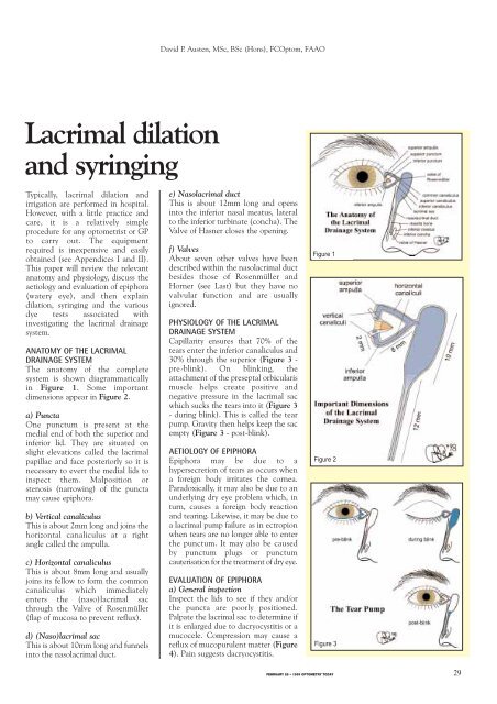
The anatomy of the complete<br />
system is shown diagrammatically<br />
in Figure 1. Some important<br />
dimensions appear in Figure 2.<br />
a) Puncta<br />
One punctum is present at the<br />
medial end of both the superior <strong>and</strong><br />
inferior lid. They are situated on<br />
slight elevations called the lacrimal<br />
papillae <strong>and</strong> face posteriorly so it is<br />
necessary to evert the medial lids to<br />
inspect them. Malposition or<br />
stenosis (narrowing) of the puncta<br />
may cause epiphora.<br />
b) Vertical canaliculus<br />
This is about 2mm long <strong>and</strong> joins the<br />
horizontal canaliculus at a right<br />
angle called the ampulla.<br />
c) Horizontal canaliculus<br />
This is about 8mm long <strong>and</strong> usually<br />
joins its fellow to form the common<br />
canaliculus which immediately<br />
enters the (naso)lacrimal sac<br />
through the Valve of Rosenmüller<br />
(flap of mucosa to prevent reflux).<br />
d) (Naso)lacrimal sac<br />
This is about 10mm long <strong>and</strong> funnels<br />
into the nasolacrimal duct.<br />
e) Nasolacrimal duct<br />
This is about 12mm long <strong>and</strong> opens<br />
into the inferior nasal meatus, lateral<br />
to the inferior turbinate (concha). The<br />
Valve of Hasner closes the opening.<br />
f) Valves<br />
About seven other valves have been<br />
described within the nasolacrimal duct<br />
besides those of Rosenmüller <strong>and</strong><br />
Horner (see Last) but they have no<br />
valvular function <strong>and</strong> are usually<br />
ignored.<br />
PHYSIOLOGY OF THE LACRIMAL<br />
DRAINAGE SYSTEM<br />
Capillarity ensures that 70% of the<br />
tears enter the inferior canaliculus <strong>and</strong><br />
30% through the superior (Figure 3 -<br />
pre-blink). On blinking, the<br />
attachment of the preseptal orbicularis<br />
muscle helps create positive <strong>and</strong><br />
negative pressure in the lacrimal sac<br />
which sucks the tears into it (Figure 3<br />
- during blink). This is called the tear<br />
pump. Gravity then helps keep the sac<br />
empty (Figure 3 - post-blink).<br />
AETIOLOGY OF EPIPHORA<br />
Epiphora may be due to a<br />
hypersecretion of tears as occurs when<br />
a foreign body irritates the cornea.<br />
Paradoxically, it may also be due to an<br />
underlying dry eye problem which, in<br />
turn, causes a foreign body reaction<br />
<strong>and</strong> tearing. Likewise, it may be due to<br />
a lacrimal pump failure as in ectropion<br />
when tears are no longer able to enter<br />
the punctum. It may also be caused<br />
by punctum plugs or punctum<br />
cauterisation for the treatment of dry eye.<br />
EVALUATION OF EPIPHORA<br />
a) General inspection<br />
Inspect the lids to see if they <strong>and</strong>/or<br />
the puncta are poorly positioned.<br />
Palpate the lacrimal sac to determine if<br />
it is enlarged due to dacryocystitis or a<br />
mucocele. Compression may cause a<br />
reflux of mucopurulent matter (Figure<br />
4). Pain suggests dacryocystitis.<br />
Figure 1<br />
Figure 2<br />
Figure 3<br />
FEBRUARY 26 • 1999 OPTOMETRY TODAY 29
<strong>Lacrimal</strong> <strong>dilation</strong> <strong>and</strong> <strong>syringing</strong><br />
b) Slit lamp examination<br />
Inspect the puncta for poor<br />
position, narrowing or blockage<br />
- pouting suggests canaliculitis.<br />
A high marginal tear strip may<br />
indicate epiphora. If fluorescein<br />
is instilled in the conjunctival<br />
sac, it should disappear within<br />
two minutes - retention<br />
suggests there is a problem with<br />
lacrimal drainage.<br />
c) Irrigation<br />
1) <strong>Lacrimal</strong> <strong>dilation</strong><br />
Several types of dilators are<br />
available, for example the<br />
double-ended stainless steel<br />
type in Figure 5 (see Appendix<br />
I) or those incorporated in<br />
punctum plug inserters. Their<br />
use may effect a cure by<br />
releasing mucous plugs or<br />
concretions. Dilation may<br />
produce temporary relief in a<br />
case of stenosis of the punctum.<br />
<strong>Lacrimal</strong> <strong>dilation</strong> is also used<br />
prior to inserting punctum<br />
plugs <strong>and</strong> <strong>syringing</strong>.<br />
Procedure<br />
1. Wash your h<strong>and</strong>s.<br />
2. Some practitioners may<br />
wish to put on surgical<br />
gloves.<br />
3. Instil a drop of anaesthetic<br />
on the inferior punctum.<br />
4. Sterilise the lacrimal<br />
dilator with a Medi-Swab.<br />
5. Insert the dilator vertically<br />
downwards up to 2mm<br />
whilst gently rotating<br />
clockwise <strong>and</strong><br />
anticlockwise (Figure 6).<br />
6. Pull the lower lid<br />
temporally to straighten<br />
the ampulla <strong>and</strong> line up<br />
the vertical <strong>and</strong> horizontal<br />
canaliculi (Figure 7).<br />
7. Rotate the dilator<br />
horizontally <strong>and</strong> insert the<br />
dilator as required.<br />
2) <strong>Lacrimal</strong> <strong>syringing</strong><br />
As well as irrigating the<br />
lacrimal system, <strong>syringing</strong> may<br />
be necessary to dislodge intracanalicular<br />
punctum plugs.<br />
Figure 4<br />
Figure 5<br />
Figure 6<br />
Figure 7<br />
Figure 8<br />
Figure 9<br />
Figure 10<br />
Procedure<br />
1. Wash your h<strong>and</strong>s.<br />
2. Some practitioners may wish to put on<br />
surgical gloves.<br />
3. Dilate the punctum <strong>and</strong> canaliculus (see<br />
under ‘<strong>Lacrimal</strong> <strong>dilation</strong>’).<br />
4. Open the sterile packets of disposable<br />
syringe <strong>and</strong> cannula <strong>and</strong> connect them<br />
together as in Figure 8.<br />
5. Remove the plunger <strong>and</strong> fill the syringe<br />
with sterile saline.<br />
6. Re-insert the plunger, <strong>and</strong> with the<br />
syringe pointing upward, squeeze out any<br />
remaining air together with some saline.<br />
7. Insert the cannula into the vertical<br />
canaliculus (Figure 9).<br />
8. Pull the lower lid temporally to straighten<br />
the ampulla <strong>and</strong> line up the horizontal<br />
canaliculus. Rotate the syringe<br />
horizontally whilst inserting until a ‘hard’<br />
or ‘soft’ stop is felt (see over), then pull<br />
back about 2mm (Figure 10).<br />
9. Press slowly <strong>and</strong> gently on the plunger.<br />
10. Ask the patient to report when they taste<br />
saline or feel it in their nose.<br />
30<br />
FEBRUARY 26 • 1999 OPTOMETRY TODAY
<strong>Lacrimal</strong> <strong>dilation</strong><br />
Figure 11<br />
Figure 12<br />
Hard stop:<br />
If the cannula touches the medial<br />
wall of the lacrimal sac <strong>and</strong> lacrimal<br />
bone, a definite end point is reached.<br />
This is a ‘hard stop’ (Figure 11) <strong>and</strong><br />
indicates that there is no complete<br />
obstruction in the canalicular<br />
system.<br />
Soft stop:<br />
If a spongy end point is felt, this is<br />
termed a ‘soft stop’ (Figure 12) <strong>and</strong><br />
indicates that the cannula has been<br />
prevented from entering the lacrimal<br />
sac. Therefore, there is a blockage in<br />
the canalicular system <strong>and</strong> there will<br />
be no distension of the lacrimal sac<br />
when the plunger is pressed.<br />
Detailed diagnosis<br />
from lacrimal <strong>syringing</strong><br />
• If saline refluxes from the inferior<br />
canaliculus, the blockage is there.<br />
• If saline refluxes from the superior<br />
canaliculus, the blockage is in the<br />
common canaliculus.<br />
• If saline passes into the nose, the<br />
problem is hypersecretion of tears or<br />
failure of the lacrimal pump or partial<br />
obstruction of the nasolacrimal system.<br />
• If saline does not reach the nose, there<br />
is a total obstruction of the<br />
nasolacrimal duct <strong>and</strong> saline may<br />
appear from the superior punctum - the<br />
saline may be purulent if infection is<br />
present - <strong>and</strong> the lacrimal sac may be<br />
distended.<br />
• An attempt may be made to close the<br />
superior punctum with a dilator or<br />
cotton bud <strong>and</strong> a further effort made to<br />
clear the obstruction.<br />
Functional obstruction<br />
Sometimes, the lacrimal drainage system<br />
may appear patent when <strong>syringing</strong> proceeds<br />
uneventfully. However, there may be a<br />
functional obstruction. This means that<br />
under the low-pressure circumstances of<br />
normal tear drainage, all or part of the<br />
lacrimal pathway may collapse. Jones dye<br />
tests may be used to distinguish between<br />
patent systems <strong>and</strong> functionally blocked<br />
ones.<br />
JONES DYE TESTS<br />
PRIMARY AND SECONDARY<br />
Procedure<br />
1. Instil one drop of fluorescein into the<br />
conjunctival sac (Figure 13).<br />
2. Put a cotton bud soaked in anaesthetic<br />
in the inferior meatus.<br />
3. If fluorescein is detected after five<br />
minutes, the system is patent (positive<br />
Primary Jones Test).<br />
4. If no fluorescein is discovered, this is a<br />
negative Primary Jones Test (Figure<br />
14) <strong>and</strong> the functional obstruction<br />
could be anywhere from the punctum<br />
to the Valve of Hasner.<br />
5. Next, wash the excess fluorescein from<br />
the conjunctival sac <strong>and</strong> syringe. If<br />
fluorescein is detected, then this shows<br />
it had entered the sac <strong>and</strong> constitutes a<br />
positive Secondary Jones Test (Figure<br />
15) <strong>and</strong> suggests a functional<br />
obstruction of the nasolacrimal duct.<br />
6. If no dye is found on the cotton bud<br />
after <strong>syringing</strong>, this is termed a negative<br />
Figure 13<br />
Figure 14<br />
Figure 15<br />
Figure 16<br />
FEBRUARY 26 • 1999 OPTOMETRY TODAY 31
<strong>Lacrimal</strong> <strong>dilation</strong><br />
Secondary Jones Test, because<br />
fluorescein had not entered the sac<br />
<strong>and</strong>, thus, there is stenosis of the<br />
puncta or canalicular system<br />
(Figure 16).<br />
7. If no saline appears in the nose,<br />
there is a complete obstruction<br />
somewhere in the lacrimal drainage<br />
system.<br />
CONCLUSION<br />
On the basis of the results obtained from<br />
the tests <strong>and</strong> procedures described above,<br />
the patient may leave with their epiphora<br />
cured. If not, at least a more informed<br />
referral may be made by describing the<br />
most likely nature <strong>and</strong> position of the<br />
obstruction.<br />
FURTHER READING<br />
1. Spalton, Hitchings, Hunter (1993) ‘Atlas of<br />
Clinical Ophthalmology’. 2nd Ed, Mosby<br />
Wolfe.<br />
2. Kanski (1994) ‘Clinical Ophthalmology’. 3rd<br />
Ed, Butterworth Heinemann.<br />
3. Casser, Fingerat, Woodcome (1997) ‘Atlas<br />
of Primary Eyecare Procedures’. 2nd Ed,<br />
Appleton & Lange.<br />
4. Schmidt (1997) ‘Lids <strong>and</strong> Nasolacrimal<br />
System’. Butterworth Heinemann.<br />
5. Last (1961) ‘Wolff’s Anatomy of the Eye<br />
<strong>and</strong> Orbit’. 5th Ed. Lewis & Co.<br />
APPENDIX I<br />
EQUIPMENT REQUIRED<br />
• <strong>Lacrimal</strong> dilator<br />
• Disposable lacrimal cannulae<br />
• 3 or 5ml disposable sterile syringes<br />
• Anaesthetic drops, e.g. Ophthaine<br />
• Tissues<br />
• Aerosol bottles of sterile saline<br />
• Disinfection for the dilators, e.g<br />
Medi-Swabs<br />
• Surgical gloves?<br />
APPENDIX II<br />
SOME EQUIPMENT SUPPLIERS<br />
John Weiss, 89-90 Alston Drive,<br />
Bradwell Abbey, Milton Keynes<br />
MK13 9HF<br />
Tel: 01908-318017 Fax: 01908-318708<br />
Castroviejo lacrimal dilator<br />
#0105040 B115<br />
<strong>Lacrimal</strong> cannulae 0108142 7276<br />
Optimed, Alveston House,<br />
11 Broad Street, Pershore<br />
Worcs, WR10 1BB<br />
Tel: 01386-561845 Fax: 01386-555177<br />
Irrigating <strong>Lacrimal</strong> Cannula<br />
26G Code No. 1276<br />
Wilders <strong>Lacrimal</strong> Dilator 13-071<br />
32<br />
FEBRUARY 26 • 1999 OPTOMETRY TODAY


