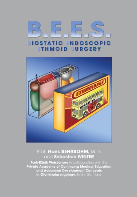
BIOSTATIC ENDOSCOPIC ETHMOID SURGERY A New Approach ...
BIOSTATIC ENDOSCOPIC ETHMOID SURGERY A New Approach ...
BIOSTATIC ENDOSCOPIC ETHMOID SURGERY A New Approach ...

You also want an ePaper? Increase the reach of your titles
YUMPU automatically turns print PDFs into web optimized ePapers that Google loves.
<strong>BIOSTATIC</strong> <strong>ENDOSCOPIC</strong><br />
<strong>ETHMOID</strong> <strong>SURGERY</strong><br />
A <strong>New</strong> <strong>Approach</strong> to Functional<br />
Endoscopic Sinus Surgery<br />
for Acute Recurrent Rhinosinusitis<br />
Prof. Hans BEHRBOHM, M.D.<br />
and Sebastian WINTER<br />
Park-Klinik Weissensee in collaboration with the<br />
Private Academy of Continuing Medical Education<br />
and Advanced Development Concepts<br />
in Otorhinolaryngology, Berlin, Germany
4<br />
Biostatic Endoscopic Ethmoid Surgery – B.E.E.S. – A <strong>New</strong> <strong>Approach</strong> to<br />
Functional Endoscopic Sinus Surgery for Acute Recurrent Rhinosinusitis<br />
Illustrations by:<br />
Katja Dalkowski, M.D.<br />
Grasweg 42, D-91054 Buckenhof,<br />
Germany<br />
E-mail: kdalkowski@online.de<br />
Andreas Mücke (bottom, p. 11)<br />
Karl-Frank-Str. 32, 12587 Berlin,<br />
Germany<br />
Please note:<br />
Medical knowledge is constantly changing. As<br />
new research and clinical experience broaden<br />
our knowledge, changes in treatment and<br />
medication may be required. The authors and<br />
editors of the material herein have consulted<br />
sources believed to be reliable in their efforts<br />
to provide information that is complete and in<br />
accordance with the standards accepted at<br />
the time of publication. However, in view of the<br />
possibility of human error by the authors,<br />
editors, or publisher of the work herein, or<br />
changes in medical knowledge, neither the<br />
authors, editors, publisher, nor any other party<br />
who has been involved in the preparation of<br />
this work, can guarantee that the information<br />
contained herein is in every respect accurate<br />
or complete, and they cannot be held responsible<br />
for any errors or omissions or for the<br />
results obtained from use of such information.<br />
The information contained within this brochure<br />
is intended for use by doctors and other health<br />
care professionals. This material is not intended<br />
for use as a basis for treatment decisions, and<br />
is not a substitute for professional consultation<br />
and/or use of peer-reviewed medical literature.<br />
Some of the product names, patents, and<br />
registered designs referred to in this booklet<br />
are in fact registered trademarks or proprietary<br />
names even though specific reference to this<br />
fact is not always made in the text. Therefore,<br />
the appearance of a name without designation<br />
as proprietary is not to be construed as a<br />
representation by the publisher that it is in the<br />
public domain.<br />
Biostatic Endoscopic Ethmoid Surgery – B.E.E.S. – A <strong>New</strong> <strong>Approach</strong> to<br />
Functional Endoscopic Sinus Surgery for Acute Recurrent Rhinosinusitis<br />
Prof. Hans BEHRBOHM, M.D.<br />
and Sebastian WINTER<br />
Park-Klinik Weissensee in collaboration with the Private Academy of Continuing<br />
Medical Education and Advanced Development Concepts in<br />
Otorhinolaryngology, Berlin, Germany<br />
Correspondence:<br />
Prof. Dr. med. Hans BEHRBOHM<br />
Kurfürstendamm 61, 10707 Berlin<br />
Phone: +49 (0)30/96283852<br />
E-mail: behrbohm@ku61.de<br />
behrbohm@park-klinik.com<br />
Website: www.ku61.de<br />
© 2009 Published by , Tuttlingen<br />
ISBN 978-3-89756-162-5, Printed in Germany<br />
P.O. Box, D-78503 Tuttlingen<br />
Phone: +49 74 61/1 45 90<br />
Fax: +49 74 61/708-529<br />
E-mail: Endopress@t-online.de<br />
Editions in languages other than English and German are in preparation.<br />
For up-to-date information, please contact<br />
publisher Tuttlingen,<br />
Germany, at the address shown above.<br />
Layout and Lithography:<br />
Verlag<br />
, Tuttlingen<br />
Printed by:<br />
Straub Druck + Medien AG<br />
D-78713 Schramberg, Germany<br />
04.09-2<br />
All rights reserved.<br />
No part of this publication may be translated, reprinted or reproduced, transmitted in any<br />
form or by any means, electronic or mechanical, now known or hereafter invented,<br />
including photocopying and recording, or utilized in any information storage or retrieval<br />
system without the prior written permission of the copyright holder.
Biostatic Endoscopic Ethmoid Surgery – B.E.E.S. – A <strong>New</strong> <strong>Approach</strong> to<br />
Functional Endoscopic Sinus Surgery for Acute Recurrent Rhinosinusitis<br />
5<br />
Contents<br />
Introduction . . . . . . . . . . . . . . . . . . . . . . . . . . . . . . . . . . . . . . . . . . . . . . . . . 6<br />
Historical Considerations . . . . . . . . . . . . . . . . . . . . . . . . . . . . . . . . . . 6<br />
Statement of Problem . . . . . . . . . . . . . . . . . . . . . . . . . . . . . . . . . . . . . 7<br />
Hypothesis . . . . . . . . . . . . . . . . . . . . . . . . . . . . . . . . . . . . . . . . . . . . . . 12<br />
Surgical Concept . . . . . . . . . . . . . . . . . . . . . . . . . . . . . . . . . . . . . . . . . . . . 14<br />
Ethmoid Infundibulotomy . . . . . . . . . . . . . . . . . . . . . . . . . . . . . . . . . . 14<br />
Bullotomy . . . . . . . . . . . . . . . . . . . . . . . . . . . . . . . . . . . . . . . . . . . . . . . 14<br />
Enlarging the Frontal Recess . . . . . . . . . . . . . . . . . . . . . . . . . . . . . . . 15<br />
Summary . . . . . . . . . . . . . . . . . . . . . . . . . . . . . . . . . . . . . . . . . . . . . . . . . . . 16<br />
Illustration of the B.E.E.S. Philosophy<br />
Main Therapeutic Options in B.E.E.S. . . . . . . . . . . . . . . . . . . . . . . . . 17<br />
Illustrative Clinical Cases . . . . . . . . . . . . . . . . . . . . . . . . . . . . . . . . . . . . . 18<br />
Catheter-Based Balloon Dilatation of the Sinus Ostia . . . . . . . . . . . . . . 19<br />
Hybrid Operations . . . . . . . . . . . . . . . . . . . . . . . . . . . . . . . . . . . . . . . . 19<br />
References . . . . . . . . . . . . . . . . . . . . . . . . . . . . . . . . . . . . . . . . . . . . . . . . . 20<br />
Recommended Instrument Set for<br />
Biostatic Endoscopic Ethmoid Surgery . . . . . . . . . . . . . . . . . . . . . . . . . . 22
6<br />
Biostatic Endoscopic Ethmoid Surgery – B.E.E.S. – A <strong>New</strong> <strong>Approach</strong> to<br />
Functional Endoscopic Sinus Surgery for Acute Recurrent Rhinosinusitis<br />
Emil Zuckerkandl (1848—1910)<br />
(Photo: Prof. Daniel Simmen, Zurich,<br />
Switzerland)<br />
Introduction<br />
Historical Considerations<br />
After initially pursuing a career as a virtuoso violinist, Emil Zuckerkandl<br />
(1849–1910) became an extraordinary professor of anatomy at 31 years of<br />
age, without having to present the customary postdoctoral credentials. In<br />
1882 Zuckerkandel became a full professor in Graz, Austria, and six years<br />
later he assumed the first anatomy chair in Vienna. The poet Arthur Schnitzler<br />
studied medicine in Vienna and shared a vivid memory of Zuckerkandl in his<br />
autobiography, Youth in Vienna, describing him as “… a pale young man<br />
with a dark goatee and black eyes. In his academic robes he closely re -<br />
sembled one of those anatomists familiar to us from the famous Rembrandt<br />
portraits, while the legendary stories of his rakish youth, filled with drinking<br />
and fencing, seemed to hover about him.” He also had a reputation for “…<br />
going straight to work from some tavern or perhaps even from the arms of a<br />
beautiful woman and launching directly into his daily routine, teaching and<br />
studying with prodigious energy far into the night.”<br />
This intense drive for scientific knowledge surely formed the basis for his<br />
pamphlet on Normal and Pathologic Anatomy of the Nasal Cavity and its<br />
Pneumatic Appendages, published in 1882. In this work, Zuckerkandl was<br />
the first author to give a detailed anatomical description of the ethmoid<br />
bone and all the paranasal sinuses, thus creating a scientific basis for<br />
understanding their anatomy. He also drew attention to specific structures<br />
and narrow passageways that contribute to the pathogenesis of rhino -<br />
sinusitis and are still relevant today – such as the ethmoid infundibulum and<br />
variants in the pneumatization and curvature of the middle nasal turbinate.<br />
He described in detail the cellular anatomy of the ethmoid labyrinth, noting<br />
that “…the ethmoid bulla is highly variable in its development, and its<br />
importance rests not only on its relationship to the middle turbinate. An<br />
ethmoid cell belonging to the lower portion of the labyrinth, the ethmoid<br />
bulla presents a convex medial surface to the nasal cavity and is bounded<br />
laterally by the lamina papyracea of the ethmoid bone or may be separated<br />
from it by another, intervening ethmoid cell…”<br />
Zuckerkandl’s writings on the variability of the middle turbinate included the<br />
following: “The variations relate both to the shape and size of the [middle]<br />
turbinate. The turbinate may be so markedly curved that it occludes the<br />
olfactory groove and engages against the nasal septum. The transformation<br />
of the anterior end of the turbinate into a large bony bulla is a common<br />
occurrence and was even described in the past century by Giovanni<br />
Santorini in his ‘Observationes anatomicae’. In cases of this kind, the<br />
turbinate contains a cavity, at times even subdivided by a septum, that<br />
communicates openly with the middle meatus.”<br />
ZUCKERKANDL E: Normal and Pathologic<br />
Anatomy of the Nasal Cavity and its<br />
Pneumatic Appendages. Vol. 2, Vienna.<br />
Wilhelm Braumüller (1892, 1893).
Biostatic Endoscopic Ethmoid Surgery – B.E.E.S. – A <strong>New</strong> <strong>Approach</strong> to<br />
Functional Endoscopic Sinus Surgery for Acute Recurrent Rhinosinusitis<br />
7<br />
In his groundbreaking work on “The Role of the Lateral Nasal Wall in the<br />
Pathogenesis, Diagnosis and Treatment of Recurrent and Chronic Rhino -<br />
sinusitis” (1987), Walter Messerklinger (1920–2001) begins by reviewing the<br />
anatomical discoveries of Zuckerkandl, Hajek, Grünwald, Peter, Killian, and<br />
Flottes on the cellular structures of the lateral nasal wall before addressing<br />
their importance in the pathogenesis of rhinogenic sinusitis. At the International<br />
Conference on Sinus Disease, Terminology, Staging and Therapy held<br />
in Princeton, <strong>New</strong> Jersey, in 1993, Stammberger et al. (1997) presented a<br />
paper titled “Anatomical Terminology and Nomenclature of Paranasal Sinus<br />
Surgery” in which they created a uniform, consistent nomenclature for the<br />
anatomy of the ethmoid bone.<br />
While discoveries on the anatomy of specific cellular structures in the<br />
ethmoid bone gave rise to important pathogenetic considerations, an<br />
endoscopic diagnostic concept, and a subsequent approach to functional<br />
endoscopic sinus surgery (F.E.S.S.), to date there have been no fundament -<br />
al studies on the stabilizing function of the principal ethmoid cells.<br />
Prof. Stammberger (2002) has repeatedly emphasized the importance of an<br />
atraumatic surgical technique, particularly in endoscopic surgery of the<br />
frontal sinuses, noting that “…an aggressive technique with the radical<br />
removal of mucosa from these small ethmoid passageways will quickly lead<br />
to scarring and stenosis. Not infrequently, traumatic surgical manipulations<br />
in this region will promote the development of frontal sinus symptoms that<br />
did not exist prior to surgery…”<br />
Walter Messerklinger (1920—2001),<br />
Chairman of the Department of Otorhinolaryngology,<br />
Graz University Hospital<br />
(1959—1990), seen at a professional gala<br />
in Vienna in 1990.<br />
Statement of Problem<br />
Lack of awareness of the biostatic importance of some important cellular<br />
structures of the ethmoid bone, such as the ethmoid bulla and middle<br />
turbinate, have led to careless handling of these structures during sinus<br />
surgery. The practice of F.E.S.S. at many centers has devolved into a<br />
“monomaniacal” clearing of ethmoid cells without taking into account the<br />
specific anatomical problems in the individual case and often without actually<br />
meeting the requirements of minimal invasiveness. The extent of<br />
ethmoid resection is often described in terms of an anterior or posterior<br />
ethmoidectomy, and endoscopes are not always used during the ethmoid<br />
dissection.<br />
Prof. Messerklinger (second from right) with<br />
colleagues in Vienna in 1990.<br />
Coronal CT scan documents high-grade<br />
stenosis of the exenterated ethmoid<br />
labyrinth 2 years after surgery for recurrent<br />
frontal sinusitis.<br />
Lateralization of the middle turbinate is a<br />
frequent consequence of ethmoidectomy.<br />
Appearance following medialization and<br />
trimming of the middle turbinate.
8<br />
Biostatic Endoscopic Ethmoid Surgery – B.E.E.S. – A <strong>New</strong> <strong>Approach</strong> to<br />
Functional Endoscopic Sinus Surgery for Acute Recurrent Rhinosinusitis<br />
The results: Occasional postoperative CT scans obtained for<br />
various indications (they are not routinely necessary) have shown<br />
the following typical problems:<br />
Significant scarring and contraction may occur after a complete<br />
ethmoidectomy.<br />
These effects can obstruct surgically created drainage routes from<br />
the frontal and maxillary sinuses.<br />
The middle turbinate is very susceptible to postoperative laterali<br />
zation.<br />
Mucosal lesions are associated with a high risk of synechia formation<br />
and ethmoid atelectasis.<br />
The ethmoid “matchbox.” The principal<br />
supporting structures of the anterior ethmoid<br />
are the ethmoid bulla (red) and the<br />
middle turbinate with its basal lamina,<br />
which separates the anterior and posterior<br />
ethmoid cells and stabilizes the width of<br />
the ethmoid bone.<br />
Other structures:<br />
brown = Agger nasi cell<br />
green<br />
= Uncinate process<br />
blue<br />
= Retrobullar cell<br />
gray and yellow = Posterior ethmoid cells<br />
shading = Ethmoid infundibulum<br />
and frontal recess.<br />
The most important postulate of F.E.S.S. in the treatment of patients with<br />
recurrent rhinosinusitis is the improvement of ventilation and drainage,<br />
which is essential for mucosal regeneration. This can be accomplished only<br />
by taking into account biostatic principles, which must be applied in order<br />
to achieve the main goal: adequate and permanent postoperative patency<br />
of the ethmoid sinuses.<br />
Establishing drainage from the frontal sinuses will be ineffective if post -<br />
operative contraction of the ethmoid causes lateralization of the middle<br />
turbinate and possible synechia formation, leading to stenosis of the<br />
drainage pathway.<br />
Biostatic principles<br />
In simplified terms, the ethmoid bone has the approximate size and<br />
shape of a matchbox stood on edge. The width of the “matchbox”<br />
is variable and depends on the degree of ethmoid pneumatization<br />
and on the variable symmetry of the ethmoid cells.<br />
The supporting structure of the ethmoid bone, which forms cavities<br />
to maintain ventilation and drainage of the maxillary and frontal<br />
sinuses, shows varying patterns of pneumatization.
Biostatic Endoscopic Ethmoid Surgery – B.E.E.S. – A <strong>New</strong> <strong>Approach</strong> to<br />
Functional Endoscopic Sinus Surgery for Acute Recurrent Rhinosinusitis<br />
9<br />
The Main Pneumatization Patterns of the Ethmoid Bone<br />
Interlamellar cells. Concha bullosa. Large ethmoid bulla.<br />
The ethmoid bone has a variable width. A complementary relationship<br />
exists between the ethmoid bulla and a possible concha bullosa in the<br />
middle turbinate. Both structures should be viewed as a functional complex<br />
whose complete removal leads to atelectasis of the ethmoid bone.<br />
• From an architectural standpoint, the ethmoid bone can be described<br />
as a suspended lightweight construction. While the massive<br />
archworks of the inner and outer tables of the skull maintain patency<br />
of the frontal infundibulum, this function is performed in the ethmoid<br />
bone by a combination of domes and horizontal struts. The struts<br />
and pillars never terminate blindly, but maintain the space and<br />
distance between the orbits on the one hand and between the skull<br />
base, vomer, and ultimately the maxilla on the other.<br />
Bony framework of the frontal infundibulum.<br />
Crista galli<br />
Perpendicular plate<br />
of ethmoid bone<br />
Anterior ethmoid cells<br />
The ethmoid bone is a suspended,<br />
lightweight frame structure that forms the<br />
ethmoid sinuses and gives central support<br />
to the midfacial region. It has a T-shaped<br />
configuration in which the vertical member<br />
is formed by the perpendicular plate of the<br />
ethmoid bone and the horizontal member is<br />
formed by the paired ethmoid labyrinths.
10<br />
Biostatic Endoscopic Ethmoid Surgery – B.E.E.S. – A <strong>New</strong> <strong>Approach</strong> to<br />
Functional Endoscopic Sinus Surgery for Acute Recurrent Rhinosinusitis<br />
We can explain the biostatic function of the ethmoid labyrinth by looking<br />
at two clinically important structures: the ethmoid bulla and the middle<br />
turbinate.<br />
Ethmoid bulla<br />
The ethmoid bone and voice<br />
The fine trabecular structure of<br />
the ethmoid bone is an important<br />
factor in the production and<br />
self-perception of vocal resonance.<br />
Voice modulation also<br />
relies upon the vibrating and<br />
damped “strings” of the ethmoid<br />
sinuses. B.E.E.S. can lead to an<br />
improvement in dynamic vocal<br />
range and voice resonance.<br />
The ethmoid bulla arises by pneumatization from the basal lamina of<br />
the second ethmoturbinate and is usually the largest of the anterior<br />
ethmoid cells. The bulla lamella forms the posterior wall of the frontal<br />
recess when it attaches to the skull base; otherwise a suprabullar<br />
recess is formed. When strongly pneumatized, the bulla may narrow<br />
the frontal recess to a ductlike structure that has contributed to<br />
the now-obsolete term “nasofrontal duct.”<br />
The middle turbinate is part of the ethmoid bone and may be pneuma<br />
tized in any of its parts, such as the head and neck. When the<br />
head of the middle turbinate is pneumatized, it forms the concha<br />
bullosa. The concha bullosa is an anatomical variant and generally<br />
does not have pathogenic significance. Interlamellar cells are a<br />
special variant caused by pneumatization of the vertical lamella of<br />
the middle turbinate. The middle turbinate is an important aero -<br />
dynamic structure for ventilating the olfactory groove and paranasal<br />
sinuses. It has three points of attachment: the anterior neck of the<br />
turbinate attaches to the lateral edge of the cribriform plate, the<br />
central part attaches to the lamina papyracea, and the posterior<br />
part inserts laterally at the level of the pterygoid process. The basal<br />
lamina of the middle turbinate is of special importance for several<br />
reasons. It forms a kind of “watershed” for the drainage of secre -<br />
tions from the anterior and posterior ethmoid, and it is an important<br />
horizontal supporting structure that contributes to the formation of<br />
the sinus cavities. By stabilizing the head of the turbinate, the basal<br />
lamina helps to maintain the patency of the ethmoid sinuses. Destabilization<br />
of the basal lamina leads to “kinking” and lateralization of<br />
the turbinate head, deflects airflow away from the olfactory groove,<br />
and causes loss of ethmoid volume.<br />
The ethmoid bulla is perched on the medial orbital wall like an architectural dome.<br />
A complete anterior ethmoidectomy promotes medialization of the lamina papyracea and<br />
lateralization of the middle turbinate. This tendency can be counteracted by preserving<br />
the lamella or upper cap of the ethmoid bulla.
Biostatic Endoscopic Ethmoid Surgery – B.E.E.S. – A <strong>New</strong> <strong>Approach</strong> to<br />
Functional Endoscopic Sinus Surgery for Acute Recurrent Rhinosinusitis<br />
11<br />
Removing the parietal mucosa from the paranasal sinuses can cause<br />
severe scarring and luminal obliteration. The Caldwell-Luc radical antro -<br />
s tomy, for example, could result in almost complete obliteration of the<br />
maxillary sinus with maxillary deformity and orbital floor depression due<br />
to typical wound healing effects in the concavities of the paranasal<br />
sinuses. Wigand (1977, 1981, 1989) made repeated references to these<br />
phenomena, which result from severe centripetal shrinkage and luminal<br />
narrowing due to appositional bone growth and scar formation. Essen -<br />
tially the same processes must take place in the ethmoid sinuses when<br />
the parietal mucosa is removed along with all internal supporting cellular<br />
structures.<br />
The ethmoid bone and olfaction<br />
The alignment of the middle<br />
turbinate is crucial for ventilation<br />
of the olfactory groove. The turbinate<br />
has the shape of an air -<br />
foil suspended between three<br />
points of attachment. For effective<br />
ventilation of the olfactory<br />
groove, a positive pressure must<br />
develop along the lateral lamella<br />
of the middle turbinate and a<br />
negative pressure along the<br />
medial lamella. The negative<br />
pressure on the “lee” side of the<br />
middle turbinate draws the<br />
inspired air up into the olfactory<br />
fossa.<br />
Wound healing and contraction effects following removal of the parietal mucosa from the<br />
ethmoid and maxillary sinuses. The effects include high-grade stenoses and lateralization<br />
of the middle turbinate and uncinate process.<br />
The peripheral human olfactory organ.<br />
fo – olfactory fossa, cg – crista galli, be – ethmoid bulla, cm – middle turbinate,<br />
o – orbit, se – ethmoid sinus, bo – olfactory bulb, fol – olfactory fibers,<br />
ro – olfactory groove with olfactory epithelium, sn – nasal septum.<br />
The principles of fluid dynamics underlying<br />
nasal airflow and olfactory perception.
12<br />
Biostatic Endoscopic Ethmoid Surgery – B.E.E.S. – A <strong>New</strong> <strong>Approach</strong> to<br />
Functional Endoscopic Sinus Surgery for Acute Recurrent Rhinosinusitis<br />
Hypothesis<br />
Today, endoscopic surgery of the paranasal sinuses has assumed a<br />
broad range of indications. Based on our own experience, we feel that<br />
strict distinctions should be drawn among the subsets of indications<br />
described below in order to fully utilize the potential of differentiated<br />
microsurgery and prevent postoperative stenosis.<br />
Coronal CT scan of a woman who underwent<br />
a previous complete right ethmoidectomy and<br />
B.E.E.S. in which the upper cap of the<br />
ethmoid bulla was preserved.<br />
Medialized middle turbinates. The bulla<br />
lamellae have been preserved on both<br />
sides.<br />
Acute recurrent rhinosinusitis is marked by inflammatory exacerbations,<br />
typically involving the frontal and maxillary sinuses. Complete<br />
anterior ethmoidectomy should never be considered a routine<br />
procedure. If exenteration of the anterior ethmoid is required, the<br />
surgeon should make every effort to preserve the lamella or the<br />
upper cap of the ethmoid bulla in order to prevent narrowing of the<br />
anterior ethmoid and frontal recess. The parietal mucosa of the<br />
lamina papyracea, anterior skull base, and middle turbinate should<br />
be preserved. Enlarging the frontal recess in a type I to IIb drainage<br />
procedure is easily accomplished under vision with a 45° endo -<br />
scope while preserving the bulla lamella. Infundibular and agger<br />
nasi cells that obstruct the frontal recess can be removed by using<br />
the “uncapping the egg” technique of Stammberger, which involves<br />
a posterior-to-anterior dissection. A maximum amount of mucosa<br />
should be preserved in the frontal recess. The basal lamina of the<br />
middle turbinate may be trephined over a circumscribed area, but it<br />
should not be fractured.<br />
Chronic rhinosinusitis with nasal polyps or sinonasal polyposis<br />
currently represents a large subset of indications for endoscopic<br />
sinus surgery. Both biomechanical and immunologic factors contribute<br />
significantly to the pathogenesis of chronic rhinosinusitis.<br />
Based on regional variations in the texture of the ethmoid sinus<br />
mucosa, such as an abundance of glands on the anterior surface of<br />
the bulla, sites of predilection exist for the development of polyps.<br />
These polyps destroy the ethmoid cells and their infrastructure. Due<br />
to pressure effects from the polyps and the absence of scar<br />
contractures with an intact parietal mucosa, sinus contraction does<br />
not occur. Typically, moreover, these patients rarely complain of<br />
headaches. Cell remnants that have already been destroyed should<br />
be removed at operation, and the parietal mucosa on the lamina<br />
papyracea, skull base and middle turbinate, for example, should be<br />
preserved.<br />
Coronal CT one year after bilateral medialization<br />
of the middle turbinates.<br />
Principle of enlarging the frontal recess,<br />
illustrated here in a patient with infundibular<br />
or agger nasi cells.<br />
The basal lamina (green) of the ethmoid<br />
bulla is preserved. The anterior ethmoidal<br />
artery usually runs approximately 2 mm<br />
behind the bulla lamella.
Biostatic Endoscopic Ethmoid Surgery – B.E.E.S. – A <strong>New</strong> <strong>Approach</strong> to<br />
Functional Endoscopic Sinus Surgery for Acute Recurrent Rhinosinusitis<br />
13<br />
b<br />
c<br />
a<br />
Legend<br />
a A “nasal polyp” may arise from the mucosa<br />
on the anterior wall of the ethmoid bulla.<br />
b Nasal polyps most commonly originate<br />
in the ethmoid sinuses and descend from<br />
there into the nasal cavity. They destroy<br />
the infrastructure of the ethmoid sinus<br />
while keeping the sinus open by their<br />
mass effect.<br />
c Polyps combined with viscous mucus<br />
are a sign of eosinophil-associated<br />
disease (usually asthma with analgesic<br />
intolerance).<br />
d Scarred, indurated nasal polyps following<br />
numerous operations.<br />
e Treatment with modern topical steroids<br />
can shrink nasal polyps to this approximate<br />
level.<br />
f Ethmoid polyps protruding into the<br />
ethmoid infundibulum.<br />
g Nasal polyp protruding from an ethmoid<br />
cell.<br />
h Typical appearance of an inverted<br />
papilloma arising from the head of the<br />
middle turbinate.<br />
d<br />
f<br />
e<br />
g<br />
h
14<br />
Biostatic Endoscopic Ethmoid Surgery – B.E.E.S. – A <strong>New</strong> <strong>Approach</strong> to<br />
Functional Endoscopic Sinus Surgery for Acute Recurrent Rhinosinusitis<br />
Surgical Concept<br />
Ethmoid Infundibulotomy<br />
The standard procedure for opening the ethmoid bone is an ethmoid<br />
infundibulotomy that includes complete and atraumatic removal of the<br />
uncinate process.<br />
The uncinate process is sharply released at its poles with endoscopic<br />
scissors. The lateral infundibular wall is removed, exposing the anterior<br />
wall of the ethmoid bulla.<br />
Ethmoid infundibulotomy:<br />
1 Medial infundibular wall,<br />
2 Sickle knife, 3 Ethmoid bulla,<br />
4 Semilunar hiatus, 5 Nasolacrimal canal,<br />
6 Middle turbinate, 7 Nasal septum,<br />
8 Agger nasi.<br />
Bullotomy<br />
The anterior wall of the ethmoid bulla is trephined with a special instrument,<br />
the bullotome, or with a straight blunt suction tip or sphenoid sinus<br />
punch (small), for example. The portions of the anterior wall or bulla that<br />
are removed will depend on the mucosal pathology.<br />
a<br />
b<br />
Trephination of the anterior wall of the<br />
ethmoid bulla is done with the bullotome<br />
(a) or a small, circular sphenoid sinus<br />
punch (b).<br />
1 Nasal septum,<br />
2 Middle turbinate,<br />
3 Circular punch,<br />
4 Ethmoid bulla,<br />
5 Frontal recess,<br />
6 Lateral nasal wall.<br />
Bullotomy with the circular punch.
Biostatic Endoscopic Ethmoid Surgery – B.E.E.S. – A <strong>New</strong> <strong>Approach</strong> to<br />
Functional Endoscopic Sinus Surgery for Acute Recurrent Rhinosinusitis<br />
15<br />
View of the skull base following bullotomy.<br />
1 Nasal septum<br />
2 Middle turbinate<br />
3 Ethmoid bulla<br />
4 Anterior ethmoidal artery<br />
5 Posterior lamella of ethmoid bulla<br />
6 Basal lamina of middle turbinate<br />
7 Frontal recess<br />
8 Terminal recess.<br />
The frontal recess is enlarged with a curette.<br />
Enlarging the Frontal Recess<br />
The frontal recess is located by following the lamella or upper cap of the<br />
ethmoid bulla. It is enlarged with the curved dissector (see instrument set<br />
in Appendix, pp. 22) or Kuhn-Bolger curette by removing infundibular or<br />
agger nasi cells.
16<br />
Biostatic Endoscopic Ethmoid Surgery – B.E.E.S. – A <strong>New</strong> <strong>Approach</strong> to<br />
Functional Endoscopic Sinus Surgery for Acute Recurrent Rhinosinusitis<br />
The floor of the frontal sinus is removed with a punch.<br />
The frontal sinus can also be opened more widely with the frontal sinus<br />
punch by removing portions of the frontal sinus floor, depending on the<br />
intended goal of the operation.<br />
Summary<br />
For endoscopic sinus surgery to be effective, the surgeon must first<br />
formulate an operative plan that is based on the individual history and a<br />
CT analysis of internal ethmoid anatomy and which takes into account<br />
the stabilizing function of the principal ethmoid structures. Surgery that is<br />
oriented entirely toward sinus exenteration cannot address the problems<br />
of chronic recurrent rhinosinusitis and may even cause further airflow<br />
compromise. Biometric data are available on this issue; they are currently<br />
undergoing mathematical analysis and will be published in the near<br />
future.
Biostatic Endoscopic Ethmoid Surgery – B.E.E.S. – A <strong>New</strong> <strong>Approach</strong> to<br />
Functional Endoscopic Sinus Surgery for Acute Recurrent Rhinosinusitis<br />
17<br />
Annotations on the<br />
B.E.E.S. Philosophy<br />
Main Therapeutic Options in B.E.E.S.<br />
a<br />
b<br />
c<br />
d<br />
e<br />
f<br />
The individual biostatics of the ethmoid bone should be analyzed by the physician, as<br />
they form the starting point for planning the operation. The coronal CT scans shown here<br />
illustrate various therapeutic options that are consistent with the biostatic philosophy of<br />
ethmoid surgery:<br />
a) Acute recurrent rhinosinusitis with inflammatory exacerbations involving the frontal and<br />
maxillary sinuses on both sides. The patient does not have nasal airway obstruction.<br />
b) Bilateral infundibulotomy.<br />
c) Bilateral infundibulotomy and partial turbinectomy.<br />
d) Bilateral infundibulotomy and anterior ethmoidectomy.<br />
e) Bilateral infundibulotomy, anterior ethmoidectomy, and partial turbinectomy.<br />
This variant predisposes to the situation in f.<br />
f) Shrinkage of the exenterated ethmoid has led to medialization of the orbit and<br />
lateralization of the middle turbinate on the right side with synechia formation and<br />
ethmoid atelectasis on the left side.
18<br />
Biostatic Endoscopic Ethmoid Surgery – B.E.E.S. – A <strong>New</strong> <strong>Approach</strong> to<br />
Functional Endoscopic Sinus Surgery for Acute Recurrent Rhinosinusitis<br />
1a<br />
2a<br />
2b<br />
Preoperative coronal CT scan.<br />
Coronal CT scan 1 year after surgery.<br />
1b<br />
Coronal CT scans, preoperative (a) and<br />
1 year postoperative (b).<br />
3a<br />
3b<br />
Coronal CT scans, preoperative (a) and<br />
1 year postoperative (b).<br />
Clinical Cases<br />
Woman 20 years of age with nasal airway obstruction and acute<br />
recurrent bilateral rhinosinusitis predominantly affecting the maxillary<br />
sinuses.<br />
Preoperative CT (Fig. 1a) shows a deviated septum with spurring<br />
toward the left side and large conchae bullosae on both sides. Slight<br />
mucosal swelling is noted in the anterior ethmoid.<br />
Operation: Surgery consists of partial bilateral middle turbinectomy,<br />
preserving portions of the upper lateral curve and upper lamina. It<br />
includes a bilateral infundibulotomy, bullotomy, supraturbinate antros -<br />
tomy, and submucous septoplasty.<br />
Postoperative CT, one year later (Fig. 1b) shows the lateral partial bilateral<br />
turbinectomy preserving the turbinate trim, position, and sites of<br />
attachment (see p. 16, removal of the frontal sinus floor with a punch).<br />
Woman 53 years of age with nasal airway obstruction and recurrent<br />
bouts of acute bilateral maxillary and frontal sinusitis.<br />
Preoperative CT (Fig. 2a) shows a septal spur on the left side, bilateral<br />
hyperplasia of the inferior turbinates, and minimal mucosal swelling in<br />
the anterior ethmoid.<br />
Operation: Surgery consists of an infundibulotomy, bullotomy, and<br />
supraturbinate antrostomy on both sides. The frontal recess is<br />
enlarged anteriorly to the bulla lamella, which is preserved. The procedure<br />
also includes a septoplasty and bilateral strip turbinectomy.<br />
Postoperative CT, one year later (Fig. 2b) documents broad, bila teral<br />
supraturbinate antrostomies with medialization of the nasal septum.<br />
The upper portion of the turbinate-bulla complex has been preserved,<br />
and the middle turbinate has been stabilized in an optimum position.<br />
Principle of stabilizing the turbinate position<br />
Postoperative CT scan (b) illustrates the principle of stabilizing the<br />
turbinate position by preserving the lamella or upper cap of the<br />
ethmoid bulla as an essential maneuver for “trimming the turbinate.”
Biostatic Endoscopic Ethmoid Surgery – B.E.E.S. – A <strong>New</strong> <strong>Approach</strong> to<br />
Functional Endoscopic Sinus Surgery for Acute Recurrent Rhinosinusitis<br />
19<br />
The guide catheter is positioned under endoscopic control.<br />
Catheter-Based Balloon Dilatation<br />
of the Sinus Ostia<br />
The technique of balloon sinuplasty originated in the U.S. It applies a<br />
proven tool for coronary vascular dilatation, first used in 1977, to the<br />
restoration of sinus drainage.<br />
In balloon sinuplasty, a guide catheter is advanced endoscopically to the<br />
stenotic ostium under visualization with a HOPKINS ® 0° telescope (4 mm<br />
diameter, length 18 cm). A flexible guidewire is then passed through the<br />
catheter into the sinus, and the balloon catheter is introduced over the<br />
guidewire. Its position can be checked fluoroscopically by two<br />
radiopaque dots at the ends of the balloon. Then the balloon is gradually<br />
inflated under manometric control to dilate the ostium.<br />
Hybrid Operations<br />
Hybrid operations are procedures in which endoscopic microsurgery of<br />
the paranasal sinuses is combined with balloon dilatation in one sitting.<br />
We have had good results with a combination of biostatic ethmoid<br />
surgery (B.E.E.S.) and balloon dilatation for the following indications.<br />
The guide catheter has been introduced<br />
into the right frontal sinus.<br />
The balloon catheter is positioned at the<br />
level of the stenotic right frontal recess.<br />
Indications for hybrid operations<br />
Acute recurrent inflammations<br />
Barosinusitis<br />
Empyema<br />
Coagulation disorders and other systemic diseases that would<br />
contraindicate other types of surgery)<br />
Restenosis<br />
Situations that warrant a combination of B.E.E.S. and balloon<br />
dilatation<br />
The balloon is inflated to dilate the frontal<br />
recess.
20<br />
Biostatic Endoscopic Ethmoid Surgery – B.E.E.S. – A <strong>New</strong> <strong>Approach</strong> to<br />
Functional Endoscopic Sinus Surgery for Acute Recurrent Rhinosinusitis<br />
References:<br />
1. BEHRBOHM, H., KASCHKE, O., NAWKA, T.: Endoskopische Diagnostik<br />
und Therapie in der HNO. Stuttgart, Gustav Fischer, 184 S.,1997<br />
2. BEHRBOHM, H., SYDOW, K., HÄRTIG, W.: Experimentelle Unter -<br />
suchungen zur Physiologie der der Nasennebenhöhlen. HNO. 39.<br />
168 – 172, 1991<br />
3. BEHRBOHM, H., TARDY Jr., M.E.: Funktionell-ästhetische Chirurgie<br />
der Nase. Stuttgart · <strong>New</strong> York, Georg Thieme, 244 S, 2003<br />
4. BREMER, B.: Der Einfluss endoskopischer Nasennebenhöhlenoperationen<br />
auf den Stimmklang bei Patienten mit Sing- und Sprechberufen.<br />
Dissertation. Humboldt-Universität, Berlin 2006.<br />
5. LANG, J.: Klinische Anatomie der Nase, Nasenhöhle und Nebenhöhlen.<br />
Aktuelle Oto-Rhino-Laryngologie, Band 11, Stuttgart · <strong>New</strong><br />
York, Georg Thieme, 136 S., 1988<br />
6. MESSERKLINGER, W.: Die normalen Sekretwege in der Nase des<br />
Menschen. Arch Klin Exp Ohren Nasen Kehlkopfheilkd. 195,<br />
138 – 151, 1969<br />
7. MESSERKLINGER, W.: Die Rolle der lateralen Nasenwand in der<br />
Pathogenese, Diagnose und Therapie der rezidivierenden und chronischen<br />
Rhinosinusitis. Laryng. Rhinol. Otol. 66, 293 – 299, 1987<br />
8. SCHNITZLER, A:. Jugend in Wien. Eine Autobiografie. Herausgegeben<br />
von Therese Nickl und Heinrich Schnitzler. Wien-München-Zürich,<br />
Fritz Molden, 1968<br />
9. SCHULTE, M.: Berta Zuckerkandl – Saloniere, Journalistin, Geheim -<br />
diplomatin. Zürich, Atrium-Verlag, 248 S., 2006<br />
10.STAMMBERGER, H., HOSEMANN, W., DRAF, W.: Anatomische Terminologie<br />
und Nomenklatur für die Nasennebenhöhlenchirurgie. Laryngo.<br />
Rhinol. Otol. 76, 435 – 449, 1997<br />
11. STAMMBERGER, H.: F.E.S.S. „Uncapping the Egg“ – der endoskopische<br />
Weg zur Stirnhöhle. Eine Operationstechnik der Grazer Schule.<br />
Tuttlingen, Endo-Press TM , 39 S., 2002<br />
12. STAMMBERGER, H.: Unsere endoskopische Operationstechnik der<br />
lateralen Nasenwand – ein endoskopisch-chirurgisches Konzept zur<br />
Behandlung entzündlicher Nasennebenhöhlenerkrankungen. Laryng.<br />
Rhinol. Otol. 64, 559 – 566, 1985<br />
13.WIGAND, W.E.: Endonasale Kieferhöhlenoperationen mit endosko -<br />
pischer Kontrolle. Laryng. Rhinol. Otol. 56, 421, 1977<br />
14.WIGAND, W.E.: Endoskopische Chirurgie der Nasennebenhöhlen und<br />
der vorderen Schädelbasis. Stuttgart, Thieme 151 S., 1989<br />
15.ZUCKERKANDL, E: Normale und pathologische Anatomie der Nasenhöhlen<br />
und ihrer pneumatischen Anhänge. Wien: Wilhelm Breumüller,<br />
197 S., 1887
Biostatic Endoscopic Ethmoid Surgery – B.E.E.S. – A <strong>New</strong> <strong>Approach</strong> to<br />
Functional Endoscopic Sinus Surgery for Acute Recurrent Rhinosinusitis<br />
21<br />
Recommended Instrument Set for<br />
Biostatic Endoscopic Ethmoid Surgery
22<br />
Biostatic Endoscopic Ethmoid Surgery – B.E.E.S. – A <strong>New</strong> <strong>Approach</strong> to<br />
Functional Endoscopic Sinus Surgery for Acute Recurrent Rhinosinusitis<br />
FESS Instruments<br />
Dissector for Dissection in the Area of Paranasal Sinuses, Skullbase and Temporal Bone<br />
BEHRBOHM Bullotome<br />
223540<br />
223535<br />
223537<br />
223530 BEHRBOHM Dissector, for dissection in the area of<br />
paranasal sinuses, skullbase and temporal bone, sharp,<br />
flat long spatula, tip angled 15°, with round handle,<br />
size 2 mm, length 17 cm<br />
223535 Same, slightly curved spatula,<br />
with round handle, size 3 mm<br />
223540 Same, round spatula, tip angled 45°,<br />
with round handle, size 3 mm<br />
223530<br />
223542<br />
223532 Dissector, for dissection in the area of paranasal sinuses,<br />
skullbase and temporal bone, curved, sharp,<br />
flat long spatula, tip angled 15°, with round handle,<br />
size 2 mm, length 17 cm<br />
223537 Same, slightly curved spatula,<br />
with round handle, size 3 mm<br />
223542 Same, round spatula, tip angled 45°,<br />
with round handle, size 3 mm<br />
223540<br />
223532<br />
529505 BEHRBOHM Bullotome,<br />
suction tube with conical tip, sharp,<br />
with cut-off hole and stylet, angular,<br />
outer diameter 5 Fr., working length 10 cm,<br />
length 17.5 cm<br />
529505
Biostatic Endoscopic Ethmoid Surgery – B.E.E.S. – A <strong>New</strong> <strong>Approach</strong> to<br />
Functional Endoscopic Sinus Surgery for Acute Recurrent Rhinosinusitis<br />
23<br />
HOPKINS ® II Telescopes<br />
for Diagnosis, Surgery and Treatment of Nose and Paranasal Sinuses, diameter 4 mm, length 18 cm<br />
7230 AA – CA<br />
7230 AA H ® Straight Forward Telescope 0°,<br />
enlarged view, diameter 4 mm, length 18 cm,<br />
autoclavable,<br />
fiber optic light transmission incorporated,<br />
color code: green<br />
7230 BA H ® Forward-Oblique Telescope 30°,<br />
enlarged view, diameter 4 mm, length 18 cm,<br />
autoclavable,<br />
fiber optic light transmission incorporated,<br />
color code: red<br />
7230 FA H ® Forward-Oblique Telescope 45°,<br />
enlarged view, diameter 4 mm, length 18 cm,<br />
autoclavable,<br />
fiber optic light transmission incorporated,<br />
color code: black<br />
7230 CA H ® Lateral Telescope 70°,<br />
enlarged view, diameter 4 mm, length 18 cm,<br />
autoclavable,<br />
fiber optic light transmission incorporated,<br />
color code: yellow<br />
7219 AA – CA<br />
7219 AA h ® Straight Forward Telescope 0°,<br />
diameter 2.7 mm, length 18 cm, autoclavable,<br />
fiber optic light transmission incorporated,<br />
color code: green<br />
7219 BA h ® Forward-Oblique Telescope 30°,<br />
diameter 2.7 mm, length 18 cm, autoclavable,<br />
fiber optic light transmission incorporated,<br />
color code: red<br />
7219 FA h ® Forward-Oblique Telescope 45°,<br />
diameter 2.7 mm, length 18 cm, autoclavable,<br />
fiber optic light transmission incorporated,<br />
color code: black<br />
7219 CA h ® Lateral Telescope 70°,<br />
diameter 2.7 mm, length 18 cm, autoclavable,<br />
fiber optic light transmission incorporated,<br />
color code: yellow
24<br />
Biostatic Endoscopic Ethmoid Surgery – B.E.E.S. – A <strong>New</strong> <strong>Approach</strong> to<br />
Functional Endoscopic Sinus Surgery for Acute Recurrent Rhinosinusitis<br />
FESS Instruments<br />
Accessories<br />
723770 STAMMBERGER Telescope Handle, flat,<br />
standard model, length 11 cm, for use with<br />
h ® Straight Forward Telescopes 0°<br />
with diameter 4 mm and length 18 cm<br />
723772 STAMMBERGER Telescope Handle, round,<br />
standard model, length 11 cm, for use with<br />
h ® Telescopes 30° – 120° with<br />
diameter 4 mm and length 18 cm<br />
723774 STAMMBERGER Telescope Handle, round,<br />
length 11 cm, for use with h ®<br />
Telescopes with diameter 1.9/2.7 mm<br />
and length 18 cm<br />
723750 A Protection Tube, for h ® Telescopes<br />
with length 11 cm<br />
723750 B Protection Tube, for h ® Telescopes<br />
with length 18 cm<br />
723005 A Trocar and Cannula for Sinuscopy,<br />
fenestrated beak, outer diameter 5 mm,<br />
length of the cannula 8.5 cm, for use with<br />
h ® Telescopes with diameter 4 mm<br />
723005 B Trocar and Cannula for Sinuscopy,<br />
oblique beak, outer diameter 5 mm,<br />
length of the cannula 8.5 cm, for use with<br />
h ® Telescopes with diameter 4 mm<br />
723103 B Trocar and Cannula for Sinuscopy,<br />
oblique beak, outer diameter 3.3 mm,<br />
length of the cannula 7.5 cm, for use with<br />
h ® Telescopes with diameter 2.7 mm
Biostatic Endoscopic Ethmoid Surgery – B.E.E.S. – A <strong>New</strong> <strong>Approach</strong> to<br />
Functional Endoscopic Sinus Surgery for Acute Recurrent Rhinosinusitis<br />
25<br />
FESS Instruments<br />
for Endoscopic Diagnosis, Surgery and Postoperative Treatment of Paranasal Sinuses and Anterior Skull Base<br />
13 cm<br />
456000 B – 456003 B<br />
456001 B – 456003 B<br />
456001 B<br />
456002 B<br />
456000 B<br />
456003 B<br />
456000 B BLAKESLEY RHINOFORCE ® II Nasal Forceps,<br />
straight, size 0, working length 13 cm<br />
456001 B Same, size 1<br />
456002 B Same, size 2<br />
456003 B Same, size 3<br />
456500 B – 456502 B<br />
456501 B<br />
456500 B<br />
456502 B<br />
456500 B BLAKESLEY-WILDE RHINOFORCE ® II Nasal<br />
Forceps, 45° upturned, size 0,<br />
working length 13 cm<br />
456501 B Same, size 1<br />
456502 B Same, size 2<br />
456801 B – 456803 B<br />
456802 B<br />
456801 B<br />
456803 B<br />
456801 B BLAKESLEY-WILDE RHINOFORCE ® II<br />
Nasal Forceps, 90° upturned, size 1,<br />
working length 13 cm<br />
456802 B Same, size 2<br />
456803 B Same, size 3<br />
13 cm<br />
456601 B<br />
456601 B<br />
456601 B<br />
456601 B BLAKESLEY-WILDE RHINOFORCE ® II<br />
Nasal Forceps, 45° upturned,<br />
handle in right horizontal position, size 1,<br />
working length 13 cm
26<br />
Biostatic Endoscopic Ethmoid Surgery – B.E.E.S. – A <strong>New</strong> <strong>Approach</strong> to<br />
Functional Endoscopic Sinus Surgery for Acute Recurrent Rhinosinusitis<br />
FESS Instruments<br />
for Endoscopic Diagnosis, Surgery and Postoperative Treatment of Paranasal Sinuses and Anterior Skull Base<br />
13 cm<br />
451000 B – 451010 B<br />
451000 B GRÜNWALD-HENKE RHINOFORCE ® II Nasal Forceps,<br />
straight, through-cutting, tissue-sparing, BLAKESLEY shape,<br />
size 0, width 3 mm, working length 13 cm<br />
451001 B Same, size 1, width 3.5 mm<br />
451002 B Same, size 2, width 4 mm<br />
451500 B GRÜNWALD-HENKE RHINOFORCE ® II Nasal Forceps,<br />
45º upturned, through-cutting, tissue-sparing, BLAKESLEY shape,<br />
size 0, width 3 mm, working length 13 cm<br />
451501 B Same, size 1, width 3.5 mm<br />
451502 B Same, size 2, width 4 mm<br />
Größe 1<br />
Größe 2<br />
452001 B MACKAY-GRÜNWALD RHINOFORCE ® II Nasal Forceps,<br />
through-cutting, tissue-sparing,<br />
straight, delicate, 8 x 3 mm, size 1,<br />
working length 13 cm<br />
452002 B Same, 11.5 x 3.5 mm, size 2<br />
Größe 1<br />
Größe 2<br />
452501 B MACKAY-GRÜNWALD RHINOFORCE ® II Nasal Forceps,<br />
through-cutting, tissue-sparing,<br />
45º upturned, delicate, 8 x 3 mm, size 1,<br />
working length 13 cm<br />
452502 B Same, 11.5 x 3.5 mm, size 2<br />
13 cm<br />
455010<br />
455010 STRUYCKEN RHINOFORCE ® II Nasal Cutting Forceps,<br />
working length 13 cm
Biostatic Endoscopic Ethmoid Surgery – B.E.E.S. – A <strong>New</strong> <strong>Approach</strong> to<br />
Functional Endoscopic Sinus Surgery for Acute Recurrent Rhinosinusitis<br />
27<br />
FESS Instruments<br />
for Endoscopic Diagnosis, Surgery and Postoperative Treatment of Paranasal Sinuses and Anterior Skull Base<br />
10 cm<br />
459012<br />
459010 STAMMBERGER RHINOFORCE ® II Antrum Punch,<br />
upside backward cutting, working length 10 cm<br />
459011 Same, right side backward cutting<br />
459012 Same, left side backward cutting<br />
10 cm<br />
459016<br />
459016 STAMMBERGER RHINOFORCE ® Antrum Punch,<br />
backward cutting, sheath 360° rotatable,<br />
with fixing screw, take apart, working length 10 cm,<br />
for use with cleaning adaptor 459015 LL<br />
459015 LL Cleaning Adaptor
28<br />
Biostatic Endoscopic Ethmoid Surgery – B.E.E.S. – A <strong>New</strong> <strong>Approach</strong> to<br />
Functional Endoscopic Sinus Surgery for Acute Recurrent Rhinosinusitis<br />
FESS Instruments<br />
for Endoscopic Diagnosis, Surgery and Postoperative Treatment of Paranasal Sinuses and Anterior Skull Base<br />
10 cm<br />
459030<br />
459030 STAMMBERGER RHINOFORCE ® II Antrum Punch,<br />
small pediatric size, slender, upside backward cutting,<br />
working length 10 cm<br />
459031 Same, right side backward cutting<br />
459032 Same, left side backward cutting<br />
10 cm<br />
459036<br />
459036 STAMMBERGER RHINOFORCE ® Antrum Punch,<br />
small pediatric size, slender, backward cutting,<br />
sheath 360° rotatable, with fixing screw, take apart,<br />
working length 10 cm,<br />
for use with cleaning adaptor 459015 LL<br />
459015 LL Cleaning Adaptor
Biostatic Endoscopic Ethmoid Surgery – B.E.E.S. – A <strong>New</strong> <strong>Approach</strong> to<br />
Functional Endoscopic Sinus Surgery for Acute Recurrent Rhinosinusitis<br />
29<br />
FESS Instruments<br />
for Endoscopic Diagnosis, Surgery and Postoperative Treatment of Paranasal Sinuses and Anterior Skull Base<br />
10 cm<br />
459051<br />
459052<br />
459051 STAMMBERGER Antrum Punch,<br />
right side downward and forward cutting,<br />
working length 10 cm<br />
459052 Same, left side downward and forward cutting<br />
13 cm<br />
449201–<br />
449203<br />
449201 RHINOFORCE ® II Nasal Scissors,<br />
straight, working length 13 cm<br />
449202 Same, curved to right<br />
449203 Same, curved to left
30<br />
Biostatic Endoscopic Ethmoid Surgery – B.E.E.S. – A <strong>New</strong> <strong>Approach</strong> to<br />
Functional Endoscopic Sinus Surgery for Acute Recurrent Rhinosinusitis<br />
FESS Instruments<br />
for Endoscopic Diagnosis, Surgery and Postoperative Treatment of Paranasal Sinuses and Anterior Skull Base<br />
651050 STAMMBERGER Punch, circular cutting,<br />
for sphenoid, ethmoid and choanal atresia,<br />
diameter 4.5 mm, working length 18 cm<br />
651055 Same, diameter 3.5 mm<br />
651055<br />
651060 STAMMBERGER Punch, circular cutting,<br />
65° upturned, for frontal sinus recess,<br />
diameter 3.5 mm, working length 17 cm<br />
651065 Same, diameter 4.5 mm<br />
651061 STAMMBERGER Punch, egg-shaped tip,<br />
circular cut, 90° cutting direction, tip diameter 3.5 mm,<br />
sheath 65° upturned, for frontal sinus recess,<br />
working length 17 cm<br />
651066 Same, diameter 4.5 mm<br />
12 cm<br />
651010<br />
651010 STAMMBERGER RHINOFORCE ® II Forceps,<br />
cupped jaws, vertical opening, 65° upturned,<br />
cupped jaws diameter 3 mm, working length 12 cm<br />
651020 Same, horizontal opening
Biostatic Endoscopic Ethmoid Surgery – B.E.E.S. – A <strong>New</strong> <strong>Approach</strong> to<br />
Functional Endoscopic Sinus Surgery for Acute Recurrent Rhinosinusitis<br />
31<br />
Nose Sinuses<br />
Microscopic/Endoscopic Surgery in the Area of Paranasal Sinuses, Skull Base and Pituitary Surgery<br />
662102-662104<br />
662102 KERRISON Micro Punch, rigid, 90° upbiting,<br />
not through-cutting, size 2 mm, working length 17 cm<br />
662104 Same, size 4 mm<br />
FESS Instruments<br />
for Endoscopic Diagnosis, Surgery and Postoperative Treatment of Paranasal Sinuses and Anterior Skull Base<br />
628001 Sickle Knife, pointed, length 19 cm<br />
628002 Same, round, double-cutting<br />
628001<br />
223300 PLESTER Sickle Knife, double-cutting,<br />
standard model, slightly curved,<br />
length 16 cm<br />
628002<br />
629820 Probe, double-ended,<br />
maxillary sinus ostium seeker,<br />
ball-shaped ends diameter 1.2 and 2 mm,<br />
length 19 cm<br />
474000 FREER Elevator, double-ended,<br />
length 20 cm<br />
628001 –<br />
628002<br />
223300<br />
629820 474000
32<br />
Biostatic Endoscopic Ethmoid Surgery – B.E.E.S. – A <strong>New</strong> <strong>Approach</strong> to<br />
Functional Endoscopic Sinus Surgery for Acute Recurrent Rhinosinusitis<br />
FESS Instruments<br />
for Endoscopic Diagnosis, Surgery and Postoperative Treatment of Paranasal Sinuses and Anterior Skull Base<br />
629826<br />
629826 KUHN Frontal Sinus Seeker, double-ended,<br />
No. 2, both sides curved 90°, one tip straight,<br />
one tip reverse angle, length 22 cm<br />
629830 Same, No. 6, both sides curved 77°<br />
628702 Antrum Curette, oblong, small size,<br />
length 19 cm<br />
628712<br />
628712 KUHN-BOLGER Frontal Sinus Curette,<br />
small, oblong, 55° curved, forward cutting,<br />
length 19 cm<br />
628714 Same, 90º curved<br />
641430 BEHRBOHM Frontal Sinus Bougie,<br />
S-shaped, size 2, outer diameter 3 mm,<br />
length 16.5 cm<br />
628714<br />
641450 Same, size 4, outer diameter 5 mm<br />
628702<br />
628712<br />
628714<br />
641430<br />
641450
Biostatic Endoscopic Ethmoid Surgery – B.E.E.S. – A <strong>New</strong> <strong>Approach</strong> to<br />
Functional Endoscopic Sinus Surgery for Acute Recurrent Rhinosinusitis<br />
33<br />
FESS Instruments<br />
for Endoscopic Diagnosis, Surgery and Postoperative Treatment of Paranasal Sinuses and Anterior Skull Base<br />
10 cm<br />
586325 –<br />
586340<br />
529305 –<br />
529309<br />
586325 v. EICKEN Antrum Cannula, LUER-Lock, long curved,<br />
outer diameter 2.5 mm, working length 11 cm, length 15 cm<br />
586330 Same, outer diameter 3 mm<br />
586340 Same, outer diameter 4 mm<br />
529305 FRAZIER Suction Tube, with mandrin and cut-off hole,<br />
with distance markings at 5 – 9 cm, 5 Fr., working length 10 cm<br />
529307 Same, 7 Fr.<br />
529309 Same, 9 Fr.
34<br />
Biostatic Endoscopic Ethmoid Surgery – B.E.E.S. – A <strong>New</strong> <strong>Approach</strong> to<br />
Functional Endoscopic Sinus Surgery for Acute Recurrent Rhinosinusitis<br />
UNIDRIVE ® ENT and UNIDRIVE ® ECO<br />
One unit – six functions<br />
• Shaver system for surgery of the paranasal<br />
sinuses and anterior skull base<br />
• Sinus Burr<br />
• Drill<br />
• STAMMBERGER-SACHSE Intranasal Drill<br />
• Micro Saw<br />
• Dermatome<br />
UNIDRIVE ® ENT<br />
The high-end solution for excellent handling<br />
and convenience in the OR<br />
UNIDRIVE ® ECO<br />
The functional and cost-effective alternative<br />
meeting the same high quality standards<br />
Special features:<br />
With touch screen<br />
• Color display<br />
• Choice between several display languages<br />
• Functions displayed in words<br />
• Clearly defined operating elements<br />
• Set values of the last session are stored<br />
• Automatic error message via text display<br />
Special features:<br />
With push-button control panel<br />
• Straightforward function selection via<br />
limited menu options<br />
• Encoded function display (numerical code)<br />
• Clearly defined operating elements<br />
• Easy to use due to push-button controls<br />
• Set values of the last session are stored<br />
• Automatic error message via numerical code
Biostatic Endoscopic Ethmoid Surgery – B.E.E.S. – A <strong>New</strong> <strong>Approach</strong> to<br />
Functional Endoscopic Sinus Surgery for Acute Recurrent Rhinosinusitis<br />
35<br />
UNIDRIVE ® ENT and UNIDRIVE ® ECO<br />
Constant motor speed<br />
• Microprocessor-controlled motor speed<br />
• Preselected parameters are maintained during drilling<br />
• Continuously adjustable speed of rotation<br />
• Maximum speed of rotation can be preset<br />
Integrated irrigation pump<br />
• Microprocessor-controlled flow rate<br />
• Quick and easy connection of the tubing set<br />
• Flow rate can be controlled from the sterile area via footswitch<br />
• Flow rate adjustable from 6–125 ml/min<br />
2 motor outputs<br />
• Simultaneous connection of 2 motors<br />
• Active output can be selected from the sterile area via footswitch<br />
Arguments in favor of both motor systems<br />
Saves time<br />
• 2 motors can be connected simultaneously<br />
no plugging or unplugging during the operation<br />
• Automatic display of error messages<br />
no time-consuming error tracing in the operating room<br />
• Exact reading and adjustment of motor speed<br />
• Preselected parameters can be stored<br />
set-point values for motor speed and flow rate do not need to be readjusted with each new procedure<br />
• Quick and easy connection of the tubing set to the pump<br />
Relieves OR personnel<br />
• The time for preparation prior to surgery is considerably reduced by standardization<br />
• Irrigation flow rate and motor speed adjustable via footswitch<br />
• Easy to use due to clearly structured design and optimized function selection<br />
• Personnel can use the time saved for other tasks<br />
• User can control multiple functions from the sterile area via footswitch<br />
Saves money<br />
• Only one unit required to perform six functions<br />
• Most of the available shaver blades, burrs and drills are reuseable<br />
enables perfect hygienic reprocessing<br />
• EC micro motor is compatible with various INTRA drill handpieces
36<br />
Biostatic Endoscopic Ethmoid Surgery – B.E.E.S. – A <strong>New</strong> <strong>Approach</strong> to<br />
Functional Endoscopic Sinus Surgery for Acute Recurrent Rhinosinusitis<br />
UNIDRIVE ® ENT and UNIDRIVE ® ECO<br />
Common technical specifications of both systems:<br />
Mode Handpiece No. Motor<br />
speed<br />
(max.) rpm<br />
Shaver mode<br />
Operation mode: oscillating<br />
Max. rev. (rpm): in conjunction with Micro Shaver Handpiece 40 7110 35 3,000*<br />
in conjunction with Paranasal Sinus Shaver Handpiece 40 711039 7,000*<br />
in conjunction with DrillCut-X Shaver Handpiece 40 711040 7,000*<br />
Sinus Burr mode<br />
Operation mode: rotating<br />
Max. rev. (rpm): in conjunction with DrillCut-X Shaver Handpiece 407110 40 12,000<br />
Drilling mode<br />
Operation mode: counter clockwise or clockwise<br />
Max. rev. (rpm): in conjunction with EC Micro Motor 20 7110 32 40,000<br />
and Connecting Cable 207110 72<br />
Micro Saw mode<br />
Max. rev. (rpm): in conjunction with EC Micro Motor 20 711032 20,000<br />
and Connecting Cable 207110 72<br />
Intranasal Drill mode<br />
Max. rev. (rpm): in conjunction with EC Micro Motor 20 711032 60,000<br />
and Connecting Cable 207110 72<br />
Dermatome mode<br />
Max. rev. (rpm): in conjunction with EC Micro Motor 20 7110 32 8,000<br />
and Connecting Cable 207110 72<br />
Power supply:<br />
Dimensions:<br />
(w x h x d)<br />
100-120, 230-240 VAC, 50/60 Hz<br />
304 x 164 x 263 mm<br />
Two outputs for parallel connection of two motors<br />
Integrated irrigation pump<br />
Flow rate:<br />
6-125 ml/min, adjustable in 8 steps<br />
* Approx. 3000 rpm is recommended as this is the most efficient suction/performance ratio.<br />
Technical differences between both systems:<br />
UNIDRIVE ® ENT<br />
UNIDRIVE ® ECO<br />
Touch Screen: 6.4" / 300 cd/m 2<br />
Weight: 6.1 kg 6.0 kg<br />
Certified to: IEC 60-1 CE acc. to MDD IEC 60601-1<br />
Selectable display English, French, German, Spanish, numerical codes<br />
languages:<br />
Italian, Portuguese, Greek, Turkish
Biostatic Endoscopic Ethmoid Surgery – B.E.E.S. – A <strong>New</strong> <strong>Approach</strong> to<br />
Functional Endoscopic Sinus Surgery for Acute Recurrent Rhinosinusitis<br />
37<br />
UNIDRIVE ® ENT<br />
20 7116 20-1<br />
40 7116 01-1 UNIDRIVE ® ENT<br />
consisting of:<br />
20 7116 20-1 UNIDRIVE ® ENT with KARL STORZ<br />
Communication Bus System<br />
®<br />
,<br />
power supply: 100 – 240 VAC, 50/60 Hz<br />
400 A Mains Cord<br />
20 0126 30 Two-Pedal Footswitch, two-stage,<br />
with proportional function<br />
20 7116 40 Silicone Tubing Set, for irrigation, sterilizable<br />
20 7116 21 Clip-Set, for use with tubing set 20 7116 40<br />
20 0901 70 SCB Connecting Cable, length 100 cm<br />
031131-01* Disposable tubing set, sterile<br />
UNIDRIVE ® ECO<br />
20 711420<br />
40 711401 UNIDRIVE ® ECO<br />
consisting of:<br />
20 711420 UNIDRIVE ® ECO,<br />
power supply: 100 – 240 VAC, 50/60 Hz<br />
400 A Mains Cord<br />
20 0126 30 Two-Pedal Footswitch, two-stage,<br />
with proportional function<br />
20 7116 40 Silicone Tubing Set, for irrigation, sterilizable<br />
20 7116 21 Clip-Set, for use with tubing set 20 7116 40<br />
* mtp medical technical promotion gmbh,<br />
Take-Off Gewerbepark 46, D-78579 Neuhausen ob Eck
38<br />
Biostatic Endoscopic Ethmoid Surgery – B.E.E.S. – A <strong>New</strong> <strong>Approach</strong> to<br />
Functional Endoscopic Sinus Surgery for Acute Recurrent Rhinosinusitis<br />
UNIDRIVE ® ENT<br />
UNIDRIVE ® ECO<br />
System Components<br />
Two-Pedal Footswitch<br />
Silicone Tubing Set<br />
20 0126 30<br />
20 7116 40<br />
UNIDRIVE ® ENT<br />
UNIDRIVE ® ECO<br />
U N I T S I D E<br />
PATIENT SIDE<br />
EC Motor<br />
with Connecting Cable<br />
STAMMBERGER-CASTELNUOVO<br />
DrillCut-X Shaver Handpiece<br />
with integrat ed suction /<br />
irrigation channel and longer<br />
shaver blade,<br />
with connecting cable<br />
STAMMBERGER, Paranasal Sinus<br />
Shaver Handpiece<br />
90° angle, with connecting cable<br />
Micro Shaver Handpiece<br />
straight, with integrated EC-Micro<br />
Motor and Connecting Cable<br />
20 7110 32<br />
20 7110 72<br />
40 7110 40<br />
40 7110 39<br />
20 7110 70<br />
40 7110 35<br />
INTRA Drill Handle<br />
Shaver Blade, straight<br />
Shaver Blade, straight<br />
252475 - 252495<br />
41201 KN<br />
40201 KN<br />
Intranasal Drill<br />
Shaver Blade, curved<br />
Shaver Blade, curved<br />
660000<br />
41202 KN<br />
40202 KN<br />
Micro Saw<br />
Sinus Burr<br />
254000 - 254300<br />
41305 DN<br />
Dermatome<br />
253000 - 253300
Biostatic Endoscopic Ethmoid Surgery – B.E.E.S. – A <strong>New</strong> <strong>Approach</strong> to<br />
Functional Endoscopic Sinus Surgery for Acute Recurrent Rhinosinusitis<br />
39<br />
Shaver Handpieces<br />
Special Features:<br />
• Strong and reliable suction<br />
• Smooth operation<br />
• Cuts the tissue without ripping; therefore less bleeding<br />
• 360° rotating shaver blade<br />
• Graduated outer sheath<br />
• All handpieces are fully autoclavable<br />
• For use with both straight or curved paranasal shaver blades and sinus burrs<br />
STAMMBERGER-CASTELNUOVO DrillCut-X Shaver Handpiece<br />
DrillCut-X Handpiece 40 7110 40<br />
• Ergonomically formed, angled handpiece,<br />
optimally fits the hand<br />
• Oscillating operation mode for shaver blades, max. 7,000 rpm<br />
• Rotating mode for sinus shavers, max. 12,000 rpm<br />
Drilling speed of 3,000 rpm is recommended as this provides the<br />
most efficient suction.<br />
• Central straight suction channel and integrated irrigation<br />
prevents ablated material from getting trapped<br />
• Very powerful motor, also suitable for removing harder<br />
material<br />
• Very quiet operation, low vibration<br />
• Special lubrication holes for easy maintenance<br />
• With LOCK for save connection of shaver blades and<br />
sinus burrs<br />
• Fully immersible and machine-washable<br />
40 7110 40 STAMMBERGER- CASTELNUOVO DrillCut-X Shaver Handpiece,<br />
for use with UNIDRIVE ® ENT
40<br />
Biostatic Endoscopic Ethmoid Surgery – B.E.E.S. – A <strong>New</strong> <strong>Approach</strong> to<br />
Functional Endoscopic Sinus Surgery for Acute Recurrent Rhinosinusitis<br />
Shaver Blades, straight<br />
for Nasal Sinuses and Skull Base Surgery<br />
For use with DrillCut-X Shaver Handpiece<br />
41201 GN<br />
Shaver Blades, straight, sterilizable<br />
for use with<br />
Detail<br />
40 7110 40<br />
DrillCut-X Shaver<br />
Handpiece<br />
Suction Shaver Blade<br />
length 12 cm<br />
41201 KN<br />
41201 KK<br />
41201 GN<br />
41201 LN<br />
41201 SN<br />
41201 KSA<br />
41201 KKSA<br />
41201 LSA<br />
serrated cutting edge,<br />
diameter 4 mm,<br />
color code: blue-red<br />
double serrated cutting edge,<br />
diameter 4 mm,<br />
color code: blue-yellow<br />
concave cutting edge, oval cutting<br />
window, diameter 4 mm,<br />
color code: blue-green<br />
concave cutting edge, rectangular<br />
cutting window, diameter 4 mm,<br />
color code: blue-black<br />
straight cutting edge,<br />
diameter 4 mm,<br />
color code: blue-blue<br />
serrated cutting edge,<br />
diameter 3 mm,<br />
color code: blue-red<br />
double serrated cutting edge,<br />
diameter 3 mm,<br />
color code: blue-yellow<br />
concave cutting edge, rectangular<br />
cutting window, diameter 3 mm,<br />
color code: blue-black
Biostatic Endoscopic Ethmoid Surgery – B.E.E.S. – A <strong>New</strong> <strong>Approach</strong> to<br />
Functional Endoscopic Sinus Surgery for Acute Recurrent Rhinosinusitis<br />
41<br />
Shaver Blades, curved<br />
for Nasal Sinuses and Skull Base Surgery<br />
For use with DrillCut-X Shaver Handpiece<br />
41204 KKB<br />
Shaver Blades, 35°/40° curve, sterilizable<br />
for use with<br />
Detail 40 7110 40<br />
DrillCut-X<br />
Shaver Handpiece<br />
Suction Shaver Blade<br />
length 12 cm<br />
41202 KN<br />
35° curve, cutting edge serrated<br />
backward, diameter 4 mm,<br />
color code: blue-red<br />
41204 KKF<br />
40° curve, cutting edge serrated<br />
forward, double serrated,<br />
diameter 4 mm,<br />
color code: blue-yellow<br />
41204 KKB<br />
40° curve, cutting edge serrated<br />
backward, double serrated,<br />
diameter 4 mm,<br />
color code: blue-yellow<br />
41204 KKFA<br />
40° curve, cutting edge serrated<br />
forward, double serrated,<br />
diameter 3 mm,<br />
color code: blue-yellow<br />
41204 KKBA<br />
40° curve, cutting edge serrated<br />
backward, double serrated,<br />
diameter 3 mm,<br />
color code: blue-yellow
42<br />
Biostatic Endoscopic Ethmoid Surgery – B.E.E.S. – A <strong>New</strong> <strong>Approach</strong> to<br />
Functional Endoscopic Sinus Surgery for Acute Recurrent Rhinosinusitis<br />
Shaver Blades, curved<br />
for Nasal Sinuses and Skull Base Surgery<br />
For use with DrillCut-X Shaver Handpiece<br />
41203 KKF<br />
Shaver Blades, 65° curve, sterilizable<br />
for use with<br />
Detail 40 7110 40<br />
DrillCut-X<br />
Shaver Handpiece<br />
Suction Shaver Blade<br />
length 12 cm<br />
41203 KNF<br />
65° curve, cutting edge serrated<br />
forward, diameter 4 mm,<br />
color code: blue-red<br />
41203 KNB<br />
65° curve, cutting edge serrated<br />
backward, diameter 4 mm,<br />
color code: blue-red<br />
41203 KKF<br />
41203 KKB<br />
41203 KKFA<br />
41203 KKBA<br />
65° curve, cutting edge serrated<br />
forward, double serrated,<br />
diameter 4 mm,<br />
color code: blue-yellow<br />
65° curve, cutting edge serrated<br />
backward, double serrated,<br />
diameter 4 mm,<br />
color code: blue-yellow<br />
65° curve, cutting edge serrated<br />
forward, double serrated,<br />
diameter 3 mm,<br />
color code: blue-yellow<br />
65° curve, cutting edge serrated<br />
backward, double serrated,<br />
diameter 3 mm,<br />
color code: blue-yellow<br />
41203 GNF<br />
65° curve, cutting edge serrated<br />
forward, oval cutting window,<br />
diameter 4 mm, length 12 cm,<br />
color code: blue-green<br />
41203 GNB<br />
65° curve, cutting edge serrated<br />
backward, oval cutting window,<br />
diameter 4 mm,<br />
color code: blue-green
Biostatic Endoscopic Ethmoid Surgery – B.E.E.S. – A <strong>New</strong> <strong>Approach</strong> to<br />
Functional Endoscopic Sinus Surgery for Acute Recurrent Rhinosinusitis<br />
43<br />
Shaver Blades, straight<br />
for Nasal Sinuses and Skull Base Surgery<br />
For use with DrillCut-X Shaver Handpiece<br />
41301 KK<br />
Shaver Blades, straight, sterile, for single use , package of 5<br />
for use with<br />
Detail 40 7110 40<br />
DrillCut-X<br />
Shaver Handpiece<br />
Suction Shaver Blade<br />
length 12 cm<br />
41301 KN<br />
41301 KK<br />
41301 GN<br />
41301 LN<br />
41301 SN<br />
41301 KSA<br />
41301 KKSA<br />
41301 LSA<br />
serrated cutting edge,<br />
diameter 4 mm,<br />
color code: blue-red<br />
double serrated cutting edge,<br />
diameter 4 mm,<br />
color code: blue-yellow<br />
concave cutting edge, oval cutting<br />
window, diameter 4 mm,<br />
color code: blue-green<br />
concave cutting edge, rectangular<br />
cutting window, diameter 4 mm,<br />
color code: blue-black<br />
straight cutting edge,<br />
diameter 4 mm,<br />
color code: blue-blue<br />
serrated cutting edge,<br />
diameter 3 mm,<br />
color code: blue-red<br />
double serrated cutting edge,<br />
diameter 3 mm,<br />
color code: blue-yellow<br />
concave cutting edge, rectangular<br />
cutting window, diameter 3 mm,<br />
color code: blue-black
44<br />
Biostatic Endoscopic Ethmoid Surgery – B.E.E.S. – A <strong>New</strong> <strong>Approach</strong> to<br />
Functional Endoscopic Sinus Surgery for Acute Recurrent Rhinosinusitis<br />
Shaver Blades, curved<br />
for Nasal Sinuses and Skull Base Surgery<br />
For use with DrillCut-X Shaver Handpiece<br />
41302 KN<br />
Shaver Blades, 35°/40° curve, sterile, for single use , package of 5<br />
for use with<br />
Detail 40 7110 40<br />
DrillCut-X<br />
Shaver Handpiece<br />
Suction Shaver Blade<br />
length 12 cm<br />
41302 KN<br />
35° curve, cutting edge<br />
serrated backward,<br />
diameter 4 mm,<br />
color code: blue-red<br />
41304 KKF<br />
40° curve, cutting edge<br />
serrated forward, double serrated,<br />
diameter 4 mm,<br />
color code: blue-yellow<br />
41304 KKB<br />
40° curve, cutting edge<br />
serrated backward, double serrated,<br />
diameter 4 mm,<br />
color code: blue-yellow<br />
41304 KKFA<br />
40° curve, cutting edge<br />
serrated forward, double serrated,<br />
diameter 3 mm,<br />
color code: blue-yellow<br />
41304 KKBA<br />
40° curve, cutting edge<br />
serrated backward, double serrated,<br />
diameter 3 mm,<br />
color code: blue-yellow
Biostatic Endoscopic Ethmoid Surgery – B.E.E.S. – A <strong>New</strong> <strong>Approach</strong> to<br />
Functional Endoscopic Sinus Surgery for Acute Recurrent Rhinosinusitis<br />
45<br />
Shaver Blades, curved<br />
for Nasal Sinuses and Skull Base Surgery<br />
For use with DrillCut-X Shaver Handpiece<br />
41303 KKB<br />
Shaver Blades, 65° curve, sterile, for single use , package of 5<br />
for use with<br />
Detail 40 7110 40<br />
DrillCut-X<br />
Shaver Handpiece<br />
Suction Shaver Blade<br />
length 12 cm<br />
41303 KNF<br />
65° curve, cutting edge<br />
serrated forward, diameter 4 mm,<br />
color code: blue-red<br />
41303 KNB<br />
65° curve, cutting edge<br />
serrated backward, diameter 4 mm,<br />
color code: blue-red<br />
41303 KKF<br />
41303 KKB<br />
41303 KKFA<br />
41303 KKBA<br />
41303 GNF<br />
65° curve, cutting edge<br />
serrated forward, double serrated,<br />
diameter 4 mm,<br />
color code: blue-yellow<br />
65° curve, cutting edge<br />
serrated backward, double serrated,<br />
diameter 4 mm,<br />
color code: blue-yellow<br />
65° curve, cutting edge<br />
serrated forward, double serrated,<br />
diameter 3 mm,<br />
color code: blue-yellow<br />
65° curve, cutting edge<br />
serrated backward, double serrated,<br />
diameter 3 mm,<br />
color code: blue-yellow<br />
65° curve, cutting edge<br />
serrated forward,<br />
oval cutting window, diameter 4 mm,<br />
length 12 cm,<br />
color code: blue-green<br />
41303 GNB<br />
65° curve, cutting edge<br />
serrated backward,<br />
oval cutting window, diameter 4 mm,<br />
color code: blue-green
46<br />
Biostatic Endoscopic Ethmoid Surgery – B.E.E.S. – A <strong>New</strong> <strong>Approach</strong> to<br />
Functional Endoscopic Sinus Surgery for Acute Recurrent Rhinosinusitis<br />
Sinus Burrs, curved<br />
n<br />
for Nasal Sinuses and Skull Base Surgery<br />
For use with DrillCut-X Shaver Handpiece<br />
41305 RN<br />
Sinus Burrs, 70°/55°/40°/15° curve, sterile, , package of 5<br />
for single use<br />
for use with<br />
Detail 40 7110 40<br />
DrillCut-X<br />
Shaver Handpiece<br />
41304 W<br />
Sinus Burrr<br />
length 12 cm<br />
40° curve, cylindric,<br />
drill diameter 3 mm,<br />
shaft diameter 4 mm,<br />
color code: red-blue<br />
41303 WN<br />
55° curve, cylindric,<br />
drill diameter 3.6 mm,<br />
shaft diameter 4 mm,<br />
color code: red-blue<br />
41305 RN<br />
41305 DN<br />
41305 D<br />
41305 DW<br />
15° curve, bud drill,<br />
drill diameter 4 mm,<br />
shaft diameter 4 mm,<br />
color code: red-black<br />
15° curve, diamond head,<br />
drill diameter 3 mm,<br />
shaft diameter 4 mm,<br />
color code: red-yellow<br />
15° curve, diamond head,<br />
drill diameter 5 mm,<br />
shaft diameter 4 mm,<br />
color code: red-yellow<br />
40° curve, diamond head,<br />
drill diameter 5 mm,<br />
shaft diameter 4 mm,<br />
color code: red-yellow<br />
41303 DT<br />
70° curve, diamond head,<br />
drill diameter 3.6 mm,<br />
shaft diameter 4 mm,<br />
color code: red-yellow
Biostatic Endoscopic Ethmoid Surgery – B.E.E.S. – A <strong>New</strong> <strong>Approach</strong> to<br />
Functional Endoscopic Sinus Surgery for Acute Recurrent Rhinosinusitis<br />
47<br />
IMAGE1 HD hub n<br />
HD hub Camera Control Unit<br />
● Genuine FULL HD (High Definition) is guaranteed<br />
by a maximum resolution and the consistent use<br />
of the native 16:9 aspect ratio throughout the<br />
entire image chain, from image capture, signal<br />
transmission to display<br />
● HD-compatible endoscopic video camera<br />
systems must be equipped with a CCD chip<br />
supporting the 16:9 input format and require that<br />
image capture is performed at a resolution of<br />
1920 x 1080 pixels<br />
The benefits of FULL HD (High Definition)<br />
for medical applications are:<br />
● 5 times higher input resolution of the camera<br />
delivers more detail and depth of focus<br />
● Using 16:9 format during image acquisition<br />
enlarges the field of view<br />
● The 16:9/16:10 format of the widescreen monitor<br />
supports ergonomic viewing<br />
● Enhanced color brilliance for optimal diagnosis<br />
● Progressive scan technology provides a steady,<br />
flicker-free display and helps eliminate eyestrain<br />
and fatigue<br />
22 2010 11U102 IMAGE1 HD hub Camera Control Unit (CCU)<br />
22 2010 11U102<br />
for use with IMAGE1 HD and standard one- and three-chip camera<br />
heads, max. resolution 1920 x 1080 pixels, with integrated KARL STORZ-SCB ®<br />
and integrated Image Processing Module, color system PAL/NTSC,<br />
power supply 100 – 240 VAC, 50/60 Hz<br />
consisting of:<br />
22 2010 20U102 IMAGE1 HD hub (with SDI) Camera Control Unit<br />
400 A Mains Cord<br />
400 B Mains Cord, US-version<br />
3 x 536 MK BNC/BNC Video Cable, length 180 cm<br />
547 S S-Video (Y/C) Connecting Cable, length 180 cm<br />
20 2032 70 Special RGB Connecting Cable<br />
2x 20 2210 70 Connecting Cable, for controlling peripheral units,<br />
length 180 cm<br />
20 0400 86 DVI Connecting Cable, length 180 cm<br />
20 0901 70 SCB Connecting Cable, length 100 cm<br />
20 2002 31U Keyboard, with English character set<br />
Specifications:<br />
Signal-to-noise ratio AGC Video output Input<br />
IMAGE1<br />
Three-chip camera systems 60 dB<br />
Microprocessorcontrolled<br />
- Composite signal to BNC socket<br />
- S-Video signal to 4-pin Mini DIN socket (2x)<br />
- RGB signal to D-Sub socket<br />
- DV signal to DV socket (only IMAGE1 with DV module)<br />
- SDI signal to BNC socket (only IMAGE1 with SDI module) (2x)<br />
- HD signal to DVI-D socket (2x)<br />
Keyboard for title generator,<br />
5-pin DIN socket<br />
Control output /input<br />
Dimensions<br />
w x h x d (mm)<br />
Weight (kg) Power supply Certified to:<br />
- KARL STORZ-SCB ® at 6-pin Mini DIN socket (2x)<br />
- 3.5 mm stereo jack plug (ACC 1, ACC 2),<br />
- Serial port at RJ-11<br />
305 x 89 x 335 2.95 100-240 VAC,<br />
50/60 Hz<br />
IEC 601-1, 601-2-18, CSA 22.2<br />
No. 601, UL 2601-1 and CE acc. to<br />
MDD, protection class 1/CF
48<br />
Biostatic Endoscopic Ethmoid Surgery – B.E.E.S. – A <strong>New</strong> <strong>Approach</strong> to<br />
Functional Endoscopic Sinus Surgery for Acute Recurrent Rhinosinusitis<br />
IMAGE1 HD<br />
HD Camera Head<br />
n<br />
22 2200 50-3 50 Hz IMAGE1 H3, Three-Chip HD Camera Head<br />
max. resolution 1920 x 1080 pixel, Progressive Scan, 50 Hz,<br />
with 2 freely programmable Camera Head buttons,<br />
with integrated Parfocal-Zoom focal length f = 14 – 30 mm (2x),<br />
for use with color system PAL<br />
22 2200 50-3/22 2201 50-3<br />
22 220150-3 60 Hz IMAGE1 H3, Three-Chip HD Camera Head<br />
max. resolution 1920 x 1080 pixel, Progressive Scan, 60 Hz,<br />
with 2 freely programmable Camera Head buttons,<br />
with integrated Parfocal-Zoom focal length f = 14 – 30 mm (2x),<br />
for use with color system NTSC<br />
Specifications:<br />
IMAGE1 HD Camera Heads<br />
50 Hz<br />
60 Hz<br />
Image sensor<br />
Pixels output signal (H x V)<br />
Resolution (pixels)<br />
Dimensions<br />
Weight<br />
Min. sensitivity<br />
Lens<br />
H3<br />
22 2200 50-3<br />
22 22201 50-3<br />
3x ¼" CCD chip<br />
1920 x 1080<br />
1920 x 1080<br />
Diameter 31-48 mm, length 114 mm<br />
210 g<br />
F1.4/1.9 lux<br />
Integrated Parfocal Zoom Lens,<br />
f = 14-28 mm<br />
Standard IMAGE1 TM camera heads may also be connected to IMAGE1 TM HD hub camera control unit (CCU).
Biostatic Endoscopic Ethmoid Surgery – B.E.E.S. – A <strong>New</strong> <strong>Approach</strong> to<br />
Functional Endoscopic Sinus Surgery for Acute Recurrent Rhinosinusitis<br />
49<br />
IMAGE1 HD<br />
HD Flat Screen<br />
n<br />
KARL STORZ<br />
HD Flat<br />
Screens<br />
Color systems<br />
PAL/NTSC<br />
Version<br />
Order No.<br />
Screen<br />
diagonal<br />
58.5 cm<br />
(23")<br />
Max. screen<br />
resolution<br />
1920 x 1200<br />
Composite signal<br />
to BNC socket<br />
S-Video to 4-pin<br />
Mini DIN socket<br />
RGB to<br />
5x BNC socket<br />
Video input<br />
VGA to 15-pin<br />
HD-D-Sub socket<br />
SDI to<br />
BNC socket<br />
HD-SDI to<br />
BNC socket<br />
DVI to<br />
DVI-D socket<br />
Wall mounted<br />
with VESA<br />
100-adaption<br />
9523 NB<br />
●<br />
●<br />
●<br />
●<br />
● ● ● ●<br />
●<br />
Desktop with<br />
pedestal<br />
9523 N<br />
9523 NB 23" KARL STORZ HD Flat Screen<br />
image format 16:10, wall-mounted with<br />
VESA 100-adaption, color systems<br />
PAL/NTSC, max. screen resolution<br />
1920 x 1200, video inputs: composite,<br />
S-Video, RGB, VGA, SDI, and DVI,<br />
brightness 500 cd/m 2 , contrast ratio 700:1,<br />
power supply 100 – 240 VAC, 50/60 Hz<br />
consisting of:<br />
9523 NG 23" HD Flat Screen<br />
9523 PS External 24 VDC Power Supply<br />
400 A Mains Cord<br />
9523 N 23" KARL STORZ HD Flat Screen<br />
image format 16:10, desktop with pedestal,<br />
color systems PAL/NTSC, max. screen<br />
resolution 1920 x 1200, video inputs:<br />
composite, S-Video, RGB, VGA, SDI, and DVI,<br />
brightness 500 cd/m 2 , contrast ratio 700:1,<br />
power supply 100 – 240 VAC, 50/60 Hz<br />
consisting of:<br />
9523 NG 23" HD Flat Screen<br />
9523 PS External 24VDC Power Supply<br />
400 A Mains Cord<br />
9419 NSF Pedestal<br />
Specifications:<br />
Brightness Max. viewing angle Video input Pixel distance Contrast ratio Input signal level<br />
500 cd/m 2 178° vertical - Composite signal to BNC socket<br />
0.258 mm 700:1 0.7 Vpp<br />
- S-Video signal to 4-pin Mini DIN socket<br />
- RGB signal to 5 x BNC sockets<br />
- SDI signal to BNC socket<br />
- HD-SDI signal to BNC socket<br />
- DVI signal to DVI-D socket<br />
Rated power Operating conditions Storage Relative humidity<br />
Dimensions<br />
in w x h x d (mm)<br />
Power supply<br />
Certified to:<br />
80 Watt<br />
0-40 °C -20-60 °C 5-85 %,<br />
non-condensing<br />
546 x 366 x 98 100-240 VAC EN 60601-1,<br />
protection class<br />
IPX 1
50<br />
Biostatic Endoscopic Ethmoid Surgery – B.E.E.S. – A <strong>New</strong> <strong>Approach</strong> to<br />
Functional Endoscopic Sinus Surgery for Acute Recurrent Rhinosinusitis<br />
Cold Light Fountains and Accessories<br />
495 NT Fiber Optic Light Cable,<br />
diameter 2.5 mm, length 180 cm<br />
495 NTW Fiber Optic Light Cable,<br />
diameter 2.5 mm, length 180 cm,<br />
with 90° deflection to the light source<br />
495 NTX Same, length 230 cm<br />
Cold Light Fountain HALOGEN 150<br />
20 1123 01 Cold Light Fountain HALOGEN 150,<br />
power supply:<br />
100/120/230/240 VAC, 50/60 Hz,<br />
including:<br />
400 A Mains Cord<br />
103 HALOGEN Spare Lamp,<br />
150 watt, 15 volt<br />
Cold Light Fountain XENON NOVA ® 175<br />
20131501 Cold Light Fountain XENON NOVA ® 175<br />
power supply:<br />
100–125 VAC/220–240 VAC, 50/60 Hz<br />
including:<br />
400 A Mains Cord<br />
20132026 XENON Spare Lamp, only,<br />
175 watt, 15 volt<br />
Cold Light Fountain XENON 300<br />
®<br />
20133101-1 Cold Light Fountain XENON 300<br />
®<br />
with built-in antifog air-pump, and integrated<br />
KARL STORZ Communication Bus System<br />
®<br />
power supply:<br />
100 –125 VAC/220 –240 VAC, 50/60 Hz<br />
including:<br />
400 A Mains Cord<br />
610 AFT Silicone Tubing Set, autoclavable,<br />
length 250 cm<br />
20 0901 70 ®<br />
Connecting Cord,<br />
length 100 cm<br />
20133027 Spare Lamp Module XENON<br />
with heat sink, 300 watt, 15 volt<br />
20133028 XENON Spare Lamp, only,<br />
300 watt, 15 volt
Biostatic Endoscopic Ethmoid Surgery – B.E.E.S. – A <strong>New</strong> <strong>Approach</strong> to<br />
Functional Endoscopic Sinus Surgery for Acute Recurrent Rhinosinusitis<br />
51<br />
KARL STORZ AIDA ® DVD-M<br />
Independent “all-in-one” System<br />
Special features:<br />
● Digital storage of still images, video and audio files<br />
(with HD Option storage of still images in<br />
FULL HD quality 1920 x 1080)<br />
● Digital alternative to video printers,<br />
video recorders and dictaphone<br />
● Compact design<br />
● Simple and intuitive operation<br />
● Allows storage on DVD, CD-ROM, USB Stick or<br />
Network, multisession and multipatient<br />
● HD-DVI (associated with the HD Option), SDI,<br />
S-Video (Y/C) and composite input<br />
● All video signals are through-patchable to the<br />
video monitor<br />
● Printing of still images with ink jet printers possible<br />
● External touch screen (accessory) confirms to<br />
EN 60601-1<br />
● Compatible to the KARL STORZ Communication<br />
Bus (SCB) and to the OR1 connect series<br />
20 2045 01-140 KARL STORZ AIDA ® DVD-M with Smartscreen,<br />
color system PAL/NTSC, power supply 100 – 240 VAC, 50/60 Hz,<br />
consisting of:<br />
20 204020-140 AIDA DVD-M with integrated DVD/CD recorder<br />
and integrated touch screen<br />
400 A Mains Cord<br />
400 B Mains Cord, US version<br />
536 MK BNC Connecting Cables, length 180 cm<br />
547 S S-VHS (Y/C) Connecting Cable, length 180 cm<br />
2x 20 0400 83 Adaptor, BNC-Cinch<br />
20 0400 84 Serial Interface Cable, length 20 cm<br />
20 0400 85 DVI Connecting Cable, length 20 cm<br />
20 0400 88 USB Extension Cable, length 7.5 m<br />
20 2045 20-1 KARL STORZ AIDA ® DVD-M without Smartscreen,<br />
color system PAL/NTSC, power supply 100 – 240 VAC, 50/60 Hz,<br />
consisting of:<br />
20 204020-140 AIDA DVD-M with integrated DVD/CD recorder<br />
400 A Mains Cord<br />
400 B Mains Cord, US version<br />
536 MK BNC Connecting Cables, length 180 cm<br />
547 S S-VHS (Y/C) Connecting Cable, length 180 cm<br />
2x 20 0400 83 Adaptor, BNC-Cinch<br />
20 0400 88 USB Extension Cable, length 7.5 m<br />
20 2000 75 KARL STORZ AIDA ® DVD-M HD Kit Option,<br />
power supply 100 – 240 VAC, 50/60 Hz,<br />
consisting of:<br />
20 2000 72 AIDA DVD-M HD Box,<br />
incl. power supply unit and mains cord<br />
20 2000 73 USB Connecting Cable, length 180 cm<br />
536 MK BNC Connecting Cable, length 180 cm<br />
20 0400 86 DVI-D Connecting Cable, length 180 cm<br />
20 2000 74 USB Hub<br />
20 204077-01 AIDA DVD-M Software Upgrade for HD compatibility<br />
2x 28003 TE Power Cord Adapter
52<br />
Biostatic Endoscopic Ethmoid Surgery – B.E.E.S. – A <strong>New</strong> <strong>Approach</strong> to<br />
Functional Endoscopic Sinus Surgery for Acute Recurrent Rhinosinusitis<br />
Basic Equipment Cart<br />
29005 LAP Basic Equipment Cart,<br />
rides on 4 antistatic dual wheels,<br />
2 equipped with locking brakes,<br />
3 fixed shelfs, one with handles,<br />
main switch at vertical beam,<br />
integrated cable conduits in vertical beams,<br />
drawer unit with lock,<br />
3 horizontal cable conduits,<br />
one with cable winding,<br />
two with 4-times electrical sub-distributer,<br />
1 set of non-sliding stands for units,<br />
1 TFT-Monitor arm (VESA 75/100),<br />
1 camera holder,<br />
8 power cords (50 cm),<br />
2 equipment rails,<br />
1 CO2-bottle holder, max. diameter 155 mm,<br />
Isolation transformer 230 VAC (50/60 Hz)<br />
with 8 sockets and earth potential and<br />
earth leakage monitor (2000 VA),<br />
Dimensions:<br />
Videocart 730 x 1470 x 716 mm (w x h x d),<br />
shelf: 630 x 480 mm (w x d),<br />
caster diameter: 150 mm<br />
29005 LAP
Biostatic Endoscopic Ethmoid Surgery – B.E.E.S. – A <strong>New</strong> <strong>Approach</strong> to<br />
Functional Endoscopic Sinus Surgery for Acute Recurrent Rhinosinusitis<br />
53<br />
Notes:
54<br />
Biostatic Endoscopic Ethmoid Surgery – B.E.E.S. – A <strong>New</strong> <strong>Approach</strong> to<br />
Functional Endoscopic Sinus Surgery for Acute Recurrent Rhinosinusitis<br />
Notes:
Excellent Technique is the<br />
Key to Long-Term Success<br />
My Recommendation:<br />
Biostatic Endoscopic<br />
Ethmoid Surgery (BEES)<br />
Claudia Pechstein, Germany.<br />
Speed Skater and Winner of<br />
Five Olympic Gold Medals.<br />
HNO 77/E/04/09/A<br />
KARL STORZ GmbH & Co. KG, Mittelstraße 8, D-78532 Tuttlingen/Germany, Phone: +49 (0)7461 708-0, Fax: +49 (0)7461 708-105, E-Mail: info@karlstorz.de<br />
KARL STORZ Endoscopy America, Inc, 2151 E. Grand Avenue, El Segundo, CA 90245-5017, USA, Phone: +1-424-218-8100, Fax: +1-800-321-1304, E-Mail: info@ksea.com<br />
KARL STORZ Endoscopia Latino-America, 815 N. W. 57 Av., Suite No. 480, Miami, FL 33126-2042, USA, Phone: +1 305 262-8980, Fax: +1 305 262-8986, E-Mail: info@ksela.com<br />
KARL STORZ Endoscopy Canada Ltd., 2345 Argentia Road, Suite 100, Mississauga, Ontario L5N 8K4, Phone: +1 905 816-8100, Fax: +1 905 858-0933, E-Mail: info@karlstorz.ca<br />
www.karlstorz.com