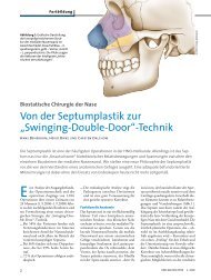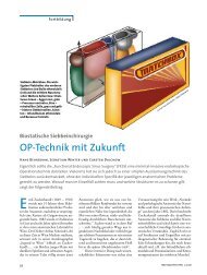
BIOSTATIC ENDOSCOPIC ETHMOID SURGERY A New Approach ...
BIOSTATIC ENDOSCOPIC ETHMOID SURGERY A New Approach ...
BIOSTATIC ENDOSCOPIC ETHMOID SURGERY A New Approach ...

You also want an ePaper? Increase the reach of your titles
YUMPU automatically turns print PDFs into web optimized ePapers that Google loves.
Biostatic Endoscopic Ethmoid Surgery – B.E.E.S. – A <strong>New</strong> <strong>Approach</strong> to<br />
Functional Endoscopic Sinus Surgery for Acute Recurrent Rhinosinusitis<br />
7<br />
In his groundbreaking work on “The Role of the Lateral Nasal Wall in the<br />
Pathogenesis, Diagnosis and Treatment of Recurrent and Chronic Rhino -<br />
sinusitis” (1987), Walter Messerklinger (1920–2001) begins by reviewing the<br />
anatomical discoveries of Zuckerkandl, Hajek, Grünwald, Peter, Killian, and<br />
Flottes on the cellular structures of the lateral nasal wall before addressing<br />
their importance in the pathogenesis of rhinogenic sinusitis. At the International<br />
Conference on Sinus Disease, Terminology, Staging and Therapy held<br />
in Princeton, <strong>New</strong> Jersey, in 1993, Stammberger et al. (1997) presented a<br />
paper titled “Anatomical Terminology and Nomenclature of Paranasal Sinus<br />
Surgery” in which they created a uniform, consistent nomenclature for the<br />
anatomy of the ethmoid bone.<br />
While discoveries on the anatomy of specific cellular structures in the<br />
ethmoid bone gave rise to important pathogenetic considerations, an<br />
endoscopic diagnostic concept, and a subsequent approach to functional<br />
endoscopic sinus surgery (F.E.S.S.), to date there have been no fundament -<br />
al studies on the stabilizing function of the principal ethmoid cells.<br />
Prof. Stammberger (2002) has repeatedly emphasized the importance of an<br />
atraumatic surgical technique, particularly in endoscopic surgery of the<br />
frontal sinuses, noting that “…an aggressive technique with the radical<br />
removal of mucosa from these small ethmoid passageways will quickly lead<br />
to scarring and stenosis. Not infrequently, traumatic surgical manipulations<br />
in this region will promote the development of frontal sinus symptoms that<br />
did not exist prior to surgery…”<br />
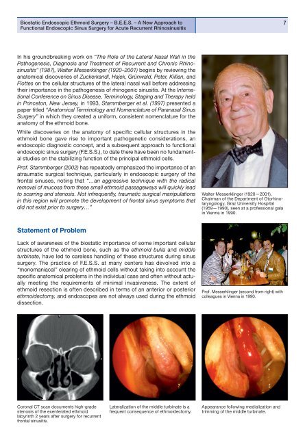
Walter Messerklinger (1920—2001),<br />
Chairman of the Department of Otorhinolaryngology,<br />
Graz University Hospital<br />
(1959—1990), seen at a professional gala<br />
in Vienna in 1990.<br />
Statement of Problem<br />
Lack of awareness of the biostatic importance of some important cellular<br />
structures of the ethmoid bone, such as the ethmoid bulla and middle<br />
turbinate, have led to careless handling of these structures during sinus<br />
surgery. The practice of F.E.S.S. at many centers has devolved into a<br />
“monomaniacal” clearing of ethmoid cells without taking into account the<br />
specific anatomical problems in the individual case and often without actually<br />
meeting the requirements of minimal invasiveness. The extent of<br />
ethmoid resection is often described in terms of an anterior or posterior<br />
ethmoidectomy, and endoscopes are not always used during the ethmoid<br />
dissection.<br />
Prof. Messerklinger (second from right) with<br />
colleagues in Vienna in 1990.<br />
Coronal CT scan documents high-grade<br />
stenosis of the exenterated ethmoid<br />
labyrinth 2 years after surgery for recurrent<br />
frontal sinusitis.<br />
Lateralization of the middle turbinate is a<br />
frequent consequence of ethmoidectomy.<br />
Appearance following medialization and<br />
trimming of the middle turbinate.